. 2018 Jun;3(6):718-731.
doi: 10.1038/s41564-018-0164-0. Epub 2018 May 24.

Antibiotic-resistant bacteria show widespread collateral sensitivity to antimicrobial peptides

Affiliations

Antibiotic-resistant bacteria show widespread collateral sensitivity to antimicrobial peptides

Viktória Lázár et al. Nat Microbiol. 2018 Jun.

Abstract

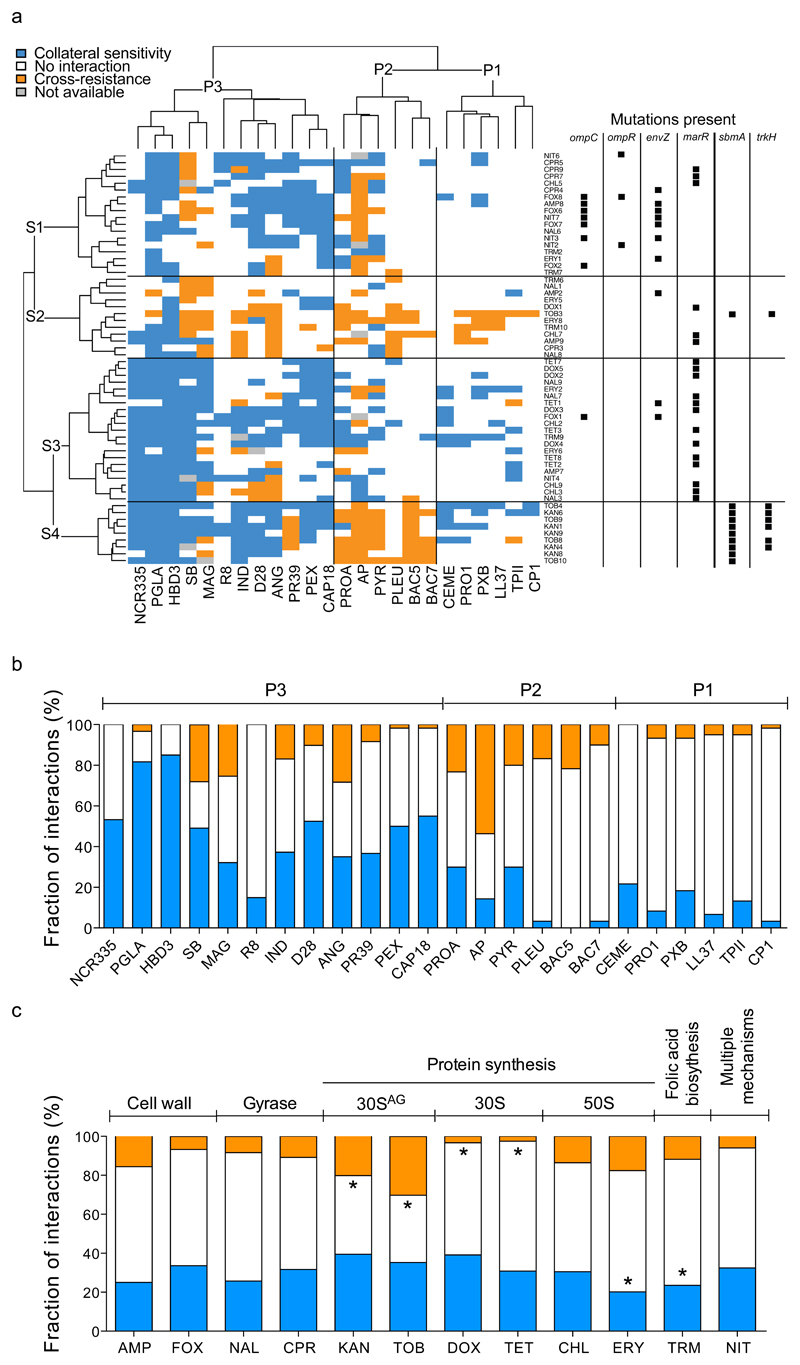
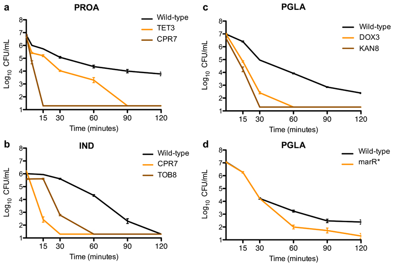
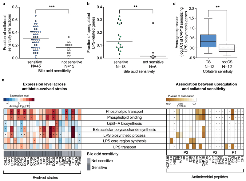
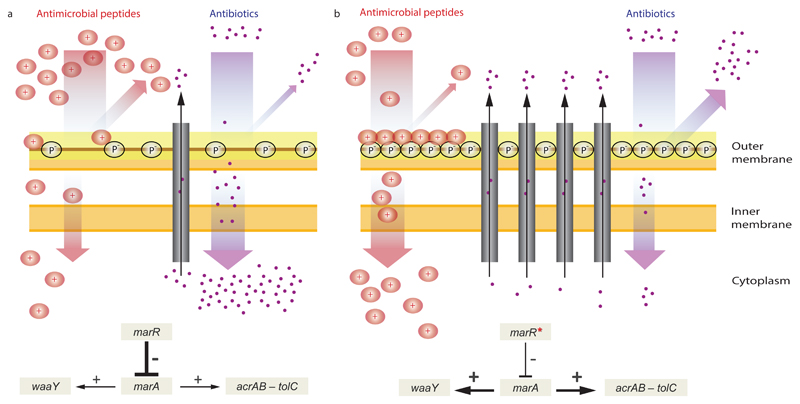
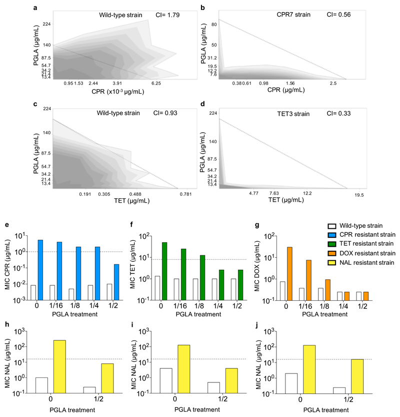
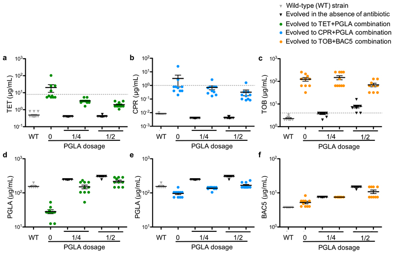
Antimicrobial peptides are promising alternative antimicrobial agents. However, little is known about whether resistance to small-molecule antibiotics leads to cross-resistance (decreased sensitivity) or collateral sensitivity (increased sensitivity) to antimicrobial peptides. We systematically addressed this question by studying the susceptibilities of a comprehensive set of 60 antibiotic-resistant Escherichia coli strains towards 24 antimicrobial peptides. Strikingly, antibiotic-resistant bacteria show a high frequency of collateral sensitivity to antimicrobial peptides, whereas cross-resistance is relatively rare. We identify clinically relevant multidrug-resistance mutations that increase bacterial sensitivity to antimicrobial peptides. Collateral sensitivity in multidrug-resistant bacteria arises partly through regulatory changes shaping the lipopolysaccharide composition of the bacterial outer membrane. These advances allow the identification of antimicrobial peptide-antibiotic combinations that enhance antibiotic activity against multidrug-resistant bacteria and slow down de novo evolution of resistance. In particular, when co-administered as an adjuvant, the antimicrobial peptide glycine-leucine-amide caused up to 30-fold decrease in the antibiotic resistance level of resistant bacteria. Our work provides guidelines for the development of efficient peptide-based therapies of antibiotic-resistant infections.


Conflict of interest statement

Competing interests

The authors declare no competing interests. BB and IN had consulting positions at SeqOmics Biotechnology Ltd. at the time the study was conceived. SeqOmics Biotechnology Ltd. was not directly involved in the design and execution of the experiments or in the writing of the manuscript. This does not alter the author’s adherence to all the Nature policies on sharing data and materials.

Figures

Figure 1
Figure 1. Susceptibility profiles of 60 laboratory-evolved antibiotic-resistant E. coli strains
a, Hierarchical clustering of 60 antibiotic-resistant strains (rows) and a set of 24 antimicrobial peptides (columns) based on the cross-resistance and collateral sensitivity interactions between them (for abbreviations of antibiotics and antimicrobial peptides see Supplementary Tables 1 and 2, respectively). Hierarchical clustering was performed separately on rows and columns, using Ward’s method. Black squares on the right side of each antibiotic-resistant strain denote previously identified mutations in antibiotic-resistance genes, that were significantly enriched in one or more strain clusters (p<0.05, two-sided Fisher’s exact test). S1 strains were enriched in envZ, ompR and ompC mutations, whereas S3 strains were enriched in marR mutations (P<0.05 for all cases, two-sided Fisher’s exact test). While S3 strains show widespread collateral sensitivity to antimicrobial peptides, especially to P1 and P3 peptides, aminoglycoside-resistant strains (S4) show extensive cross-resistance to proline-rich peptides (P2) (p<0.0001, two-sided Fisher’s exact test). b, Efficiency of antimicrobial peptides against antibiotic-resistant bacteria expressed as the percentage of strains showing collateral sensitivity (blue), no interaction (white), or cross-resistance (orange) against each peptide. A total of 56 to 60 strains per antimicrobial peptide was employed for the analysis. c, Relative frequency of collateral sensitivity and cross-resistance interactions towards antimicrobial peptides upon adaptation to single antibiotics (n=5 strains per antibiotic). The frequency of interactions for each peptide was calculated by counting the number of cross-resistance (orange), no interaction (white) and collateral sensitivity (blue) interactions displayed by all strains adapted to a given antibiotic. Antibiotic modes-of-action are shown on the top of the figure. 30SAG refers to aminoglycosides. Asterisks (*) mark significant deviations from hypergeometric distribution models calculated from all the interactions of all peptide-strain combinations separately for cross-resistance and collateral sensitivity, respectively. Strains adapted to DOX and TET were depleted, whereas strains adapted to TOB and KAN were enriched in cross-resistance interactions towards peptides (p=0.005, p<0.008, p=0.003 and p<0.001, respectively, two-sided Fisher's combined probability test). Furthermore, strains adapted to ERY and TRM were significantly depleted in collateral sensitivity interactions (p=0.003 and p<0.001 respectively, two-sided Fisher's combined probability test).
Figure 2
Figure 2. Survival of collateral-sensitive antibiotic-resistant strains under lethal antimicrobial peptide stress
a-c, The wild-type and antibiotic-resistant strains were exposed to high concentrations of antimicrobial peptides: (a) the tetracycline-resistant TET3 and the ciprofloxacin-resistant CPR7 strains were exposed to 15-fold (3000 μg/mL) MIC of protamine (PROA), (b) the ciprofloxacin-resistant CPR7 and the tobramycin-resistant TOB8 were exposed to 10-fold MIC (125 μg/mL) of indolicidin (IND), and (c) the doxycycline-resistant DOX3 and the kanamycin-resistant KAN8 were exposed to 15-fold MIC (1500 μg/mL) of PGLA. All antibiotic-resistant strains exhibited collateral sensitivity towards the applied peptide. d, A strain containing a single point mutation in marR (marR*) was also exposed to 15-fold MIC of PGLA. This strain exhibits resistance to multiple antibiotics and collateral sensitivity to many of the peptides tested, including PGLA (Table 1). Cells were incubated with the particular peptide for 120 minutes. Samples were taken at defined time points and plated in LB agar plates. Percentage of survival was calculated by counting the colony forming units (CFU). Each data point shows the mean ± standard error of the mean of 3 biological replicates.
Figure 3
Figure 3. Altered membrane composition in antibiotic-resistant bacteria contributes to increased sensitivity to antimicrobial peptides
a, Antibiotic-resistant strains sensitive towards a membrane-damaging agent (bile acid) show especially large numbers of collateral sensitivity interactions to antimicrobial peptides. For antibiotic abbreviations see Supplementary Table 1. Strains sensitive to bile acid show significantly more collateral sensitivity interactions to peptides than those showing wild-type bile acid sensitivity (not sensitive group) (p<10-3, two-sided generalized linear mixed model with binomial response distributions, see Materials and Methods section). Relative frequency was calculated by dividing the number of the collateral sensitivity interactions by the number of all tested peptides (N=24). b, Resistant strains with sensitivity to bile acid have significantly more LPS-related synthesis genes being transcriptionally upregulated than those showing wild-type bile acid sensitivity (p<10-3, two-sided GLMM with binomial response distributions). Relative frequency was calculated by dividing the number of the upregulated genes by the number of all LPS-related genes (N=100). c, Left heatmap shows the average log2(fold change) of genes related to selected membrane-associated GO processes. Many antibiotic-resistant strains are enriched in significantly up- or downregulated genes (fold change>2 or <0.5, FDR-corrected p-value<0.05, two-sided Fisher’s exact test), associated with membrane-related functions. Significant enrichments (p<0.05) are marked with an asterisk (*). Strains sensitive to a given peptide show significant upregulations in specific GO groups compared to non-sensitive strains (right heatmap, two-sided Student’s t–test; for further details, see Supplementary Figure 5). Peptides with either too few or too many collateral sensitivity interactions (n<4 or n>21, respectively) were excluded from the statistical analysis based on sample size calculation with alpha=0.1, power=0.8, delta=2, SD=1, and are indicated with a minus sign (-). Sample size used in this analysis is provided in Supplementary Table 3 d, Upregulation of LPS-related genes sensitize to CAP18. CAP18-sensitive antibiotic-resistant strains (CS, n=12) have significantly higher expression levels of CAP18 sensitizing genes within the ‘LPS biosynthetic process’ GO category than non-sensitive strains (not CS, n=12) (p=0.008, two-sided Wilcoxon rank-sum test). Boxplots show the median, first and third quartiles, with whiskers showing the 5th and 95th percentile. Significant difference (p<0.01) is marked with asterisks (**).
Figure 4
Figure 4. A putative mechanism underlying collateral sensitivity of antibiotic-resistant bacteria to cationic antimicrobial peptides
a, The wild-type MarR represses marA, which leads to the reduced expression of the AcrAB-TolC efflux pump and hence the cytosolic accumulation of antibiotics. b, Upon a canonical resistance mutation in the marR gene (marR*), repression of marA is substantially decreased leading to the upregulation of the AcrAB-TolC efflux pump, and hence an increased resistance to multiple antibiotics. On the other hand, MarA simultaneously promote the upregulation of WaaY, a kinase responsible for phosphorylation of the inner core of lipopolysaccharides (LPS), which increases the net negative surface charge of the bacterial outer membrane. This in turn enhances susceptibility to membrane-interacting cationic antimicrobial peptides (CAP+). In addition, phosphorylation of the LPS core may also enhance the permeability barrier of the outer membrane by cross-linking of neighboring LPS molecules. Such changes promote both 1) a decreased uptake and increased efflux of antibiotics, thereby contributing to antibiotic resistance, and 2) a higher affinity of the positively charged antimicrobial peptides to the negative phosphoryl groups of the LPS core, leading to enhanced sensitivity to antimicrobial peptides.
Figure 5
Figure 5. Interaction of PGLA and antibiotics, when applied in combination
Antibiotic-PGLA interactions were determined in E. coli K12 BW25133 wild-type and corresponding antibiotic-resistant strains. For antibiotic abbreviations see Supplementary Table 1. Figures a-d show the combination effect of PGLA and ciprofloxacin (CPR) or tetracycline (TET) on the wild-type strain (a and c), ciprofloxacin-resistant strain (CPR7) (b) and tetracycline-resistant strain (TET3) (d). While the combination shows strong antagonism (a) or no interaction (c) in the wild-type strain, the interaction shifted to strong synergism in the resistant strain (b and d). Dashed line represents no interaction calculated based on the Loewe additivity model (see Materials and Methods). Growth rate is represented in the combination space by the shade of the grey color with darker shades denoting higher growth rates Figures e-j show the effect of subinhibitory concentrations of PGLA on antibiotic activity. Ciprofloxacin-resistant CPR7 (e), tetracycline-resistant TET3 (f) and doxycycline-resistant DOX3 (g) strains, derived from E. coli K12 BW25133 were treated with subinhibitory concentrations of PGLA, while measuring the MIC for the given antibiotic to which they were adapted. The concentrations of PGLA used were 1/16, 1/8, 1/4 and 1/2 of its MIC against the wild-type strain. The minimal inhibitory concentration of nalidixic acid (NAL) was measured in E. coli clinical isolates 0370 (h), 3539 (i) and CFT073 (j), and their corresponding nalidixic acid-resistant strains in the presence of 1/2 of the MIC for PGLA. None of the PGLA concentrations, when applied alone, affected the growth of the wild-type or the resistant strains (the only exception being the 40% growth rate reduction of the tetracycline (TET) resistant strain in response to ½ MIC PGLA). Dashed lines represent the clinical breakpoints for the antibiotics in E. coli (not available for doxycycline (DOX)). Data in this figure is representative of at least 2 biological replicates.
Figure 6
Figure 6. Minimum inhibitory concentrations (MICs) of laboratory-evolved lines adapted to antibiotics in the absence and the presence of subinhibitory dosage of antimicrobial peptides
MIC was measured following a laboratory evolution of the wild-type E. coli strain to tetracycline (TET, green), ciprofloxacin (CPR, blue) and tobramycin (TOB, orange) in the absence or in the presence of ¼ or ½ of the MIC of the antimicrobial peptides PGLA (a, b, d, e) or BAC5 (c, f) against the wild-type strain. MICs of the wild-type and both PGLA and BAC5 evolved lines (in the absence of antibiotic) are represented by grey and white colored bars, respectively. Each data point represents the MIC value of one of each ten parallel-evolved lines. Error bars represent the mean ± standard error of the mean for each experimental condition. Dashed lines represent clinical breakpoints for TET, CPR or TOB in E. coli. Both the CPR-PGLA and the TET-PGLA combinations, which are representatives of collateral sensitive interactions (Figure 1), significantly slowed down the evolution of resistance towards the given antibiotic when administered together. Reassuringly, the control combination (BAC5-TOB), representing a cross-resistance interaction, did not reduce the rate of TOB resistance evolution (P=0.0834, 1-way ANOVA).

References

    1. Imamovic L, Sommer MO. Use of collateral sensitivity networks to design drug cycling protocols that avoid resistance development. Sci Transl Med. 2013;5:204ra132. - PubMed
    1. Lázár V, et al. Genome-wide analysis captures the determinants of the antibiotic cross-resistance interaction network. Nat Commun. 2014;5:4352. - PMC - PubMed
    1. Lázár V, et al. Bacterial evolution of antibiotic hypersensitivity. Molecular Systems Biology. 2013;9:700–700. - PMC - PubMed
    1. Toprak E, et al. Evolutionary paths to antibiotic resistance under dynamically sustained drug selection. Nat Genet. 2012;44:101–105. - PMC - PubMed
    1. Palmer AC, Kishony R. Understanding, predicting and manipulating the genotypic evolution of antibiotic resistance. Nat Rev Genet. 2013;14:243–248. - PMC - PubMed

Publication types

MeSH terms

Substances