

Case Reports
doi: 10.1155/2018/4192657.
eCollection 2018.
Mistaken Diabetic Ulcers: A Case of Bilateral Foot Verrucous Carcinoma
Affiliations
- PMID: 29796321
- PMCID: PMC5896404
- DOI: 10.1155/2018/4192657
Item in Clipboard
Case Reports
Mistaken Diabetic Ulcers: A Case of Bilateral Foot Verrucous Carcinoma
Case Rep Dermatol Med.
.
Erratum in
-
Corrigendum to "Mistaken Diabetic Ulcers: A Case of Bilateral Foot Verrucous Carcinoma".Case Rep Dermatol Med. 2018 Apr 15;2018:6405129. doi: 10.1155/2018/6405129. eCollection 2018. Case Rep Dermatol Med. 2018. PMID: 29849220 Free PMC article.
Abstract
Verrucous carcinoma (VC) is a rare, low-grade, and well-differentiated variant of squamous cell carcinoma. These tumors are slow-growing and exophytic and have a negligible incidence of metastasis. Treatment is complete surgical resection, ideally by Mohs micrographic surgery, to ensure adequate clear margins. Cutaneous VC predominantly occurs on the plantar surface of the foot and rarely occurs in multiple sites. This case study describes the fourth reported occurrence of bilateral VC of the feet in a woman with chronic diabetic foot ulcers. The case provides further support for persistent wounds contributing to the development of this lesion and describes their role in the characteristic delay in diagnosis of VC.
Figures
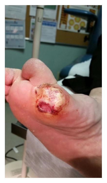
Exophytic lesion at the base of the first MTP joint of the right foot within a chronic diabetic ulcer. Pathology confirmed this lesion to be verrucous carcinoma.
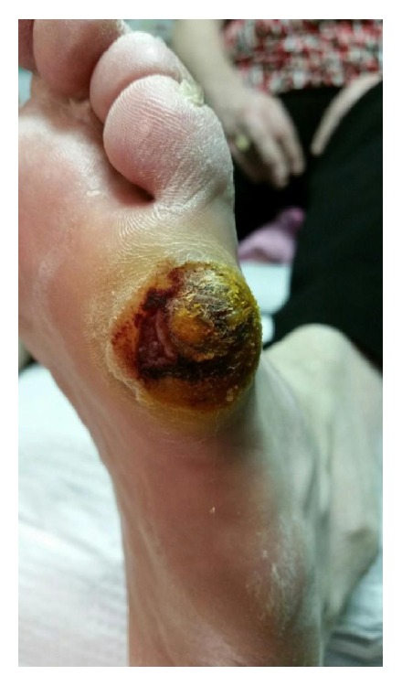
Painful exophytic lesion on the plantar surface of the left 5th MTP joint, which, after multiple biopsies, was confirmed by pathology to be verrucous carcinoma.
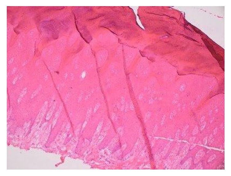
Representative medium power image of the verrucous carcinoma demonstrates prominent surface hyperkeratosis and marked epidermal acanthosis, with a combined exophytic and endophytic growth pattern. Cytologic atypia is only mild to moderate. Rather than the infiltrative pattern typically seen at the deep edge of conventional invasive squamous cell carcinoma, verrucous carcinoma demonstrates insidious pushing-type invasion by expansile and irregular rete pegs (4x, H&E stain).
References
-
- Ackerman L. V. Verrucous carcinoma of the oral cavity. Surgery. 1948;23(4):670–678. - PubMed
Publication types
LinkOut - more resources
Full Text Sources
Other Literature Sources
