Review
. 2018 May 12:5:11.
doi: 10.1186/s40662-018-0104-3. eCollection 2018.

Emerging Applications of Optical Coherence Tomography Angiography (OCTA) in neurological research

Affiliations
Review

Emerging Applications of Optical Coherence Tomography Angiography (OCTA) in neurological research

Liang Wang et al. Eye Vis (Lond). .

Abstract

Purpose: To review the clinical and research value of optical coherence tomography angiography (OCTA) in the field of neurology.

Methods: Current literature involving OCTA were reviewed through PubMed using the search terms "optical coherence tomography angiography", with "multiple sclerosis", "Alzheimer's disease", "optic neuropathy", or other closely-related terms.

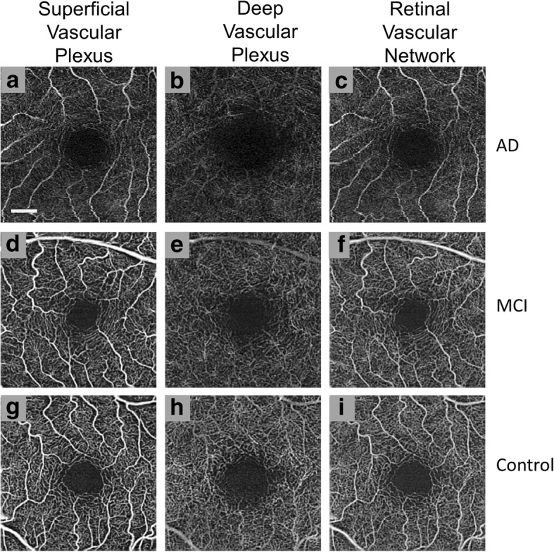
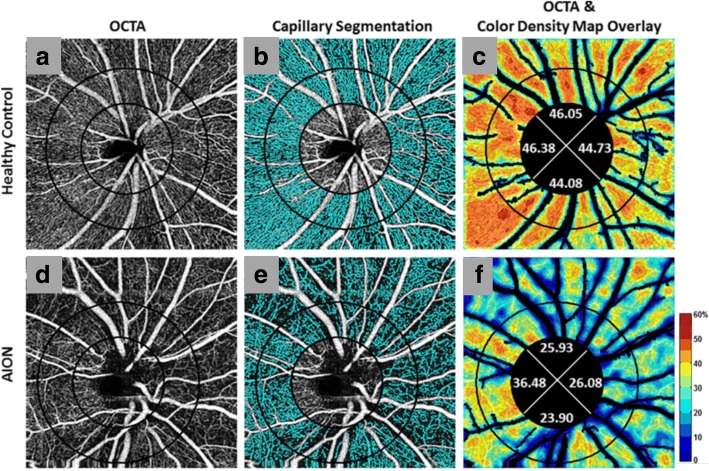
Results: OCTA has been applied in research to advance our understanding of the pathobiology of neurological disorders. OCTA-derived blood flow and vessel density measures are altered in multiple sclerosis (MS), Alzheimer's disease (AD), and various optic neuropathies (ON) in varying regions of the posterior segment vasculature of the eye. These emerging research findings support the occurrence of retinal vascular alterations across a host of neurological disorders and raise the possibility that vasculopathy can be clinically relevant since it contributes to the pathobiology of several neurological disorders.

Conclusion: OCTA may be beneficial for neurological research. Additional investigations using OCTA in neurological disorders will help to further validate its clinical and research utilities in terms of characterizing the role of vasculopathy in neurological disorders.

Keywords: Alzheimer’s disease; Multiple sclerosis; Neurology; Optic neuropathy; Optical coherence tomography angiography.


Conflict of interest statement

Not applicable.The authors declare that they have no competing interests.

Figures

Fig. 1
Fig. 1
Wide field montage of healthy retinal microvasculature. Using Cirrus™ optical coherence tomography angiograph with optical microangiography, 42 scans were taken and combined to create a 12 × 16 mm2 image (panel a). The images are portrayed in different colors representing varying retinal depth. Red depicts the retinal nerve fiber layer, green the ganglion cell and inner plexiform layers, and blue the inner nuclear and outer plexiform layers. White boxes outline (b) optic nerve head, (c) fovea, and (d) temporal region. (Reprinted from Zhang et al. Wide-field imaging of retinal vasculature using optical coherence tomography-based microangiography provided by motion tracking. JBO, 20, 066009 (2015), DOI: 10.1117/1.JBO.20.6.066008) [43]
Fig. 2
Fig. 2
General scanning protocol for optical coherence tomography angiography (OCTA). (a) Repeated B-scans are taken on the “x” fast axis at each of the “y” slow scan axis points to detect relative flow signal. (b) Top view of the same general scan pattern as (a) with repeated B-scans taken on the fast axis “x” along each “y” location of the slow axis. Sample scans were obtained with Angiovue™ OCTA (a, b)
Fig. 3
Fig. 3
Anatomy of the posterior segment vasculature. Using Angiovue™ optical coherence tomography angiography (OCTA), the vascular plexus from the internal limiting membrane to the Bruch’s membrane of a healthy subject was non-invasively visualized in 3 × 3 mm (a), 6 × 6 mm (b), and 8 × 8 mm (c) angiograms. Intravenous Fluorescein Angiography (IVFA) cropped to 8 × 8 mm (d) shows less microvasculature detail than that of OCTA angiogram (a-c). The inner retinal vascular plexus is further separated into the superficial inner vascular plexus (e), which supplies the retinal nerve fiber layer and ganglion cell layer and the deep inner vascular plexus (f), which supplies the inner plexiform layer, inner nuclear layer, and outer plexiform layer [76]. No distinct vasculature can be detected in the outer retina (g) and choriocapillaris (h) using 3 × 3 mm OCTA. (Reprinted and modified from de Carlo et al. A review of optical coherence tomography angiography (OCTA), Int J Retina Vitreous, 1,5 (2015), DOI: 10.1186/s40942-015-0005-8) [76]
Fig. 4
Fig. 4
Optical coherence tomography angiography (OCTA) of the optic nerve head (ONH) in a representative Multiple Sclerosis (MS) patient. As determined with split-spectrum amplitude decorrelation angiography (SSADA), images (N = nasal, T = temporal) show apparent qualitative reduction of the ONH microvascular density in the peripapillary area (between circles) predominantly in the temporal region in both MS eyes with a history of ON (MSON) (a) and MS eyes without a history of ON (MSNON) (b) eyes in comparison to a healthy control example (c). Bar = 0.5 mm
Fig. 5
Fig. 5
Optical coherence tomography angiography (OCTA) of the parafoveal region in Mild Cognitive Impairment (MCI) and Alzheimer’s Disease (AD). As determined using Cirrus ™, retinal microvasculature for patients with AD (a, b, and c), MCI (d, e, and f), and healthy controls (g, h, and i) are shown. For the large vessels, no significant differences in density were observed, but some degree of increased tortuosity was seen in the superficial vascular plexus (SVP) (a) in comparison to normal controls (g). The microvasculature of the deep vascular plexus (DVP) (b) had a significant decrease in density with a visually larger foveal avascular zone when compared with normal controls (h) and MCI patients (e). The overall retinal vascular network contains both the SVP and DVP (c, f, and i). Bar = 0.5 mm [31]. (Images provided courtesy of Dr. Hong Jiang, MD, PhD of Bascom Palmer Eye Institute at the University of Miami)
Fig. 6
Fig. 6
Optical coherence tomography angiography (OCTA) with peripapillary capillary density maps in Non-arteritic Anterior Ischemic Optic Neuropathy (NAION). Images were derived using Angiovue™ with SSADA and they depict optic disc-centered angiograms (first column) (a, d) with perfused microvessels marked in cyan (second column). The major vessels were not included in the capillary segmentation in panels b and e. Original OCTA overlaid with corresponding color-coded density map (legend: higher density % up to 60% = redder) and capillary density percentage for four quadrants (third column) (c, f). Patients with NAION had a smaller perfusion region when compared with normal controls as seen with the visible loss of microvessels (cyan) in NAION (e) in comparison to healthy controls (b) and the decreased capillary density percentages (less red overall) for all quadrants in NAION (f) in comparison to healthy controls (c). (Reprinted from Fard et al. Pattern of peripapillary capillary density loss in ischemic optic neuropathy compared to that in primary open-angle glaucoma. PLoS One, 13:e0189237 (2018), DOI: 10.1371/journal.pone.0189237) [33]

References

    1. Carpineto P, Mastropasqua R, Marchini G, Toto L, Di Nicola M, Di Antonio L. Reproducibility and repeatability of foveal avascular zone measurements in healthy subjects by optical coherence tomography angiography. Br J Ophthalmol. 2016;100(5):671–676. doi: 10.1136/bjophthalmol-2015-307330. - DOI - PubMed
    1. Huang Y, Zhang Q, Thorell MR, An L, Durbin MK, Laron M, et al. Swept-source OCT angiography of the retinal vasculature using intensity differentiation-based optical microangiography algorithms. Ophthalmic Surg Lasers Imaging Retina. 2014;45(5):382–389. doi: 10.3928/23258160-20140909-08. - DOI - PMC - PubMed
    1. Nehemy MB, Brocchi DN, Veloso CE. Optical Coherence Tomography Angiography Imaging of Quiescent Choroidal Neovascularization in Age-Related Macular Degeneration. Ophthalmic Surg Lasers Imaging Retina. 2015;46(10):1056–7. - PubMed
    1. Rispoli M, Savastano MC, Lumbroso B. Capillary network anomalies in branch retinal vein occlusion on optical coherence tomogrpahy angiography. Retina. 2015;35(11):2332–2338. doi: 10.1097/IAE.0000000000000845. - DOI - PMC - PubMed
    1. de Carlo TE, Chin AT, Joseph T, Baumal CR, Witkin AJ, Duker JS, et al. Distinguishing Diabetic Macular Edema From Capillary Nonperfusion Using Optical Coherence Tomography Angiography. Ophthalmic Surg Lasers Imaging Retina. 2016;47(2):108–14. - PubMed

LinkOut - more resources