. 2018 Oct 9;20(11):1494-1504.
doi: 10.1093/neuonc/noy082.

Oncolytic virotherapy in glioblastoma patients induces a tumor macrophage phenotypic shift leading to an altered glioblastoma microenvironment

Affiliations

Oncolytic virotherapy in glioblastoma patients induces a tumor macrophage phenotypic shift leading to an altered glioblastoma microenvironment

Wouter B L van den Bossche et al. Neuro Oncol. .

Abstract

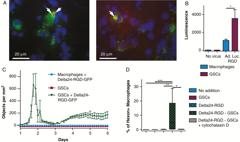
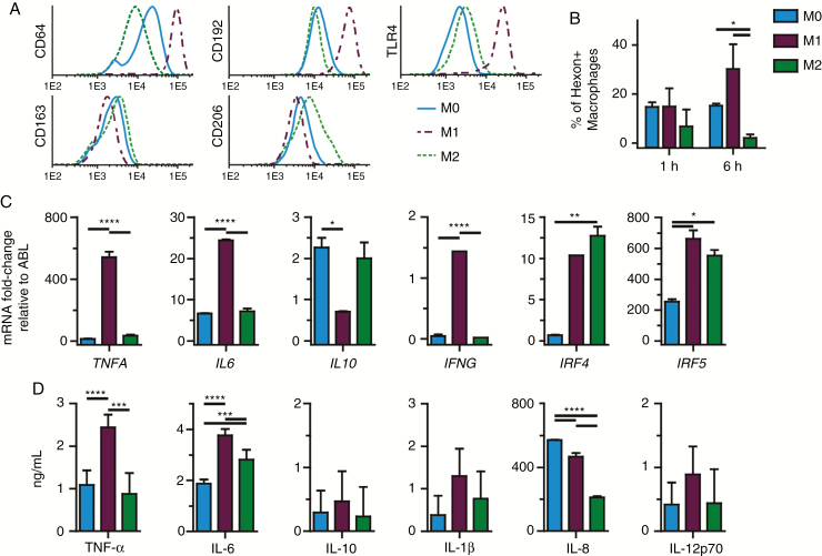
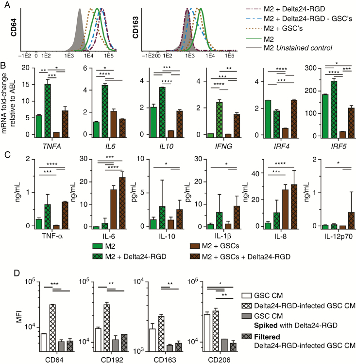
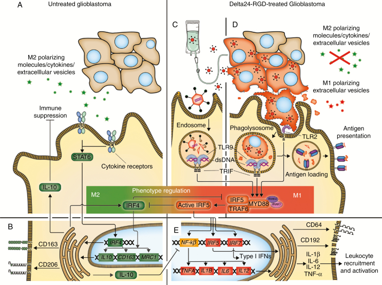
Background: Immunosuppressive protumoral M2 macrophages are important in pathogenesis, progression, and therapy resistance in glioblastoma (GBM) and provide a target for therapy. Recently oncolytic virotherapy in murine models was shown to change these M2 macrophages toward the pro-inflammatory and antitumoral M1 phenotype. Here we study the effects of the oncolytic virotherapy Delta24-RGD in humans, using both in vitro models and patient material.

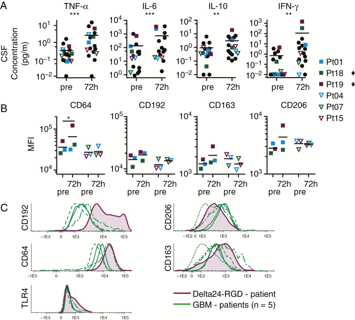
Methods: Human monocyte-derived macrophages were co-cultured with Delta24-RGD-infected primary glioma stem-like cells (GSCs) and were analyzed for their immunophenotype, cytokine expression, and secretion profiles. Cerebrospinal fluid (CSF) from 18 Delta24-RGD-treated patients was analyzed for inflammatory cytokine levels, and the effects of these CSF samples on macrophage phenotype in vitro were determined. In addition, tumor macrophages in resected material from a Delta24-RGD-treated GBM patient were compared with 5 control GBM patient samples by flow cytometry.

Results: Human monocyte-derived M2 macrophages co-cultured with Delta24-RGD-infected GSCs shifted toward an M1-immunophenotype, coinciding with pro-inflammatory gene expression and cytokine production. This phenotypic switch was induced by the concerted effects of a change in tumor-produced soluble factors and the presence of viral particles. CSF samples from Delta24-RGD-treated GBM patients revealed cytokine levels indicative of a pro-inflammatory microenvironment. Furthermore, tumoral macrophages in a Delta24-RGD-treated patient showed significantly greater M1 characteristics than in control GBM tissue.

Conclusion: Together these in vitro and patient studies demonstrate that local Delta24-RGD therapy may provide a therapeutic tool to promote a prolonged shift in the protumoral M2 macrophages toward M1 in human GBM, inducing a pro-inflammatory and potentially tumor-detrimental microenvironment.


Figures

Fig. 1
Fig. 1
Tumor macrophages acquire hexon accumulations by phagocytosis of virus-infected tumor cells. (A) Delta24-RGD–treated patient tumor-resection material, stained for CD68 (green) and hexon (red). White bar indicates 20 μM. (B) Mean ± SEM luminescence of human macrophages and GSCs 24 h after mock or Ad.Luc.RGD infection. (C) GFP-positive objects per mm2 in 5-day culture of Delta24-RGD-GFP–infected macrophages and GSCs. (D) Mean ± SEM percentage of hexon-positive macrophages after 24 h of culture with GSCs, Delta24-RGD, or Delta24-RGD–infected GSCs with or without cytochalasin. (B–D) Data represent at least 2 independent experiments. *P < 0.05; **P < 0.01; ***P < 0.005 (one-way ANOVA, Tukey post-hoc test).
Fig. 2
Fig. 2
Immunophenotyping, gene expression and cytokine production demonstrate in vitro macrophage polarization. (A) CD64, CD192, TLR4, CD163, and CD206 expression on cultured human macrophages after 24 h either unpolarized (M0) or polarized with IFN-γ or IL-4. (B) Mean ± SEM of hexon-positive macrophages per M0, M1, and M2 phenotype, indicative of phagocytosis efficiency at 1 h and 6 h (Student’s t-test). (C) Mean ± SEM of mRNA fold-change relative to ABL of TNFA, IL6, IL10, IFNG, IRF4, and IRF5 in M0 and M1 or M2 macrophages, 24 h after polarization. (D) Mean ± SEM of cytokine concentration of TNF-α, IL-6, IL-10, IL-1β, IL-8, and IL-12p70 in culture supernatants of M0, M1, and M2 macrophages. (B–D) Data represent at least 2 independent experiments. *P < 0.05; **P < 0.01; ***P < 0.005; ****P < 0.001 (one-way ANOVA, Tukey post-hoc test).
Fig. 3
Fig. 3
Delta24-RGD in vitro changes human macrophages’ genotype, immunophenotype, and cytokine profile. (A) CD64 and CD163 expression on M2 macrophages after 72 h of culturing with Delta24-RGD, GSCs, or Delta24-RGD–infected GSCs. (B) Mean ± SEM of mRNA fold-change relative to ABL of TNFA, IL6, IL10, IFNG, IRF4, and IRF5 in M2 macrophages after 72 h of culturing with Delta24-RGD, GSCs, or Delta24-RGD–infected GSCs. (C) Mean ± SEM cytokine concentration of TNF-α, IL-6, IL-10, IL-1β, IL-12p70 in supernatant of M2 macrophages co-cultured for 72 h with Delta24-RGD, GSCs, or Delta24-RGD–infected GSCs. (D) Mean fluorescence intensity (MFI) of CD64, CD192, CD163, CD206 on M0 macrophages cultured for 72 h with conditioned medium (CM) from Delta24-RGD–infected or control GSCs either with Delta24-RGD or filtered with a 100 kD filter. (B–D) Data represent at least 2 independent experiments. *P < 0.05; **P < 0.01; ***P < 0.005; ****P < 0.001 (one-way ANOVA, Tukey post-hoc).
Fig. 4
Fig. 4
CSF cytokine profile and macrophage phenotype shift to pro-inflammatory under influence of Delta24-RGD treatment. (A) CSF cytokine concentration of TNF-α, IL-6, IL-10, and IFN-γ in 20 Delta24-RGD–treated patients (18 individual patients, 15 pairs) before and 72 h after viral infusion. Each dot represents one patient. Squares represent patients with increased cytokine concentrations. Open triangles represent patients with little cytokine changes upon treatment. Black line is the mean, **P < 0.01; ***P < 0.005 (Wilcoxon signed rank test). (B) CD64, CD163, CD192, and CD206 expression on macrophages cultured for 72 h with patient’s CSF both pretreatment and 72 h after start of treatment. Black line is the mean, each symbol represents mean of 2 separate experiments. The symbol ♦ marks patients with statistical significant changes, *P < 0.05 (one-way ANOVA, Tukey post-hoc). (C) CD206, CD163, CD192, CD64, and TLR4 expression on GBM macrophages, in untreated (N = 5) and Delta24-RGD–treated (N = 1) patients, *P < 0.05 (Student’s t-test).
Fig. 5
Fig. 5
Macrophage phenotype regulation in Delta24-RGD therapy of glioblastoma. (A) Glioblastoma cells secrete M2 polarizing molecules, cytokines, and extracellular vesicles that primarily affect the macrophage IRF4-STAT6. (B) This induces M2-related gene transcription and M2 membrane marker upregulation. Furthermore IL-10, which suppresses leukocytes and the M1-associated NF-κβ pathway, and the MyD88–IRF5 complex inhibitor IRF4 are produced. (C) Delta24-RGD administration by direct infusion or released by infected tumor cells (as particles or in extracellular vesicles) leads to viral double-stranded DNA detection by TLR9 in macrophage endosomes, activating IRF5 via the MyD88–IRF5 complex. (D) Delta24-RGD infection of tumor cells inhibits M2 polarizing molecule production, allowing M1 polarization of macrophages and promoting tumor cell phagocytosis. Tumor cell digestion in the phagolysosome leads to virus detection by TLR2 and TLR9, activating IRF5 via the MyD88–IRF5 complex. Furthermore, digested infected tumor cells are loaded onto human leukocyte antigen class II for antigen presentation. (E) Active IRF5 inhibits IRF4 and the subsequent M2-related pathways and promotes NF-κβ, IRF5, and IRF7 transcription, leading to CD64 and CD192 upregulation, type I IFN production, and M1-associated gene transcription, together inducing inflammation, leukocyte recruitment, and activation.

Similar articles

Cited by

References

    1. Hughes R, Qian BZ, Rowan C, et al. . Perivascular M2 macrophages stimulate tumor relapse after chemotherapy. Cancer Res. 2015;75(17):3479–3491. - PMC - PubMed
    1. Du R, Lu KV, Petritsch C, et al. . HIF1alpha induces the recruitment of bone marrow-derived vascular modulatory cells to regulate tumor angiogenesis and invasion. Cancer Cell. 2008;13(3):206–220. - PMC - PubMed
    1. Edin S, Wikberg ML, Dahlin AM, et al. . The distribution of macrophages with a M1 or M2 phenotype in relation to prognosis and the molecular characteristics of colorectal cancer. PLoS One. 2012;7(10):e47045. - PMC - PubMed
    1. Movahedi K, Laoui D, Gysemans C, et al. . Different tumor microenvironments contain functionally distinct subsets of macrophages derived from Ly6C(high) monocytes. Cancer Res. 2010;70(14):5728–5739. - PubMed
    1. Poli A, Wang J, Domingues O, et al. . Targeting glioblastoma with NK cells and mAb against NG2/CSPG4 prolongs animal survival. Oncotarget. 2013;4(9):1527–1546. - PMC - PubMed

Publication types