. 2018 Sep 6;46(15):7924-7937.
doi: 10.1093/nar/gky394.

Stability of local secondary structure determines selectivity of viral RNA chaperones

Affiliations

Stability of local secondary structure determines selectivity of viral RNA chaperones

Jack P K Bravo et al. Nucleic Acids Res. .

Abstract

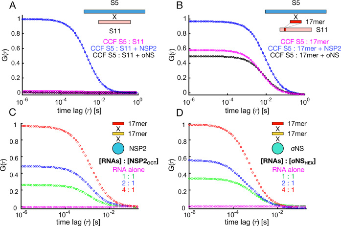
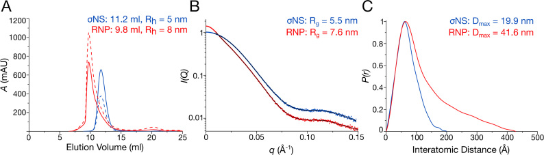
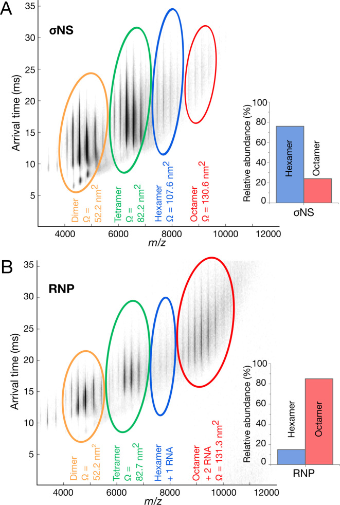
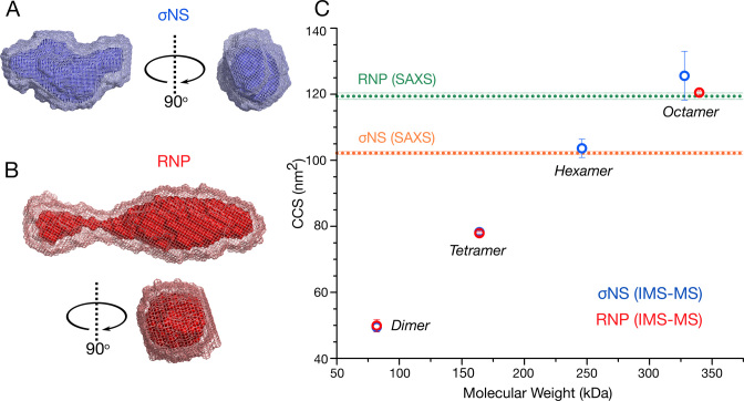
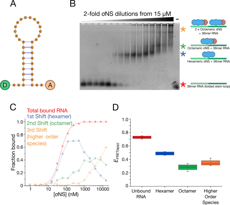
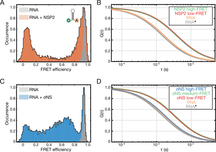
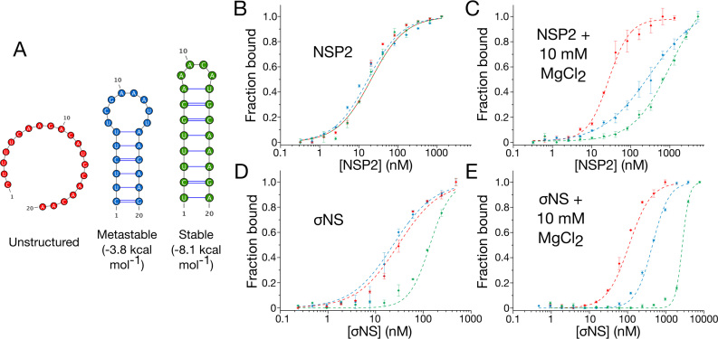
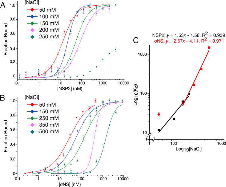
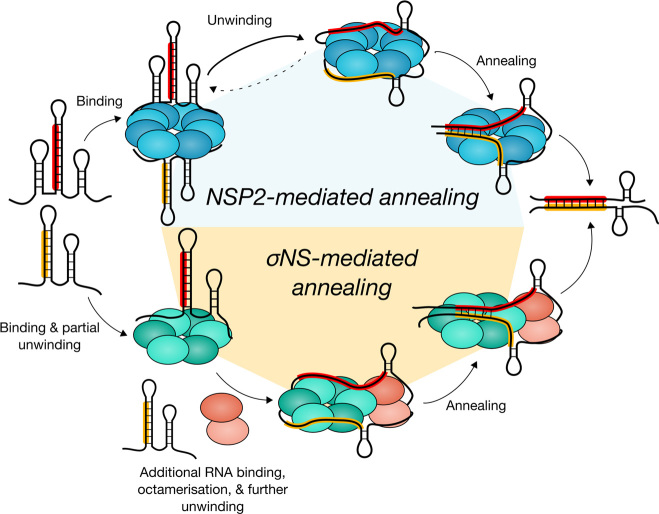
To maintain genome integrity, segmented double-stranded RNA viruses of the Reoviridae family must accurately select and package a complete set of up to a dozen distinct genomic RNAs. It is thought that the high fidelity segmented genome assembly involves multiple sequence-specific RNA-RNA interactions between single-stranded RNA segment precursors. These are mediated by virus-encoded non-structural proteins with RNA chaperone-like activities, such as rotavirus (RV) NSP2 and avian reovirus σNS. Here, we compared the abilities of NSP2 and σNS to mediate sequence-specific interactions between RV genomic segment precursors. Despite their similar activities, NSP2 successfully promotes inter-segment association, while σNS fails to do so. To understand the mechanisms underlying such selectivity in promoting inter-molecular duplex formation, we compared RNA-binding and helix-unwinding activities of both proteins. We demonstrate that octameric NSP2 binds structured RNAs with high affinity, resulting in efficient intramolecular RNA helix disruption. Hexameric σNS oligomerizes into an octamer that binds two RNAs, yet it exhibits only limited RNA-unwinding activity compared to NSP2. Thus, the formation of intersegment RNA-RNA interactions is governed by both helix-unwinding capacity of the chaperones and stability of RNA structure. We propose that this protein-mediated RNA selection mechanism may underpin the high fidelity assembly of multi-segmented RNA genomes in Reoviridae.


Figures

Figure 1.
Figure 1.
Probing RNA interactions mediated by RV NSP2 and ARV σNS. (A) Inter-segment RNA–RNA interactions probed by FCCS. Normalized cross-correlation functions (CCF) are shown for interacting S5 and S11 RV RNAs. Equimolar mixtures of RV RNAs S5 and S11 (55 nM each) were incubated in the presence of 5 μM NSP2 (blue), or σNS (black), and diluted to achieve 1 nM RNA concentration. Under these conditions protein-free and σNS-bound S5 and S11 RNAs do not interact (magenta and black, respectively). (B) Inter-molecular RNA interactions between a full-length S5 RNA and an unstructured 17-mer RNA derived from S11 RNA (Supplementary Table S1). ssRNAs were incubated as described in (A), analyzed by FCCS, yielding CCFs in the presence of NSP2 (blue) or σNS (black). (C and D) Simultaneous binding of distinct 17-mer ssRNAs by NSP2 and σNS protein oligomers. Equimolar mixtures of Cy3- and Cy5-labeled non-complementary RNAs (Supplementary Table S1) were incubated alone, and at variable RNA : protein oligomer ratios (hexameric σNS, σNSHEX and octameric NSP2, NSP2OCT). CCF amplitudes were normalized by their respective ACFs, and the resulting amplitudes were then normalized to the highest CCF observed for 4:1 [RNA] : protein oligomer ratio, revealing co-diffusion of protein-bound distinct ssRNAs.
Figure 2.
Figure 2.
RNA binding results in assembly of a larger σNS oligomer. (A) Size-exclusion chromatography elution profiles of σNS apoprotein (blue) and σNS–RNP complex (red). Absorbances at 260 and 280 nm are shown as dashed and continuous lines, respectively. DLS-derived hydrodynamic radii (Rh) are shown for each species. A second, minor peak corresponds to free, excess RNA. (B) SAXS curves for σNS apoprotein (blue) and RNP complex (red), with respective fits shown in black. Scattering profiles are shown as the logarithm of the scattering intensity, I, as a function of the momentum transfer, q = 4πsin(θ)/λ. Radii of gyration (Rg) values of both species are displayed. (C) Normalized pair-wise distance distribution functions, P(r), calculated from the scattering curves of σNS apoprotein (blue) and RNP complex (red) showing an increase in maximum dimension (Dmax).
Figure 3.
Figure 3.
σNS RNP complex is predominantly octameric. (A and B) Native ESI-MS of σNS apoprotein (A) and RNP complex (B). Averaged collision cross-sections, CCS (Ω) are shown in nm2 for each species. Inset: relative abundances of hexameric and octameric σNS oligomers. Smaller protein oligomers observed in both spectra are due to dissociation of higher order species during the ionization process.
Figure 4.
Figure 4.
σNS undergoes a hexamer-to-octamer transition upon binding RNA. (A and B) SAXS-derived ab initio models of hexameric σNS and σNS–RNP complex. Twenty best models for each σNS apoprotein species (A) and σNS–RNP (B) were generated as described in ‘Materials and Methods’ section, averaged using DAMAVER (light mesh) and filtered using DAMFILT (superimposed dark surface). (C) CCS of σNS oligomers detected by ESI-IMS-MS. σNS apoprotein species are shown in blue and σNS–RNP are shown in red. Dashed horizontal lines denote CCS values estimated for the SAXS models of σNS apoprotein and the RNP complex, shown in (A) and (B), respectively. Masses of each oligomer and their charge states are summarized in Supplementary Tables S4 and 5.
Figure 5.
Figure 5.
σNS oligomers have different helix-unwinding activities. (A) Structure of the dual-labeled RNA stem-loop with 5′-donor (‘D’) and 3′-acceptor (‘A’) fluorophores, used for helix-unwinding assays monitored by FRET. (B and C) EMSA of the dual-labeled stem-loop bound to σNS oligomers. Multiple shifts occur (blue, green and yellow asterisks) as σNS concentration increases. (D) Helix-unwinding activities of different σNS oligomers formed at increasing σNS concentration. Apparent in-gel FRET efficiencies (EFRET(app)) of the unbound, hexamer-bound and octamer-bound RNA stem-loops were estimated for each band-shift shown in (B).
Figure 6.
Figure 6.
Helix-destabilizing activities of σNS and NSP2, examined by single-pair FRET (spFRET). (A) Histogram of spFRET efficiency of the dual-labeled RNA stem-loop (10 pM, shown in gray), and in the presence of 25 nM NSP2 (orange). (B) Species-selective correlation analysis of the high-FRET (green autocorrelation function, ACF) and low-FRET (red ACF) populations, and freely diffusing RNA (orange). A typical ACF of a freely diffusing stem-loop is shown in gray (RNA*). Note rightward shift in ACFs of protein-bound stem-loops due to slower diffusion. (C) Histogram of spFRET efficiency of the dual-labeled RNA stem-loop (see panel A), alone (gray) and in the presence of 25 nM σNS (blue). (D) Species-selective correlation analysis of the high-FRET (blue ACF), intermediate FRET (green ACF) and low-FRET (red ACF) populations, and freely diffusing folded RNA (high-FRET, orange). A typical ACF of a freely diffusing stem-loop is shown in gray (RNA*). Only intermediate and low-FRET species are bound to σNS.
Figure 7.
Figure 7.
Stability of RNA structure determines preferential binding by NSP2 and σNS. (A) Fluorescently-labeled unstructured (red), metastable (blue) and stable (green) 20-mer RNAs used for fluorescence anisotropy binding assays. (B and D) NSP2 binds unstructured and stable RNAs with similar affinities. In contrast, stable secondary structure impedes σNS binding. (C and E) Mg2+-dependent stabilization of RNA structure impairs binding of ssRNAs by both NSP2 and σNS. Note the apparent affinity of both proteins for unstructured 20-mer remains largely unchanged upon addition of 10 mM MgCl2 Due to NSP2 aggregation at higher concentrations, protein titrations were only performed with [NSP2] up to 2 μM (C).
Figure 8.
Figure 8.
NSP2 and σNS display different electrostatic contributions to RNA binding. (A and B) Salt-dependence of NSP2 (A) and σNS (B) binding to unstructured 20-mer ssRNA measured by fluorescence anisotropy. (C) Linear correlation between log(KD) and log([NaCl]) for both NSP2 (black) and σNS (red). Derived mean Kd ± sd (N = 3) values are summarized in Supplementary Table S3. The number of salt bridges contributing to RNA binding is estimated from fitted slopes, corresponding to <2 for NSP2 and 3–4 for σNS.
Figure 9.
Figure 9.
NSP2 and σNS employ different mechanisms to promote RNA–RNA interactions. NSP2 (blue) and σNS (green) can bind multiple RNAs per oligomer. NSP2 octamer binding results in efficient RNA unwinding, thereby promoting duplex formation between complementary sequences (highlighted in red and yellow) within interacting genomic segment ssRNAs. In contrast, efficient RNA unwinding by σNS requires a hexamer-to-octamer transition triggered by additional RNA binding. Failure of σNS oligomers to fully disrupt complementary sequences sequestered within RNA secondary structure results in abrogation of strand-annealing activity.

Similar articles

Cited by

References

    1. Desselberger U. Rotaviruses. Virus Res. 2014; 190:75–96. - PubMed
    1. Mertens P. The dsRNA viruses. Virus Res. 2004; 101:3–13. - PubMed
    1. McDonald S.M., Nelson M.I., Turner P.E., Patton J.T.. Reassortment in segmented RNA viruses: mechanisms and outcomes. Nat. Rev. Microbiol. 2016; 14:448–460. - PMC - PubMed
    1. McDonald S.M., Patton J.T.. Assortment and packaging of the segmented rotavirus genome. Trends Microbiol. 2011; 19:136–144. - PMC - PubMed
    1. Sung P.Y., Roy P.. Sequential packaging of RNA genomic segments during the assembly of bluetongue virus. Nucleic Acids Res. 2014; 42:13824–13838. - PMC - PubMed

Publication types

MeSH terms