Comparative Study
. 2018 Oct;80(10):e22875.
doi: 10.1002/ajp.22875. Epub 2018 May 24.

Oxytocin- and arginine vasopressin-containing fibers in the cortex of humans, chimpanzees, and rhesus macaques

Affiliations
Comparative Study

Oxytocin- and arginine vasopressin-containing fibers in the cortex of humans, chimpanzees, and rhesus macaques

Christina N Rogers et al. Am J Primatol. 2018 Oct.

Abstract

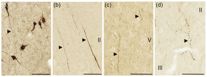
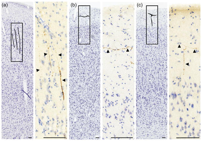
Oxytocin (OT) and arginine-vasopressin (AVP) are involved in the regulation of complex social behaviors across a wide range of taxa. Despite this, little is known about the neuroanatomy of the OT and AVP systems in most non-human primates, and less in humans. The effects of OT and AVP on social behavior, including aggression, mating, and parental behavior, may be mediated primarily by the extensive connections of OT- and AVP-producing neurons located in the hypothalamus with the basal forebrain and amygdala, as well as with the hypothalamus itself. However, OT and AVP also influence social cognition, including effects on social recognition, cooperation, communication, and in-group altruism, which suggests connectivity with cortical structures. While OT and AVP V1a receptors have been demonstrated in the cortex of rodents and primates, and intranasal administration of OT and AVP has been shown to modulate cortical activity, there is to date little evidence that OT-and AVP-containing neurons project into the cortex. Here, we demonstrate the existence of OT- and AVP-containing fibers in cortical regions relevant to social cognition using immunohistochemistry in humans, chimpanzees, and rhesus macaques. OT-immunoreactive fibers were found in the straight gyrus of the orbitofrontal cortex as well as the anterior cingulate gyrus in human and chimpanzee brains, while no OT-immunoreactive fibers were found in macaque cortex. AVP-immunoreactive fibers were observed in the anterior cingulate gyrus in all species, as well as in the insular cortex in humans, and in a more restricted distribution in chimpanzees. This is the first report of OT and AVP fibers in the cortex in human and non-human primates. Our findings provide a potential mechanism by which OT and AVP might exert effects on brain regions far from their production site in the hypothalamus, as well as potential species differences in the behavioral functions of these target regions.

Keywords: axons; cingulate; insula; neuropeptides; primates.


Figures

FIGURE 1
FIGURE 1
Mid-sagittal view of anatomical regions included in this analysis for (a), rhesus macaques, (b), chimpanzees, and (c), humans. Regions inside the rectangles represent the blocks examined in each species. Important target regions are labeled. See Figures 4–6 for a coronal view including insular regions not visible in a mid-sagittal view
FIGURE 2
FIGURE 2
Morphology of AVP-ir fibers in the cortex closely resemble those in the hypothalamus. (a) Labeled hypothalamic cell bodies and fibers in the human supraoptic nucleus. (b) Cortical fibers in human insula. (c) Cortical fibers in chimpanzee insula. (d) Cortical fibers in macaque subgenual cingulate gyrus. OT and AVP fibers in all species showed the same morphological features. Roman numerals indicate cortical layers. Scale bar = 100 μm
FIGURE 3
FIGURE 3
AVP-ir fibers in the human insula. Nissl sections are shown at lower magnification. AVP fibers are shown at higher magnification, enhanced for contrast, and brightness with Aperio ImageScope software. (a) Radially oriented fibers in cortical layer II. (b) Fibers in the human insula oriented tangentially in cortical layer I. (c) A radial fiber that appears to end with tangential branches in cortical layer I. Scale bar = 100 μm
FIGURE 4
FIGURE 4
Nissl section illustrating a selection of regions where OT and AVP fibers were found in human cortex. The midline is to the left; the dashed line at the dorsal edge indicates where the section was cut through the corpus callosum, internal capsule, and superior parts of frontal cortex. Tissue with OT fibers are marked by letters within circles; letters within rectangles represent AVP fibers. a–h) High magnification view of OT and AVP fibers. Letters on section view correspond to histology panels. Fiber images are from nearby sections (within 1 mm). FI, frontoinsular cortex; AON, olfactory nucleus; SG, straight gyrus; Cd, caudate. Scale bar = 100 μm
FIGURE 5
FIGURE 5
Nissl section illustrating a selection of regions where OT and AVP fibers were found in chimpanzee cortex. Tissue with OT fibers are marked by letters within circles; letters within rectangles represent AVP fibers. a–e) High magnification view of OT and AVP fibers. Letters on section view correspond to histology panels. Fiber images are from nearby sections (within 1 mm). FI, frontoinsular cortex; AON, olfactory nucleus; SG, straight gyrus; Cd, caudate. Scale bar = 100 μm
FIGURE 6
FIGURE 6
Nissl section illustrating a selection of regions where AVP fibers were found in macaque cortex. Letters within rectangles represent AVP fibers. a,b) High magnification view of AVP fibers. Letters on section view correspond to histology panels. Fiber images are from nearby sections (within 1 mm). AON, olfactory nucleus; Cd, caudate. Scale bar = 100 μm

References

    1. Adolphs R. The neurobiology of social cognition. Current Opinion in Neurobiology. 2001;11(2):231–239. doi: 10.1016/S0959-4388(00)00202-6. - DOI - PubMed
    1. Albers HE. Species, sex and individual differences in the vasotocin/vasopressin system: Relationship to neurochemical signaling in the social behavior neural network. Frontiers in Neuroendocrinology. 2015;36:49–71. doi: 10.1016/j.yfrne.2014.07.001. - DOI - PMC - PubMed
    1. Allman JM, Tetreault NA, Hakeem AY, Manaye KF, Semendeferi K, Erwin JM, … Hof PR. The von Economo neurons in the frontoinsular and anterior cingulate cortex. Annals of the New York Academy of Sciences. 2011;1225:59–71. doi: 10.1111/j.1749-6632.2011.06011.x. - DOI - PMC - PubMed
    1. Apps MA, Rushworth MF, Chang SW. The anterior cingulate gyrus and social cognition: Tracking the motivation of others. Neuron. 2016;90(4):692–707. doi: 10.1016/j.neuron.2016.04.018. - DOI - PMC - PubMed
    1. Barton RA. Olfactory evolution and behavioral ecology in primates. American Journal of Primatology. 2006;68(6):545–558. doi: 10.1002/ajp.20251. - DOI - PubMed

Publication types

LinkOut - more resources