. 2019 Apr;45(3):262-277.
doi: 10.1111/nan.12501. Epub 2018 Jun 26.

Differential insular cortex subregional vulnerability to α-synuclein pathology in Parkinson's disease and dementia with Lewy bodies

Affiliations

Differential insular cortex subregional vulnerability to α-synuclein pathology in Parkinson's disease and dementia with Lewy bodies

Y Y Fathy et al. Neuropathol Appl Neurobiol. 2019 Apr.

Abstract

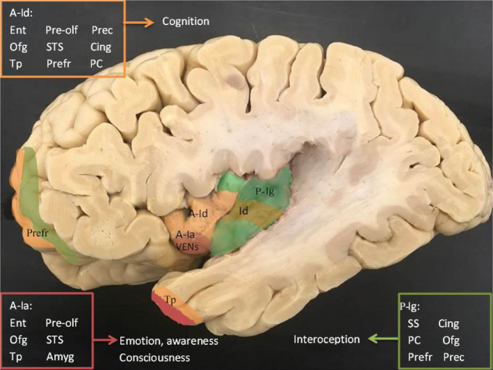
Aim: The insular cortex consists of a heterogenous cytoarchitecture and diverse connections and is thought to integrate autonomic, cognitive, emotional and interoceptive functions to guide behaviour. In Parkinson's disease (PD) and dementia with Lewy bodies (DLB), it reveals α-synuclein pathology in advanced stages. The aim of this study is to assess the insular cortex cellular and subregional vulnerability to α-synuclein pathology in well-characterized PD and DLB subjects.

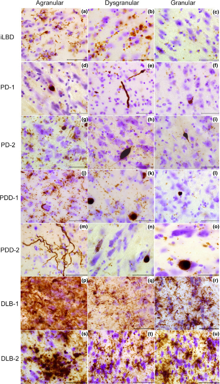
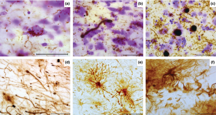
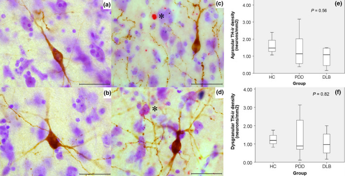
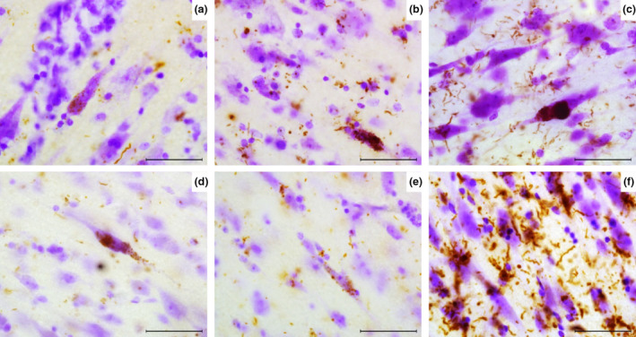
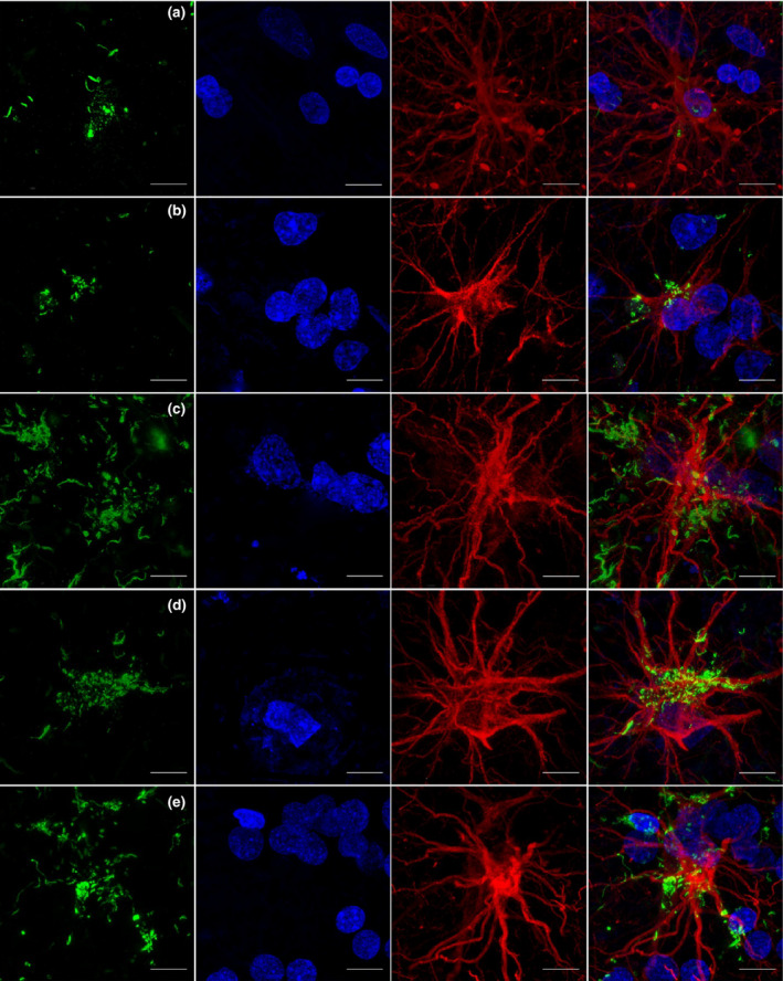
Methods: We analysed postmortem insular tissue from 24 donors with incidental Lewy body disease, PD, PD with dementia (PDD), DLB and age-matched controls. The load and distribution of α-synuclein pathology and tyrosine hydroxylase (TH) cells were studied throughout the insular subregions. The selective involvement of von Economo neurons (VENs) in the anterior insula and astroglia was assessed in all groups.

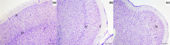
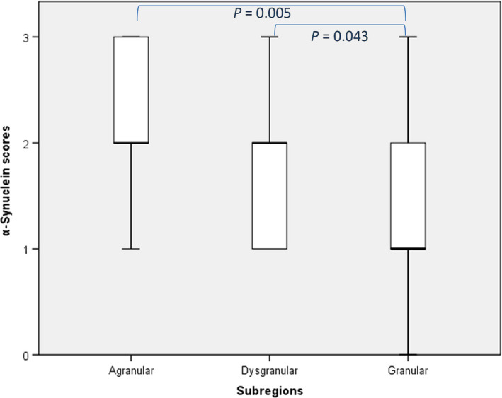
Results: A decreasing gradient of α-synuclein pathology load from the anterior periallocortical agranular towards the intermediate dysgranular and posterior isocortical granular insular subregions was found. Few VENs revealed α-synuclein inclusions while astroglial synucleinopathy was a predominant feature in PDD and DLB. TH neurons were predominant in the agranular and dysgranular subregions but did not reveal α-synuclein inclusions or significant reduction in density in patient groups.

Conclusions: Our study highlights the vulnerability of the anterior agranular insula to α-synuclein pathology in PD, PDD and DLB. Whereas VENs and astrocytes were affected in advanced disease stages, insular TH neurons were spared. Owing to the anterior insula's affective, cognitive and autonomic functions, its greater vulnerability to pathology indicates a potential contribution to nonmotor deficits in PD and DLB.

Keywords: Parkinson's disease; alpha synuclein; astrocytes; insular cortex; von Economo neurons; vulnerability.


Figures

Figure 1
Figure 1
Macroscopy of the insular cortex subregions and corresponding connections. The insular cortex is seen within the sylvian fissure. The agranular insula (A‐Ia) is seen ventro‐anteriorly (red), is connected to the olfacotory cortex, orbitofrontal, amygala and temporopolar region. While the dysgranular insula (A‐Id) is seen dorsally (orange) and is connected to various limbic and neocortical regions. Although the granular insula (P‐Ig) (green) is mostly present within the posterior insula. It is preferentially connected to the somatosensory cortex, parietal cortex and cingulate. Some regions are coloured to outline connections to the insular subregions including the prefrontal cortex and temporopolar cortex. Amyg, amygdala; Cing, cingulate gyrus; Ent, entorhinal cortex; Ofg, orbitofrontal gyrus; PC, parietal cortex; Prec, precentral sulcus; Prefr, prefrontal cortex; Pre‐olf, prepiriform part of olfactory cortex; SS, somatosensory cortex; STS, superior temporal sulcus; Tp, temporopolar cortex.
Figure 2
Figure 2
Definition of insular subregions in 60 μm thick sections. (a) Granular insular grey matter shows uniform and well defined granular layers II and IV in an iLBD case. (b) Dysgranular insula shows less dense and granular layers II and IV. (c) Agranular insula grey matter is shown lacking layers II and IV. iLBD, incidental Lewy body disease; II, layer two; III, layer three; IV, layer four. Magnification: 25 ×, scale bar 500 μm.
Figure 3
Figure 3
Distribution pattern of α‐synuclein in insular subregions. iLBD shows mild LNs and astroglial α‐synuclein inclusions in layer I of agranular insula (a), few glial inclusions in dysgranular insula (b), and sparse dot‐like aggregates in granular insula (c). PD‐1 agranular insula shows a LB‐like inclusion and dot‐like aggregates (d), the dysgranular insula shows bulgy LNs in layer I (e), and the granular insula shows an intracellular LB inclusion (f). PD‐2 shows many LNs inclusions in agranular insula and glial α‐synuclein (g) and less but bulgy LN in dysgranular (h) and granular regions (i). In PDD‐2 severe astroglial α‐synuclein inclusions are shown in agranular insula (j) few LBs and LNs in dysgranular insula (k). The granular insula shows dot‐like aggregates and astroglial α‐synuclein (l). In PDD‐1 agranular insula, very long LNs and some dot‐like aggregates are seen in layer I (m). Dysgranular insula in PDD‐1 shows granular cytoplasmic inclusions in neurons and a LB (n) while the granular insula shows less aggregates and a LB in the infragranular layer (o). In DLB‐1, severe α‐synuclein inclusions are seen in agranular insula throughout all layers (p). Severe astroglial inclusions are seen in the supragranular layers of dysgranular and granular insula (q,r). In DLB‐2, a cluster of dystrophic LNs and glial inclusions are shown in layer II of the agranular insula (s). The dysgranular insula contains LNs and dot‐like structures (t) also abundant in the granular insula superficial layers (u). DLB, dementia with Lewy bodie; iLBD, incidental Lewy body disease; LB, Lewy bodies; LN, Lewy neurites; PD, Parkinson's disease; PDD, Parkinson's disease dementia. Magnification: 630 × , scale bar 50 μm.
Figure 4
Figure 4
Semiquantitative analysis of α‐synuclein pathology across the insular subregions. The local density of α‐synuclein wass assessed at 200 ×  magnification. A significant difference in subregional distribution of α‐synuclein pathology was observed [χ2 (2, N = 63) = 9099, P = 0.011]. Pairwise comparison between different subregions showed a significant difference between the agranular and granular subregions as well as dysgranular and granular subregions (= 0.005 and 0.043, respectively).
Figure 5
Figure 5
Cell specific morphology and inclusions in Lewy body diseases. In PDD‐2 with astroglial tauopathy, infragranular layer of agranular insula show astroglial‐to‐neuronal α‐synuclein inclusions (a), an elongated α‐synuclein positive process with bulbous endings (possibly glial) surrounded by astroglial α‐synuclein inclusions (*) and LNs (b). DLB‐2 shows LB inclusions, LNs and astroglial α‐synuclein (*) within the deep infragranular layers (c). Loose GFAP + astrocytic processes are shown containing bulbous end feet and donut‐shaped structures in the supragranular layers of the agranular insula in DLB‐2 (d). PDD‐1 with astroglial tauopathy shows small and dysmorphic astrocytes containing multiple varicosities within their processes, possibly representing fuzzy astrocytes (e). A GFAP + astrocyte is shown surrounded by disorganized processes in DLB‐2 (f). Magnification: 630 × , scale bar 50 μm. DLB, dementia with Lewy bodie; LB, Lewy bodies; LN, Lewy neurites; PDD, Parkinson's disease dementia; GFAP, glial fibrillary acidic protein.
Figure 6
Figure 6
Morphological characteristics of tyrosine hydroxylase immunoreactive (TH‐ir) neurons, distribution pattern, and relationship with α‐synuclein deposits in the Insular cortex subregions. TH‐ir neurons were predominant in layers V and VI, and were mostly bipolar in morphology and few multipolar (a,b). No α‐synuclein deposits (*) were present within the TH‐ir neurons (brown) or their neurites; these TH‐ir neurons were often found surrounded by beaded dopaminergic fibres (c,d). There were no significant differences in TH‐ir neurons between groups in the agranular and dysgranular subregions (e,f). Magnification: 630 × , scale bar: 50 μm.
Figure 7
Figure 7
α‐synuclein deposits in VENs. (a) PD‐2 shows granular LB inclusions (brown) along a VEN. (b) LB in VEN and surrounding astrocytes in PD‐2. (c) PDD‐2 shows a VEN containing a large LB and multiple granular inclusions within the cell body, astroglial α‐synuclein inclusions are also seen. (d) PDD‐1 shows LB in the soma and dendrite of a VEN. (e) α‐synuclein inclusions are shown in a fork cell in PDD‐1. (f) DLB‐2 agranular insula shows many deposits surrounding pyramidal neurons and rod shaped VEN. Magnification: 630 × , scale bar 50 μm. PDD, Parkinson's disease dementia; LB, Lewy bodies; DLB, dementia with Lewy bodie; VENs, von Economo neurons.
Figure 8
Figure 8
Relationship between α‐synuclein immunoreactivity and astrocytes in insular cortex in PD(D) and DLB. α‐synuclein (green) is present within varicose projection astrocyte (GFAP, red) cell body in deep layer in the anterior insula in PD‐2 (a). In PDD‐2, an astrocyte is shown containing α‐synuclein aggregates and surrounded by a cluster of nuclei in the anterior insula (b). In DLB‐3, a protoplasmic astrocyte is shown surrounded by multiple α‐synuclein aggregates but no inclusions were present within the astrocyte (c). DLB‐1 shows α‐synuclein deposits surrounding the cell body of an interlaminar astrocyte in layer I (d) and similar inclusions are shown within a protoplasmic astrocyte, its processes, and the surrounding clustered nuclei in the posterior insula in DLB‐2 (e). Magnification 100 × , Scale bar: 10 μm. DLB, dementia with Lewy bodie; PDD, Parkinson's disease dementia; GFAP, glial fibrillary acidic protein.

References

    1. Fahn S. Description of Parkinson's disease as a clinical syndrome. Ann N Y Acad Sci 2003; 991: 1–14 - PubMed
    1. Aarsland D, Andersen K, Larsen JP, Lolk A, Nielsen H, Kragh‐Sørensen P. Risk of dementia in Parkinson's disease: a community‐based, prospective study. Neurology 2001; 56: 730–6 - PubMed
    1. Goedert M, Spillantini MG, Del Tredici K, Braak H. 100 years of Lewy pathology. Nat Rev Neurol 2013; 9: 13–24 - PubMed
    1. Kalia LV, Lang AE. Parkinson's disease. Lancet 2015; 386: 896–912 - PubMed
    1. McKeith I, Mintzer J, Aarsland D, Burn D, Chiu H, Cohen‐Mansfield J, Dickson D, Dubois B, Duda JE, Feldman H, Gauthier S, Halliday G, Lawlor B, Lippa C, Lopez OL, Carlos Machado J, O'Brien J, Playfer J, Reid W. International psychogeriatric association expert meeting on DLB. Dementia with Lewy bodies. Lancet Neurol 2004; 3: 19–28 - PubMed

Publication types

Substances