Review
. 2018 May 25;122(11):1608-1624.
doi: 10.1161/CIRCRESAHA.118.313058.

Role of Resident Stem Cells in Vessel Formation and Arteriosclerosis

Affiliations
Review

Role of Resident Stem Cells in Vessel Formation and Arteriosclerosis

Li Zhang et al. Circ Res. .

Abstract

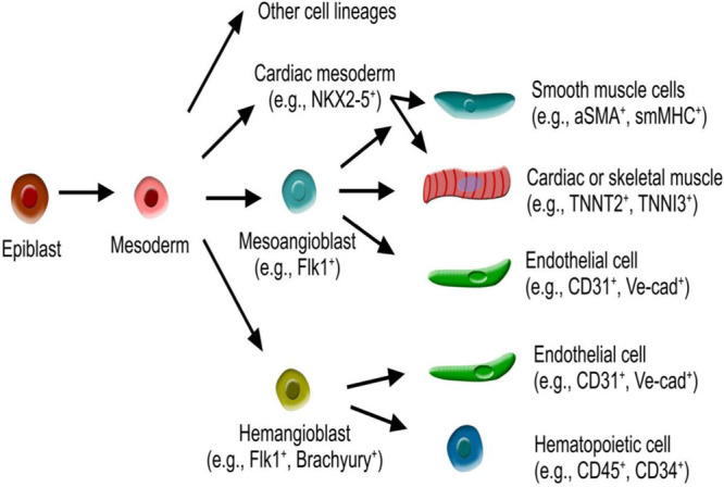
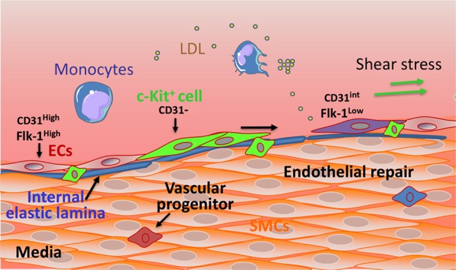
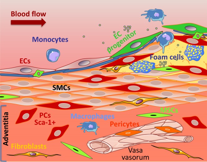
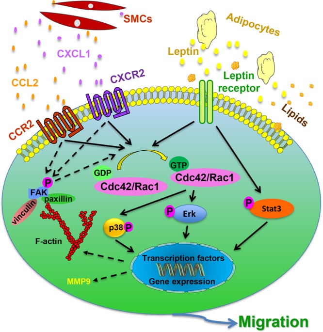
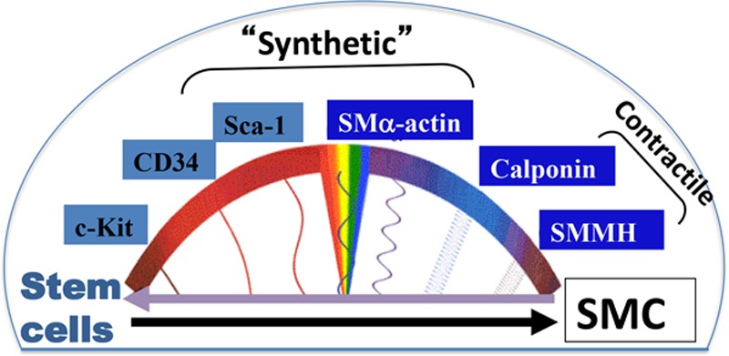
Vascular, resident stem cells are present in all 3 layers of the vessel wall; they play a role in vascular formation under physiological conditions and in remodeling in pathological situations. Throughout development and adult early life, resident stem cells participate in vessel formation through vasculogenesis and angiogenesis. In adults, the vascular stem cells are mostly quiescent in their niches but can be activated in response to injury and participate in endothelial repair and smooth muscle cell accumulation to form neointima. However, delineation of the characteristics and of the migration and differentiation behaviors of these stem cells is an area of ongoing investigation. A set of genetic mouse models for cell lineage tracing has been developed to specifically address the nature of these cells and both migration and differentiation processes during physiological angiogenesis and in vascular diseases. This review summarizes the current knowledge on resident stem cells, which has become more defined and refined in vascular biology research, thus contributing to the development of new potential therapeutic strategies to promote endothelial regeneration and ameliorate vascular disease development.

Keywords: cell lineage; myocytes, smooth muscle; neointima; stem cells; vascular diseases.


Figures

Figure 1.
Figure 1.
Schematic figure showing mesoangioblast and hemangioblast-derived from mesoderm. Mesoangioblasts are multipotent and give rise to endothelial cells, cardiac or skeletal muscle, and smooth muscle cells. Hemangioblasts are bipotent and give rise to endothelial cells and hematopoietic cells. aSMA indicates smooth muscle actin-α; smMHC, smooth muscle myosin heavy chain; TNNI, troponin I; TNNT, troponin T; and Ve-cad, VE-cadherin.
Figure 2.
Figure 2.
Developmental fate map of vascular smooth muscle. The different colors represent the different embryonic origins for smooth muscle cells (SMCs), as indicated in the figure. Aortic SMCs originate from 3 distinct developmental lineages. The secondary heart field (red) contributes to SMCs of the basal aortic root, the neural crest (blue) gives rise to SMCs of the ascending aorta and aortic arch, the paraxial/somitic mesoderm (yellow) contributes to SMCs of the descending aorta. The lineage boundaries depicted are shown to approximation and may change with aging.
Figure 3.
Figure 3.
Proposed roles for vascular wall resident endothelial progenitors in endothelial generation. The vessel wall comprises an inner layer (intima), a thick media layer, and an outer layer (adventitia). The intima is composed of the elastic lamina (blue) and a monolayer of endothelial cells (ECs). The intima encompasses mature and terminally differentiated cells (ECs: CD31High/Flk-1High) and also a niche of endothelial progenitors, such as c-Kit+ cells (green), which can proliferate, migrate, and differentiate into ECs. Endothelial progenitors (EPCs) have clonogenic and endothelial generative potential and express stem cell markers, such as c-Kit, associated with low expression of CD31. EPCs are progenitors of a transient amplifying population of cells (purple) which display an intermediate expression of CD31 and a low expression of Flk-1. In response to EC turnover and under regulatory signals, the endothelial progenitors can be activated and undergo migration, proliferation, and differentiation to replace lost ECs. Flk indicates fetal liver kinases; LDL, low-density lipoprotein; and SMC, smooth muscle cell.
Figure 4.
Figure 4.
Proposed roles for vascular wall resident progenitor cells (PCs) in lesion formation. The adventitia is a dynamic layer in active communication with the other vessel wall layers, and it contains various cell types, including PCs (red), mesenchymal stem cells (MSCs in green), macrophages (blue), fibroblasts (yellow), and pericytes (orange) surrounding the adventitial vasa vasorum, among others. Adventitial PCs, expressing the stem cell marker Sca-1 (red), have the ability to migrate to the lesions and to differentiate into smooth muscle cells (SMCs), thus contributing to the intimal SMCs pool. EC indicates endothelial cell.
Figure 5.
Figure 5.
Proposed mechanism of vascular wall resident Sca-1+ progenitor cell migration in response to chemokines and lipoproteins. Sca-1+ vascular progenitor cells are resident in the vessel wall. Chemokines, such as CCL2 (chemokine [C-C motif] ligand) and CXCL1 (chemokine [C-X-C motif] ligand), are released by activated smooth muscle cells (SMCs) and act on the Sca-1+ cells by binding to their respective receptors, CCR2 (C-C chemokine receptor type) and CXCR2. As a result, downstream pathways are activated, which include GTPAses Rac1 and Cdc42, p38 is phosphorylated and cytoskeleton-related proteins upregulation (FAK [focal adhesion kinase], vinculin, paxillin). Adipocytes also release signals, such as leptin, which bind to the corresponding receptors expressed in Sca-1+ cells. Consequently, Rac1 and Cdc42 are activated, leading to ERK (extracellular signal–regulated kinase) phosphorylation. Leptin also activates Stat3 pathway and the expression of cytoskeleton-related proteins. Together, the activation of the mentioned signaling pathways leads to either transcription of migration-related genes or direct activation of the cytoskeleton to promote cell migration. Furthermore, lipid particles, such as low-density lipoprotein or cholesterol, may be incorporated by Sca-1+ cells and lead to increased migration by activating relevant migration pathways and inducing the expression of matrix degradation proteins by the expression of matrix metalloproteinase 9.
Figure 6.
Figure 6.
Smooth muscle cell (SMC) heterogeneity and stem cell differentiation. During vascular development or in response to injury, the media SMCs exhibit a synthetic, proliferative, and migratory phenotype, where they may express some stem cell markers (Sca-1) and also the marker SMα-actin. This is accompanied by a decrease in the expression of SMC differentiation markers, such as calponin and SMMHC (smooth muscle myosin heavy chain). Stem cells (Sca-1+, c-Kit+, CD34+) can differentiate into SMCs in response to vascular injury, and thus, their expression of stem cell markers is decreased, whereas the expression of SMC markers is increased (SMα-actin, calponin, SMMHC). Mature and fully differentiated SMCs express high levels of calponin and SMMHC and show reduced or negligent levels of stem cell markers. These cells present a more elongated morphology and a contractile phenotype, with a low proliferative and migratory rate. Stem/progenitor cell markers can potentially be expressed in dedifferentiated SMCs, which have gone through a dedifferentiation process in atherosclerosis, and which contribute to vessel wall remodeling.

Similar articles

Cited by

References

    1. Flamme I, Frölich T, Risau W. Molecular mechanisms of vasculogenesis and embryonic angiogenesis. J Cell Physiol. 1997;173:206–210. doi: 10.1002/(SICI)1097-4652(199711)173:2<206::AID-JCP22>3.0.CO;2-C. - PubMed
    1. Carmeliet P. Angiogenesis in health and disease. Nat Med. 2003;9:653–660. doi: 10.1038/nm0603-653. - PubMed
    1. Kovacic JC, Moore J, Herbert A, Ma D, Boehm M, Graham RM. Endothelial progenitor cells, angioblasts, and angiogenesis–old terms reconsidered from a current perspective. Trends Cardiovasc Med. 2008;18:45–51. doi: 10.1016/j.tcm.2007.12.002. - PubMed
    1. Ross R. The pathogenesis of atherosclerosis: a perspective for the 1990s. Nature. 1993;362:801–809. doi: 10.1038/362801a0. - PubMed
    1. Libby P, Hansson GK. Inflammation and immunity in diseases of the arterial tree: players and layers. Circ Res. 2015;116:307–311. doi: 10.1161/CIRCRESAHA.116.301313. - PMC - PubMed

Publication types

MeSH terms