Grading of carotid artery stenosis with computed tomography angiography: whether to use the narrowest diameter or the cross-sectional area
- PMID: 29799095
- PMCID: PMC6108978
- DOI: 10.1007/s13244-018-0622-5
Grading of carotid artery stenosis with computed tomography angiography: whether to use the narrowest diameter or the cross-sectional area
Abstract
Objectives: To compare the estimation of carotid artery stenosis by computed tomography angiography (CTA) based on cross-sectional area versus the smallest diameter measurement, and test the accuracy of both CTA measurements using color Doppler ultrasonography (CDUS) as a reference method.
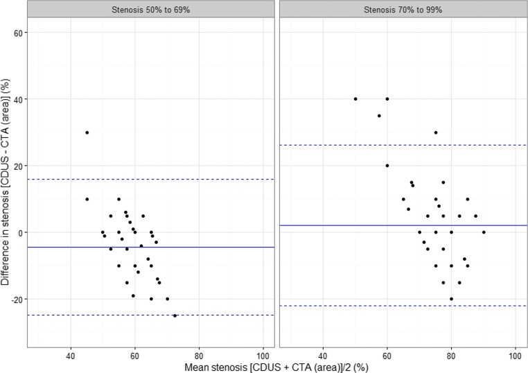
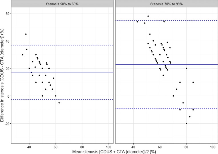
Methods: For 113 carotid arteries with stenosis ≥50% we analysed the differences in the estimated stenosis level between both CTA methods and CDUS using the Bland-Altman approach. Further, we calculated sensitivity, specificity and plotted receiver operating characteristic (ROC) curves for both CTA methods.
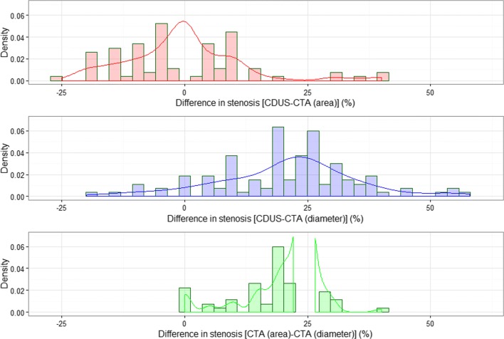
Results: The mean difference between CDUS and CTA (area) measurements was -0.4% (p = 0.68); between CDUS and CTA (diameter), 20.7% (p < 0.001); and between CTA (area) and CTA (diameter), 21.2% (p < 0.001). Sensitivity and specificity for the CTA (area) method were 81% and 77%, and for CTA (diameter) were 23% and 100%. The area under the curve (AUC) for CTA (diameter) was 0.62 (0.57, 0.66), and for CTA (area) 0.79 (0.71-0.87). The equality test for the two AUCs was <0.0001.
Conclusions: CTA (diameter)-based measurements significantly underestimated the degree of carotid stenosis. We recommend the CTA (area) method because of its higher predictive power for a correct stenosis classification and a lack of significant difference in the estimated stenosis level, compared to CDUS.
Main messages: • Cross-sectional area measurement considers asymmetric shape of the residual vessel lumen. • CTA (diameter) method on average significantly underestimates the true level of stenosis. • CTA (area) method correctly classifies vessels needing surgical intervention.
Keywords: Atherosclerosis; CT angiography; Carotid artery stenosis; Color Doppler ultrasonography; Medical imaging.
Figures


Similar articles
-
COMPARISON OF CAROTID STENOSIS GRADING BY CT ANGIOGRAPHY AND DOPPLER ULTRASONOGRAPHY: HOW THE STATISTICAL METHODS APPLIED INFLUENCE THE RESULTS.Acta Clin Croat. 2022 Feb;60(3):457-466. doi: 10.20471/acc.2021.60.03.17. Acta Clin Croat. 2022. PMID: 35282499 Free PMC article.
-
Cross-sectional area for the calculation of carotid artery stenosis on computed tomographic angiography.J Vasc Surg. 2013 Sep;58(3):659-65. doi: 10.1016/j.jvs.2013.02.031. Epub 2013 Apr 20. J Vasc Surg. 2013. PMID: 23611710
-
Diagnostic accuracy of colour Doppler ultrasonography, CT angiography and blood-pool-enhanced MR angiography in assessing carotid stenosis: a comparative study with DSA in 170 patients.Radiol Med. 2012 Feb;117(1):54-71. doi: 10.1007/s11547-011-0651-3. Epub 2011 Mar 7. Radiol Med. 2012. PMID: 21424318 English, Italian.
-
Coronary CT angiography versus intravascular ultrasound for estimation of coronary stenosis and atherosclerotic plaque burden: a meta-analysis.J Cardiovasc Comput Tomogr. 2013 Jul-Aug;7(4):256-66. doi: 10.1016/j.jcct.2013.08.006. Epub 2013 Aug 23. J Cardiovasc Comput Tomogr. 2013. PMID: 24148779 Review.
-
Diagnostic Performance of Hybrid Cardiac Imaging Methods for Assessment of Obstructive Coronary Artery Disease Compared With Stand-Alone Coronary Computed Tomography Angiography: A Meta-Analysis.JACC Cardiovasc Imaging. 2018 Apr;11(4):589-599. doi: 10.1016/j.jcmg.2017.05.020. Epub 2017 Aug 16. JACC Cardiovasc Imaging. 2018. PMID: 28823745 Free PMC article.
Cited by
-
Challenges and Pitfalls in CT-Angiography Evaluation of Carotid Bulb Stenosis: Is It Time for a Reappraisal?Life (Basel). 2022 Oct 22;12(11):1678. doi: 10.3390/life12111678. Life (Basel). 2022. PMID: 36362834 Free PMC article.
-
Systematic review and meta-analysis of the diagnostic value of computed tomography angiography for severe internal carotid artery stenosis.BMC Med Imaging. 2024 Aug 14;24(1):215. doi: 10.1186/s12880-024-01390-6. BMC Med Imaging. 2024. PMID: 39143526 Free PMC article.
-
Duplex ultrasound for diagnosing symptomatic carotid stenosis in the extracranial segments.Cochrane Database Syst Rev. 2022 Jul 11;7(7):CD013172. doi: 10.1002/14651858.CD013172.pub2. Cochrane Database Syst Rev. 2022. PMID: 35815652 Free PMC article.
-
Overcoming calcium blooming and improving the quantification accuracy of percent area luminal stenosis by material decomposition of multi-energy computed tomography datasets.J Med Imaging (Bellingham). 2020 Sep;7(5):053501. doi: 10.1117/1.JMI.7.5.053501. Epub 2020 Oct 1. J Med Imaging (Bellingham). 2020. PMID: 33033732 Free PMC article.
-
COMPARISON OF CAROTID STENOSIS GRADING BY CT ANGIOGRAPHY AND DOPPLER ULTRASONOGRAPHY: HOW THE STATISTICAL METHODS APPLIED INFLUENCE THE RESULTS.Acta Clin Croat. 2022 Feb;60(3):457-466. doi: 10.20471/acc.2021.60.03.17. Acta Clin Croat. 2022. PMID: 35282499 Free PMC article.
References
LinkOut - more resources
Full Text Sources
Other Literature Sources