Review
. 2019 Apr;25(2):155-166.
doi: 10.1177/1073858418775027. Epub 2018 May 25.

Fluid Dynamics Inside the Brain Barrier: Current Concept of Interstitial Flow, Glymphatic Flow, and Cerebrospinal Fluid Circulation in the Brain

Affiliations
Review

Fluid Dynamics Inside the Brain Barrier: Current Concept of Interstitial Flow, Glymphatic Flow, and Cerebrospinal Fluid Circulation in the Brain

Tsutomu Nakada et al. Neuroscientist. 2019 Apr.

Abstract

The discovery of the water specific channel, aquaporin, and abundant expression of its isoform, aquaporin-4 (AQP-4), on astrocyte endfeet brought about significant advancements in the understanding of brain fluid dynamics. The brain is protected by barriers preventing free access of systemic fluid. The same barrier system, however, also isolates brain interstitial fluid from the hydro-dynamic effect of the systemic circulation. The systolic force of the heart, an essential factor for proper systemic interstitial fluid circulation, cannot be propagated to the interstitial fluid compartment of the brain. Without a proper alternative mechanism, brain interstitial fluid would stay stagnant. Water influx into the peri-capillary Virchow-Robin space (VRS) through the astrocyte AQP-4 system compensates for this hydrodynamic shortage essential for interstitial flow, introducing the condition virtually identical to systemic circulation, which by virtue of its fenestrated capillaries creates appropriate interstitial fluid motion. Interstitial flow in peri-arterial VRS constitutes an essential part of the clearance system for β-amyloid, whereas interstitial flow in peri-venous VRS creates bulk interstitial fluid flow, which, together with the choroid plexus, creates the necessary ventricular cerebrospinal fluid (CSF) volume for proper CSF circulation.

Keywords: AQP-4; Virchow-Robin space; aquaporin; choroid plexus; tight junction.


Conflict of interest statement

Declaration of Conflicting Interests: The author(s) declared no potential conflicts of interest with respect to the research, authorship, and/or publication of this article.

Figures

Figure 1.
Figure 1.
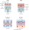
Blood-brain barrier. Common capillaries have a leaky endothelium due to the presence of fenestrations. The water channel aquaporin-1 (AQP-1) is also abundantly expressed. Accordingly, fluid from the intracapillary space moves freely into the interstitial fluid space (right). In contrast, brain capillaries lack fenestrations and have tight junctions. Expression of AQP-1 is actively suppressed. This makes the endothelium virtually non-permeable to water, constituting the highly selective blood-brain barrier (BBB) for substrates to enter the interstitial fluid space. Blue fluid indicates the fluid inside the Brain Barrier. Modified from Kitaura and others (2009).
Figure 2.
Figure 2.
blood–cerebrospinal fluid (CSF) barrier. The choroid plexus has fenestrated capillaries. Nevertheless, the apical side of choroid plexus epithelium is tightly connected with tight junctions, making the blood-CSF barrier (BCSFB). Ependymal cells are joined with gap junctions, and CSF communicates relatively freely with interstitial fluid. Blue fluid indicates the fluid inside the Brain Barrier.
Figure 3.
Figure 3.
Outer brain barrier. Capillaries within the dura matter and subdural space are fenestrated. Arachnoid barrier cells are tightly connected with tight junctions, making the outer brain barrier (OBB). Arteries and veins within the subarachnoid space are well sealed by the tunica, and capillaries inside the arachnoid barrier cells (ABCs) have tight endothelium. Cerebrospinal fluid (CSF) of the subarachnoid space communicates relatively freely with interstitial fluid across the pia matter. Blue fluid indicates the fluid inside the Brain Barrier. TJ, tight junction.
Figure 4.
Figure 4.
Main locations for aquaporin (AQP). Expression of AQP is uniquely polarized. The primary distribution of AQP-4 is confined to astrocyte endfeet at the subpial glia limitans externa (GLE) and peri-capillary space, and glial side of ependymal cell membrane. AQP-1 is found on the apical (cerebrospinal fluid [CSF]) side of choroid plexus epithelium. Virtually all AQP-4 in the brain are expressed on membrane localized inside the Brain Barrier, confirming that AQP-4 does not play a direct role for water communication between the outside and inside of the Brain Barrier. Blue fluid indicates fluid inside the Brain Barrier. VRS, Virchow-Robin space; TJ, tight junction; GJ, gap junction, ABC, arachnoid barrier cell.
Figure 5.
Figure 5.
Schematic presentation of astrocyte aquaporin-4 (AQP-4) system. The AQP-4 system provides water influx into the peri-capillary Virchow-Robin space (VRS). Necessary water enters astrocytes through AQP-4 at the glia limitans externa (GLE). This system promotes appropriate interstitial fluid circulation, including bulk flow through the VRS (interstitial flow). Modified from Suzuki and others (2017).
Figure 6.
Figure 6.
Schematic presentation of interstitial fluid dynamics. In order to have a hydrodynamic condition similar to that of the systemic environment with its fenestrated capillaries, water influx has to be provided into the peri-capillary VRS. The astrocyte aquaporin-4 (AQP-4) system effects this by removing water out of the subpial space and infusing water into peri-capillary VRS (double dotted line). The system creates the proper hydrodynamic environment for interstitial circulation as well as glymphatics akin to systemic lymphatics. Blue fluid indicates the fluid inside the Brain Barrier. ABC, arachnoid barrier cell; GLE, glia limitans externa; VRS, Virchow-Robin space; TJ, tight junction.
Figure 7.
Figure 7.
Unified view. The only driving force for the entire brain fluid dynamics system is the astrocyte aquaporin-4 (AQP-4) system. Water influx through AQP-4 at the peri-capillary VRS produces interstitial fluid flow within the VRS in both arterial and venous directions, in addition to providing proper interstitial circulation. Interstitial flow in the peri-arterial VRS is retrograde and is sufficient to create bulk flow. Nevertheless, together with intermittent closure of the flow passage by arterial pulsation, this minor flow helps prevent backflow of interstitial fluid to clear β-amyloid into the subarachnoid space (glymphatic). In contrast, interstitial flow in the peri-venous VRS is orthograde and constitutes bulk flow that has long been known to brain scientists. This bulk interstitial flow in the peri-venous VRS together with CSF produced by choroid plexus continuously provide water influx into the ventricles, creating CSF circulation. Water can enter inside of the Brain Barrier by non-specific water permeability through the plasma membrane. Metabolic water is another source of parenchymal interstitial fluid. Claudin-2 of the tight junctions in the outer brain barrier (OBB) may provide an additional water source to the subarachnoid space.

References

    1. Abbott NJ. 2004. Evidence for bulk flow of brain interstitial fluid: significance for physiology and pathology. Neurochem Int 45:545–52. - PubMed
    1. Amiry-Moghaddam M, Williamson A, Palomba M, Eid T, de Lanerolle NC, Nagelhus EA, and others. 2003. Delayed K+ clearance associated with aquaporin-4 mislocalization: phenotypic defects in brains of α-syntrophin-null mice. Proc Natl Acad Sci U S A 100:13615–20. - PMC - PubMed
    1. Asgari M, de Zélicourt D, Kurtcuoglu V. 2016. Glymphatic solute transport does not require bulk flow. Sci Rep 6:38635. - PMC - PubMed
    1. Badaut J, Brunet JF, Grollimund L, Hamou MF, Magistretti PJ, Villemure JG, and others. 2003. Aquaporin 1 and aquaporin 4 expression in human brain after subarachnoid hemorrhage and in peritumoral tissue. Acta Neurochir 86:495–8. - PubMed
    1. Benga G, Popescu O, Borza V, Pop VI, Muresan A, Mocsy I, and others. 1986. Water permeability of human erythrocytes. Identification of membrane proteins involved in water transport. Eur J Cell Biol 41:252–62. - PubMed

Publication types

LinkOut - more resources