Review
. 2018 May 25;6(2):31.
doi: 10.3390/vaccines6020031.

Characterization of Hemagglutinin Antigens on Influenza Virus and within Vaccines Using Electron Microscopy

Affiliations
Review

Characterization of Hemagglutinin Antigens on Influenza Virus and within Vaccines Using Electron Microscopy

John R Gallagher et al. Vaccines (Basel). .

Abstract

Influenza viruses affect millions of people worldwide on an annual basis. Although vaccines are available, influenza still causes significant human mortality and morbidity. Vaccines target the major influenza surface glycoprotein hemagglutinin (HA). However, circulating HA subtypes undergo continual variation in their dominant epitopes, requiring vaccines to be updated annually. A goal of next-generation influenza vaccine research is to produce broader protective immunity against the different types, subtypes, and strains of influenza viruses. One emerging strategy is to focus the immune response away from variable epitopes, and instead target the conserved stem region of HA. To increase the display and immunogenicity of the HA stem, nanoparticles are being developed to display epitopes in a controlled spatial arrangement to improve immunogenicity and elicit protective immune responses. Engineering of these nanoparticles requires structure-guided design to optimize the fidelity and valency of antigen presentation. Here, we review electron microscopy applied to study the 3D structures of influenza viruses and different vaccine antigens. Structure-guided information from electron microscopy should be integrated into pipelines for the development of both more efficacious seasonal and universal influenza vaccine antigens. The lessons learned from influenza vaccine electron microscopic research could aid in the development of novel vaccines for other pathogens.

Keywords: cryo-EM; design; electron microscopy; influenza; structure; vaccines.


Conflict of interest statement

The authors declare no conflicts of interest.

Figures

Figure 1
Figure 1
Influenza virus organization and hemagglutinin (HA) structure. (A) schematic of an influenza virus particle. Viral glycoproteins are hemagglutinin (HA, green); neuraminidase (NA, yellow); matrix 2 (M2) (purple). The membrane is shown in light blue. Genomic ribonucleoprotein complexes (RNP) filaments are inside with a trimeric viral polymerase complex (pink) at the end of each RNP; (B) the most populous glycoprotein on the virion surface is HA, which is accompanied by lesser amounts of NA, and a minority of M2; (C,D) influenza virus particles negatively stained with PTA; (C) Influenza virus, A/Victoria/3/75 (H3N2), displaying filamentous morphology. Scale bar, 50 nm; (C, inset) individual glycoprotein spikes on the virion surface are indicated with arrows. Scale bar, 25 nm; (D) A/Victoria/3/75 virions with spherical morphologies. Scale bar, 50 nm; (EH) trimeric HA ectodomains solved by protein X-ray crystallography. HA1 is shown in red and HA2 is blue with Fabs in cyan. (E) H3 HA (PDB 4O5N); (F) H1 HA (PDB 3LZG); (G) H1 HA in complex with Fab CH65 bound to receptor binding site (PDBID 5UGY); and (H) H1 HA in complex with Fab CR6261 bound to the stem region (PDB 3GBN). Scale bar, 5 nm. Panel images are originals created for this review.
Figure 2
Figure 2
Influenza virions imaged by cryo-electron microscopy (cryo-EM). (A) capsule shaped influenza virions. Scale same as (B); (B) spherical and capsule shaped influenza virions. 100 nm scale bar; (C) a filamentous virus particle several microns in length among smaller spherical virions. 100 nm scale bar. (D,E) slices through 3D maps (i.e., tomograms) of a spherical virion (panel D) and a capsule-like virion (panel E). Arrows in panel (D) indicate an HA molecule with an apparent peanut shape (white arrow) and an NA molecule with an apparent mushroom shape (black arrow). Scale bars, 50 nm; (F) model of the surface distribution of HA (green) and NA (gold) on an influenza virus particle with membrane shown in blue. Scale bar, 20 nm. Virus analyzed is influenza X-31 H3N2. Panels (AC) are originals and panels (DF) were adapted from Harris et al. [4].
Figure 3
Figure 3
Three-dimensional structures of HA embedded in the viral membrane by cryo-electron tomography (cryo-ET). (A) side view of the 3D structure of H1 HA embedded in the viral membrane derived from cryo-electron tomography of 2009 H1N1 pandemic influenza virus. The 3D map is shown as a solid isosurface (green) with the membrane region colored blue; (B) docking of HA1 (red) and HA2 (blue) ectodomain coordinates (PDB 3LZG) into the density map of trimeric HA, shown as wire mesh; (C) side view of the density map of the complex formed by C179 antibody with trimeric HA. The HA-C179 map is represented as a solid isosurface (blue); (D) molecular model of the H1 HA-C179 complex by docking H1 HA (PDB 3LZG) and C179 Fab coordinates (PDB 4HLZ) into the HA-C179 map (wire-mesh); (E,F) top and side views, respectively, for a model of IgG molecules bound to C179 stem epitopes on viral HA. H1 coordinates (PDB 3LZG) were docked into the HA-C179 density map along with three surrogate IgG molecules (PDB 1IGY) based on Fab density protruding from the HA-C179 map. HA and Fabs are shown as ribbons, with HA1 in red, HA2 in blue, helix A of HA2 in green, the Fc region of the antibody in yellow. The non-bound Fab arm is magenta, and the Fab arm used to dock the IgG into the map in cyan. Scale bars, 5 nm; (G) 3D model of the distribution of unbound HA (green) and C179-bound HA (blue) spikes on a virus surface. Membrane is displayed as brick red. Scale bar, 20 nm. Virus is pandemic 2009 H1N1 influenza virus. Panels (AG) were based on and adapted from Harris et al. [45]. Panels (AD) used deposited 3D maps from the Electron Microscopy Data Bank (EMDB) (H1, EMD-5682; H1-C179, EMD-5684).
Figure 4
Figure 4
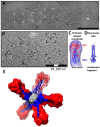
Characterization of HA complexes used as vaccine immunogens by electron microscopy. (A) molecular complexes imaged by negative staining EM for a commercial influenza HA subunit-based vaccine for the 2017–2018 influenza season (Fluzone). Density contrast is represented as white. Brackets denote some select HA complexes. An arrow denotes an HA molecule. Scale bar, 100 nm; (B) HA complexes of recombinant H7 HA imaged by cryo-electron microscopy. Density contrast is represented as black. Brackets denote some select HA complexes. Scale bars, 100 nm and 20 nm; (C) 3D map of H7 HA derived from cryo-EM images of HA complexes. The map is shown as a wire mesh with docked H7 ectodomain coordinates (PDB 4DJ6) in a pre-fusion state and compared with (D) post-fusion HA coordinates (PDB 1QU1). HA1 is red with HA2 in blue. (E) 3D model of a HA-complex. HA molecules are from coordinates (3LZG) and are shown as molecular surfaces. One constituent HA molecule is shown as a ribbon diagram with a transparent surface. Panels (A) and (E) are originals and panels (BD) were adapted from McCraw et al. [39].
Figure 5
Figure 5
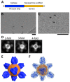
Design concept for the HA-stem nanoparticle and analysis by cryo-electron microscopy. (A) schematic organization of an engineered fusion protein made of an epitope fused to a nanoparticle scaffold. In this example the epitope is the conserved stem region of H1 HA (blue) and the nanoparticle scaffold is ferritin (orange); (B) cryo-EM of HA-stem nanoparticle. The region that is in brackets is enlarged in panel (C); (C) HA-stem nanoparticle by cryo-EM with some particles denoted by arrows. Scale bars, 50 nm; (D) some examples of reference-free 2D class averages from cryo-EM that display 2-fold, 3-fold, and 4-fold symmetry views consistent with ferritin octahedral symmetry; (E) 3D structure of HA-stem nanoparticle solved by single particle cryo-EM. The 3D map is shown as a surface rendering. The map is the deposited map from the 3DEM database (EMD-6332). The regions corresponding to spikes are colored blue and the base orange; (F) 3D map of the HA-stem nanoparticle shown as a transparent isosurface with docked coordinates. Coordinates for the designed fusion protein were computationally derived using HA and ferritin sequences [11]. Coordinates corresponding to regions for the HA-stem are in blue and ferritin regions are in orange. The shapes of the HA2-stem regions are consistent with a pre-fusion state of HA2 being displayed on the nanoparticle without the presence of HA1 heads. The original construct is named H1-SS-np and is referred to here as the HA-stem nanoparticle.
Figure 6
Figure 6
Schematic of structure-guided immunogen design integrated into a vaccine development pipeline. (Design) A conserved epitope from a surface antigen (green) is designed into a fusion protein in order for epitope display on a nanoparticle. Design uses bioinformatics taking into account conserved sequences and epitopes along with structural and molecular modeling of nanoparticle structures in silico. (Produce) To produce nanoparticles, protein encoding DNAs are synthesized and screened for protein expression and nanoparticle formation. (Purify) Nanoparticle production and purification is scaled for further characterization. (Characterize) Immunoassays and biophysical techniques such as electron microscopy are used. 3D structures can be produced by cryo-EM to assess particle integrity, along with epitope display and conformation. (Immunize, Challenge) Immunogenicity and challenge studies using nanoparticles as immunogens can be carried out in animal models, such as mice and ferrets with progression into human studies. Double arrows denote the general iterative nature among steps in the process of structure-guided antigen design within a vaccine pipeline.

References

    1. Bishop D.H., Roy P., Bean W.J., Jr., Simpson R.W. Transcription of the influenza ribonucleic acid genome by a virion polymerase. 3. Completeness of the transcription process. J. Virol. 1972;10:689–697. - PMC - PubMed
    1. Compans R.W., Caliguiri L.A. Isolation and properties of an RNA polymerase from influenza virus-infected cells. J. Virol. 1973;11:441–448. - PMC - PubMed
    1. Noda T., Sagara H., Yen A., Takada A., Kida H., Cheng R.H., Kawaoka Y. Architecture of ribonucleoprotein complexes in influenza a virus particles. Nature. 2006;439:490–492. doi: 10.1038/nature04378. - DOI - PubMed
    1. Harris A., Cardone G., Winkler D.C., Heymann J.B., Brecher M., White J.M., Steven A.C. Influenza virus pleiomorphy characterized by cryoelectron tomography. Proc. Natl. Acad. Sci. USA. 2006;103:19123–19127. doi: 10.1073/pnas.0607614103. - DOI - PMC - PubMed
    1. Schulze I.T. The structure of influenza virus. I. The polypeptides of the virion. Virology. 1970;42:890–904. doi: 10.1016/0042-6822(70)90338-7. - DOI - PubMed