Review
. 2018 May 25;23(6):1272.
doi: 10.3390/molecules23061272.

The ABP Dendrimer, a Drug-Candidate against Inflammatory Diseases That Triggers the Activation of Interleukin-10 Producing Immune Cells

Affiliations
Review

The ABP Dendrimer, a Drug-Candidate against Inflammatory Diseases That Triggers the Activation of Interleukin-10 Producing Immune Cells

Séverine Fruchon et al. Molecules. .

Abstract

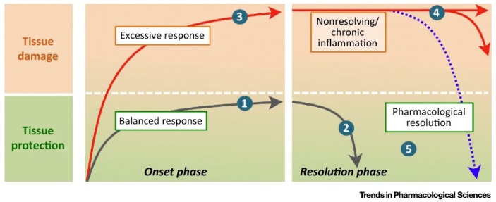
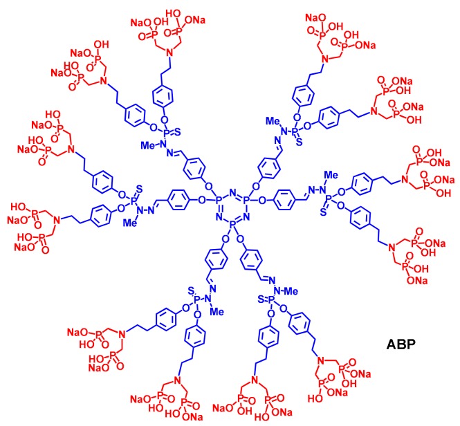
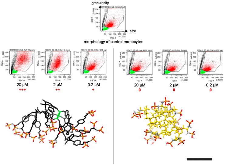
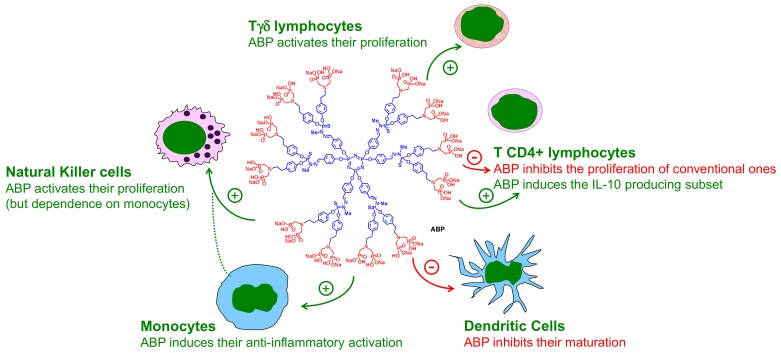
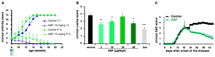
The ABP dendrimer, which is built on a phosphorus-based scaffold and bears twelve azabisphosphonate groups at its surface, is one of the dendrimers that has been shown to display immuno-modulatory and anti-inflammatory effects towards the human immune system. Its anti-inflammatory properties have been successfully challenged in animal models of inflammatory disorders. In this review, we trace the discovery and the evaluation of the therapeutic effects of the ABP dendrimer in three different animal models of both acute and chronic inflammatory diseases. We emphasize that its therapeutic effects rely on the enhancement of the production of Interleukin-10, the paradigm of anti-inflammatory cytokines, by different subsets of immune cells, such as monocytes/macrophages and CD4+ T lymphocytes.

Keywords: azabisphosphonate; chronic inflammatory diseases; inflammation; interleukin-10; monocytes/macrophages; phosphorus-based dendrimers; resolution.


Conflict of interest statement

R.P. is co-founder and shareholder of IMD-Pharma.

Figures

Figure 1
Figure 1
Time phase engagement of proresolving mediators. Inflammation is a physiological response that, when effectively controlled in extent and time, leads to tissue protection without causing tissue damage (profile 1). An effectively mounted inflammatory response will also imply the activation of pathways intended to safely terminate the inflammatory response by ‘cleaning up’ the insulted tissue and promoting healing (profile 2). In some cases, however, an exaggerated (unchecked) response to inflammatory stimuli can have detrimental consequences and result in substantial tissue harm, as in profile 3. A failure in proresolving pathways can extend in time the actions of proinflammatory mechanisms resulting in prolonged (nonresolving) or chronic inflammation (profile 4). It is reasoned that activation of endogenous circuits of resolution through novel resolution-based therapeutics can restore tissue structure and function (return to homeostasis) (profile 5) (taken from [2]) Reproduced with permission from Perretti et al., Trends in Pharmacological Science; published by Elsevier Ltd., 2015.
Figure 2
Figure 2
Structure of the ABP dendrimer. The cyclotriphosphazene core (N3P3) and the PMMH branches (including the point of divergence) are in blue. The twelve tyramine-based (in blue) ABP surface groups are in red.
Figure 3
Figure 3
(A) Sequential images (64 first seconds) from confocal videomicroscopy of purified monocytes (with cytoplasmic labelling by orange 5-(and-6)-(4-chloromethyl(benzoyl)amino) tetramethylrhodamine [CMTMR]) incubated with the ABP dendrimer emitting green fluorescence, added at second 1. (B) Membranous and internal location at 15 min, but only in the intracellular location at 120 min of the ABP dendrimer (white arrows) seen in confocal microscopy; white bars indicate 10 µM (adapted from [19]). Reproduced with permission from Poupot et al., FASEB Journal; published by the Federation of American Societies for Experimental Biology, 2006.
Figure 4
Figure 4
(A) Flow cytometry analysis of the phenotype of human monocytes activated in vitro by the ABP dendrimer (black line) compared to M2 (green line) and M1 monocytes (blue line). Left graph: surface expression of CD206 (the mannose receptor MRC1), which is an anti-inflammatory marker; central graph: surface expression of CD64 (a FcγRI receptor), an inflammatory marker; right graph: surface expression of CD13 (the aminopeptidase N), an inflammatory marker. In grey, the negative control of each surface marker (see [22]). (B) Flow cytometry quantification (as mean intensity fluorescence, mfi, ± SD) of intracellular IL10 in CD4+ T lymphocytes induced by human monocytes activated by the ABP dendrimer (black bar), by M2 monocytes (green bar) and by M1 monocytes (blue bar) in in vitro co-cultures. ** p < 0.001, one-way ANOVA. (see [22]). Reproduced with permission from Fruchon et al., Journal of Leukocyte Biology; published by the Society for Leukocyte Biology, 2009.
Figure 5
Figure 5
Relation between the bioactivity and the 3D structure of dendrimers. The bioactivity of dendrimers on human monocytes was analyzed by flow cytometry. Each dot in the plots is indicative of morphological changes (size—the Forward SCatter (FSC) parameter on the x axis—and granulosity—the Side SCatter (SSC) parameter on the y axis) undergone by purified human monocytes (red points gated in the polygon) in the presence of the dendrimers at 20, 2, and 0.2 µM. Green points are the remaining lymphocytes after purification, black points are dead or dying cells. Under the dot plots, the 3D conformation of dendrimers in water solution. +++ indicates strong morphological changes (increase in size and granulosity) when compared to control monocytes, ++ indicates mild changes, + indicates slight changes, 0 indicates no change. On the left: directional conformation of the bio-active ABP dendrimer which is the paradigm of anti-inflammatory ABP-capped dendrimers; on the right: non-directional conformation of an inactive PAMAM dendrimer on the second-generation capped with eight ABP groups at its surface. The black bar represents 1 nm. The phosphonate groups appear in red (oxygen atoms) and orange (phosphorus atoms) (see [28]). Reproduced with permission from Caminade et al., Nature Communications; published by Nature Publishing Group, 2015.
Figure 6
Figure 6
Overview of the different immuno-modulatory effects displayed by the ABP dendrimer towards different sub-populations of human mononuclear immune cells of the peripheral blood: Tγδ lymphocytes [32], T CD4+ lymphocytes [22,35], Dendritic Cells [36], Monocytes [22], Natural Killer cells [30,32].
Figure 7
Figure 7
Proof of efficacy of the ABP dendrimer in animal models of inflammatory diseases. (A) In a spontaneous mouse model of experimental arthritis, the ABP dendrimer was administered either intravenously (I.V.) or orally (P.O.) at the dose of 10 mg/kg/week starting at the age of eight weeks (blue and green arrows). The clinical arthritis score was monitored weekly ending twelve weeks after the first injection of the ABP dendrimer (maximal score is 12). (B) In a rat model of uveitis, the ABP dendrimer was administered loco-regionally (intra-vitreal) once at doses between 2 and 60 µg/eye, the same day than the induction of the disease. The clinical uveitis score was monitored 24 h later (the maximal score is 5). Dex stands for the gold standard dexamethasone at 20 µg/eye in this preclinical model. Histogram bars represent the mean clinical score + SEM. * p < 0.05, ** p < 0.01, *** p < 0.001 versus vehicle. (C) In a mouse model of Experimental Auto-immune Encephalomyelitis (EAE), the ABP dendrimer was administered intravenously at the dose of 10 mg/kg/3 days starting 18 days after the onset of the disease (green arrow). The clinical EAE score was monitored daily (maximal score is 5).

Similar articles

Cited by

References

    1. Murray P.J. Immune regulation by monocytes. Semin. Immunol. 2017 doi: 10.1016/j.smim.2017.12.005. - DOI - PubMed
    1. Perretti M., Leroy X., Bland E.J., Montero-Melendez T. Resolution pharmacology: Opportunities for therapeutic innovation in inflammation. Trends Pharmacol. Sci. 2015;36:737–755. doi: 10.1016/j.tips.2015.07.007. - DOI - PubMed
    1. Sugimoto M.A., Sousa L.P., Pinho V., Perretti M., Teixeira M.M. Resolution of inflammation: What controls its onset? Front. Immunol. 2016;7:160. doi: 10.3389/fimmu.2016.00160. - DOI - PMC - PubMed
    1. Ortega-Gomez A., Perretti M., Soehnlein O. Resolution of inflammation: An integrated view. EMBO Mol. Med. 2013;5:661–674. doi: 10.1002/emmm.201202382. - DOI - PMC - PubMed
    1. Fiorentino D.F., Bond M.W., Mosmann T.R. Two types of mouse T helper cell. IV. Th2 clones secrete a factor that inhibits cytokine production by Th1 clones. J. Exp. Med. 1989;170:2081–2095. doi: 10.1084/jem.170.6.2081. - DOI - PMC - PubMed

MeSH terms