. 2018 May 25;13(5):e0198140.
doi: 10.1371/journal.pone.0198140. eCollection 2018.

A panel of correlates predicts vaccine-induced protection of rats against respiratory challenge with virulent Francisella tularensis

Affiliations

A panel of correlates predicts vaccine-induced protection of rats against respiratory challenge with virulent Francisella tularensis

Roberto De Pascalis et al. PLoS One. .

Abstract

There are no defined correlates of protection for any intracellular pathogen, including the bacterium Francisella tularensis, which causes tularemia. Evaluating vaccine efficacy against sporadic diseases like tularemia using field trials is problematic, and therefore alternative strategies to test vaccine candidates like the Francisella Live Vaccine Strain (LVS), such as testing in animals and applying correlate measurements, are needed. Recently, we described a promising correlate strategy that predicted the degree of vaccine-induced protection in mice given parenteral challenges, primarily when using an attenuated Francisella strain. Here, we demonstrate that using peripheral blood lymphocytes (PBLs) in this approach predicts LVS-mediated protection against respiratory challenge of Fischer 344 rats with fully virulent F. tularensis, with exceptional sensitivity and specificity. Rats were vaccinated with a panel of LVS-derived vaccines and subsequently given lethal respiratory challenges with Type A F. tularensis. In parallel, PBLs from vaccinated rats were evaluated for their functional ability to control intramacrophage Francisella growth in in vitro co-culture assays. PBLs recovered from co-cultures were also evaluated for relative gene expression using a large panel of genes identified in murine studies. In vitro control of LVS intramacrophage replication reflected the hierarchy of protection. Further, despite variability between individuals, 22 genes were significantly more up-regulated in PBLs from rats vaccinated with LVS compared to those from rats vaccinated with the variant LVS-R or heat-killed LVS, which were poorly protective. These genes included IFN-γ, IL-21, NOS2, LTA, T-bet, IL-12rβ2, and CCL5. Most importantly, combining quantifications of intramacrophage growth control with 5-7 gene expression levels using multivariate analyses discriminated protected from non-protected individuals with greater than 95% sensitivity and specificity. The results therefore support translation of this approach to non-human primates and people to evaluate new vaccines against Francisella and other intracellular pathogens.


Conflict of interest statement

The authors have declared that no competing interest exist.

Figures

Fig 1
Fig 1. Rat splenocytes control intramacrophage Francisella growth that is accompanied by IFN-γ and NO production.
BMMΦ from Fischer 344 rats were infected with LVS (Macs), and co-cultured with naïve splenocytes or with immune splenocytes derived from LVS-vaccinated rats. Decreasing numbers of splenocytes were added to a constant number of confluent LVS-infected macrophages, where 1 = 5 x 10⁶, 1/2 = 2.5 x 10⁶, 1/4 = 1.25 x 10⁶, 1/8 = 0.625 x 10⁶, 1/16 = 0.312 x 10⁶, 1/32 = 0.156 x 10⁶ splenocytes. After three days of co-culture, BMMΦ were lysed to evaluate the recovery of intracellular bacteria (Panel A). Values shown are the mean numbers of CFU/ml ± SD of viable bacteria from triplicate wells. Supernatants were collected for analyses of IFN-γ by ELISA (Panel B) and NO by Griess reaction (Panel C), and concentrations of each were calculated using standard curves as references. Values shown are the mean concentration ± standard deviation of triplicate wells. Brackets indicate a significant difference (P < 0.05) between the recoveries of bacteria from macrophages in co-cultures or production of IFN-γ and nitric oxide.
Fig 2
Fig 2. Rat PBLs from differentially vaccinated rats control intramacrophage Francisella growth in a pattern that reflects vaccine efficacy.
Groups of 6 Fischer 344 rats were vaccinated with LVS, LVS-R, HK-LVS, or PBS (naïve) for a total of 31–37 rats per group, and challenged 5 weeks later i.t. with 103 CFU F. tularensis. Percent survival after one month is shown with data combined from five independent challenge experiments (Panel A). * indicates significantly greater survival compared to naïve rats or rats vaccinated with LVS-R and HK-LVS; # indicates significantly greater survival compared to naïve rats. There was no significant difference between survival of LVS-R and HK-LVS vaccinated rats. In parallel, BMMΦ from Fischer 344 rats were infected with LVS (Macs alone), and co-cultured with PBLs obtained 5 weeks after vaccination from 3 remaining naïve or vaccinated Fischer 344 rats, as indicated. After two days of co-culture, BMMΦ were lysed to evaluate the recovery of intracellular bacteria. Values shown are the mean numbers of CFU/ml ± SD of viable bacteria for five independent experiments of similar design and outcomes (Panel B). Brackets indicate a significant difference (P < 0.05) between the recoveries of bacteria from macrophages in co-cultures. There was no significant difference between the recoveries of bacteria from co-cultures using LVS-R or HK-LVS-immune PBLs.
Fig 3
Fig 3. Control of intramacrophage LVS growth depends on T cells but not B cells.
BMMΦ from Fischer 344 rats were infected with LVS and co-cultured with total, CD45R+ (B lymphocytes) or CD45R- splenocytes (non-B lymphocytes) obtained from naïve or vaccinated Fischer 344 rats, as indicated. Splenocytes were analyzed by flow cytometry to evaluate purity of B and T cells, as indicated. After three days of co-culture, BMMΦ were lysed to evaluate the recovery of intracellular bacteria (Panel A). Supernatants were collected for analyses of IFN-γ by ELISA (Panel B) and NO by Griess reaction (Panel C), and their concentrations were calculated using standard curves as references. Values shown are the mean concentration ± standard deviation of triplicate wells. Brackets indicate significant differences between the indicated groups (P < 0.05). Data shown are from one representative experiments of two of similar design and outcome.
Fig 4
Fig 4. Gene expression of potential correlates of protection is differentially up-regulated in rat PBLs and splenocytes after vaccination with LVS-derived vaccines.
Total RNA was purified from PBLs (Panel A) or splenocytes (Panel B) obtained at the indicated time points after vaccination from three naïve rats or from three rats immunized with LVS or LVS-R. RNA was used for semi-quantitative analyses of gene expression using the indicated sets of primers/probes. Values shown are average fold changes ± standard deviation of the indicated genes compared to those from naïve rats. Asterisks indicate significant differences in gene expression (P < 0.05) between LVS- and LVS-R-derived cells.
Fig 5
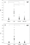
Fig 5. LVS-vaccinated rats exhibit humoral immune responses.
Sera from individual rats were obtained 2–3 weeks after vaccination and analyzed for anti-LVS total IgG (Panel A) and IgM (Panel B) antibodies. Sera from four sets of vaccinations were tested for a total of 27–35 sera for each vaccine group. Shown are the OD values obtained at a 1:1600 dilution for each individual serum; lines indicate median values for the group. Brackets indicate significant differences (P < 0.05) between the indicated antibody responses.
Fig 6
Fig 6. Bacterial replication measurements and relative gene expression measurements discriminate LVS-vaccinated rats from LVS-R-vaccinated, HK-LVS vaccinated, and naïve rats.
BMMΦ from Fischer 344 rats were infected with LVS and co-cultured with splenocytes obtained from naïve Fischer 344 rats or rats vaccinated with LVS, LVS-R, or HK-LVS. Splenocytes from 22 rats from each group were analyzed individually in studies comprised of 6–8 separate experiments. After two days of co-culture, splenocytes were recovered and used to purify total RNA, then BMMΦ were lysed to evaluate the recovery of intracellular bacteria. Semi-quantitative gene expression analyses of total RNA from recovered splenocytes were performed using the indicated sets of primers/probes, chosen among those that best reflected the hierarchy of in vivo efficacy. Values shown indicate median (white line), 1st and 3rd quartiles (lower and upper box), and maximum and minimum values (upper and lower line). Bacterial growth and gene expression were significantly different for each variable in splenocytes after vaccination with LVS compared to that of naïve, LVS-R, and HK-LVS groups (p < 0.05).
Fig 7
Fig 7. Sensitivity and specificity increase by using groups of 5–7 variables.
Eleven variables (CFU and ΔCt data) from the complete data set were used alone or in multiple combinations to generate 2047 experimental models; each model therefore included between 1 and 11 variables. The sensitivity and specificity for a subset of all the models were calculated and plotted (Panel A). This subset was generated by dividing the experimental models into eleven groups according to the number of variables used, and by selecting the top eleven scorers for each group. Plots using numbers or letters indicate the number of variables used in each model, where t and e correspond to 10 and 11 variables, respectively. The area within the rectangle in the upper right hand corner of Panel A corresponds to the highest values of sensitivity and specificity and is enlarged for presentation in Panel B.

References

    1. Petersen JM, Molins CR. Subpopulations of Francisella tularensis ssp. tularensis and holarctica: identification and associated epidemiology. Future Microbiol. 2010;5(4):649–61. doi: 10.2217/fmb.10.17 . - DOI - PubMed
    1. Staples JE, Kubota KA, Chalcraft LG, Mead PS, Petersen JM. Epidemiologic and molecular analysis of human tularemia, United States, 1964–2004. Emerg Infect Dis. 2006;12(7):1113–8. doi: 10.3201/eid1207.051504 . - DOI - PMC - PubMed
    1. Snoy PJ. Establishing efficacy of human products using animals: the U.S. Food and Drug Administration’s "animal rule". Vet Pathol. 2010;47(5):774–8. doi: 10.1177/0300985810372506 . - DOI - PubMed
    1. Eigelsbach HT, Downs CM. Prophylactic effectiveness of live and killed tularemia vaccines. Journal of Immunology. 1961;87:415–25. - PubMed
    1. Pasetti MF, Cuberos L, Horn TL, Shearer JD, Matthews SJ, House RV, et al. An improved Francisella tularensis live vaccine strain (LVS) is well tolerated and highly immunogenic when administered to rabbits in escalating doses using various immunization routes. Vaccine. 2008;26(14):1773–85. doi: 10.1016/j.vaccine.2008.01.005 . - DOI - PMC - PubMed

Publication types

Substances