. 2018 Sep 1;114(11):1547-1563.
doi: 10.1093/cvr/cvy112.

Hypertension and increased endothelial mechanical stretch promote monocyte differentiation and activation: roles of STAT3, interleukin 6 and hydrogen peroxide

Affiliations

Hypertension and increased endothelial mechanical stretch promote monocyte differentiation and activation: roles of STAT3, interleukin 6 and hydrogen peroxide

Roxana Loperena et al. Cardiovasc Res. .

Abstract

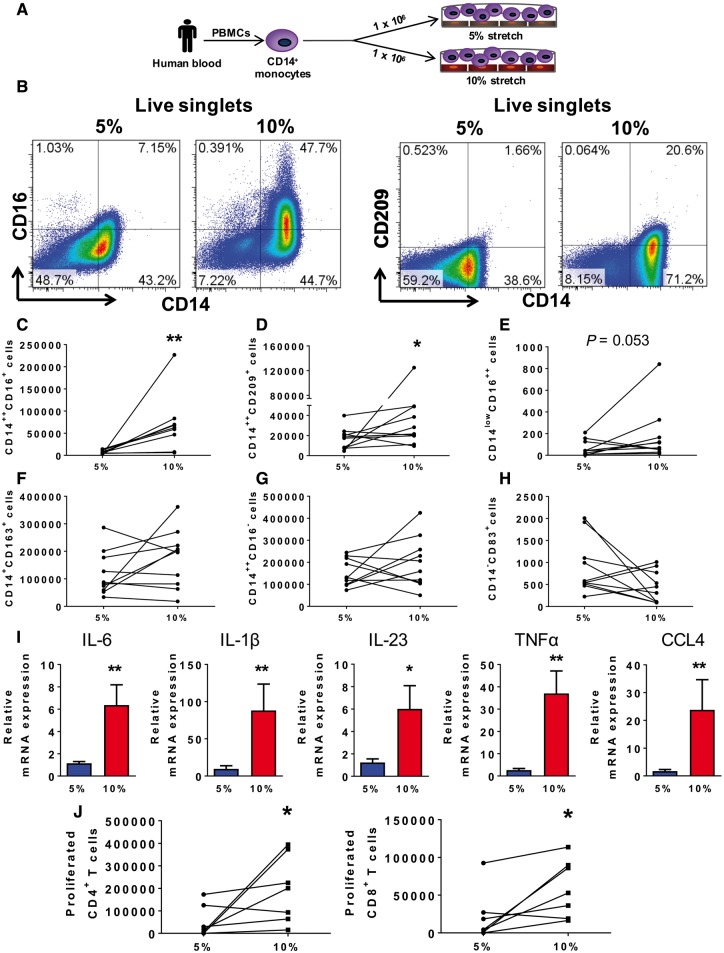
Aims: Monocytes play an important role in hypertension. Circulating monocytes in humans exist as classical, intermediate, and non-classical forms. Monocyte differentiation can be influenced by the endothelium, which in turn is activated in hypertension by mechanical stretch. We sought to examine the role of increased endothelial stretch and hypertension on monocyte phenotype and function.

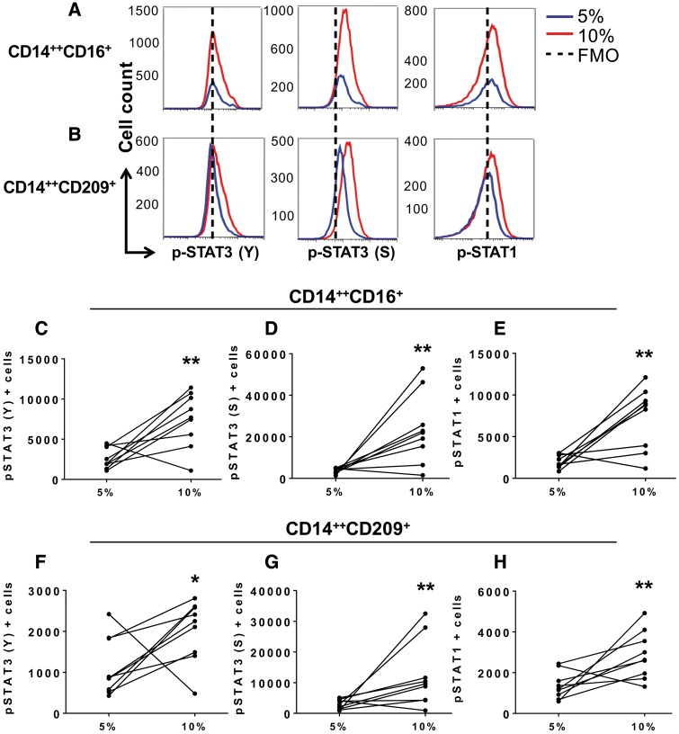
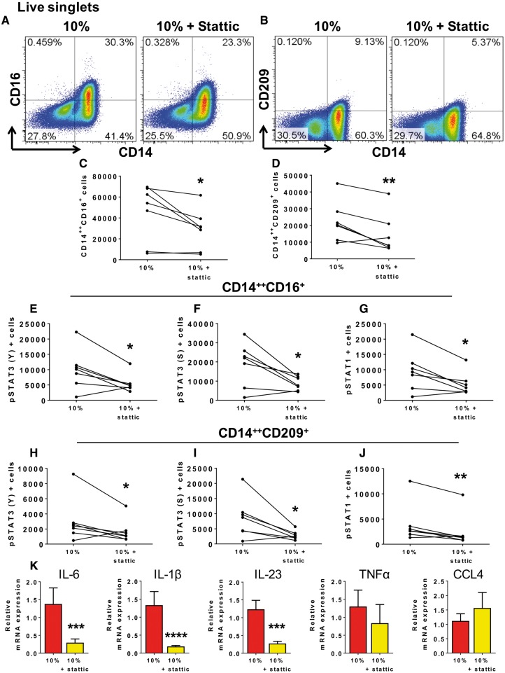
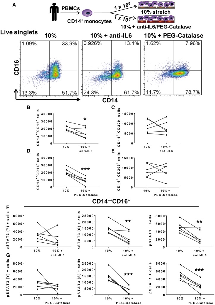
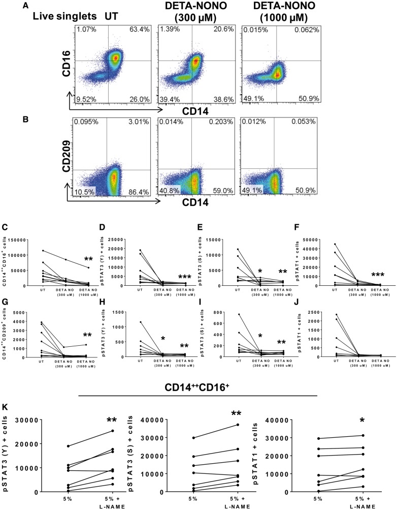
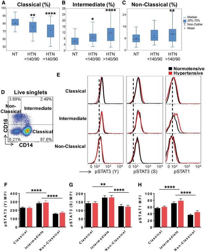
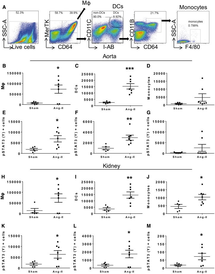
Methods and results: Human monocytes were cultured with confluent human aortic endothelial cells undergoing either 5% or 10% cyclical stretch. We also characterized circulating monocytes in normotensive and hypertensive humans. In addition, we quantified accumulation of activated monocytes and monocyte-derived cells in aortas and kidneys of mice with Angiotensin II-induced hypertension. Increased endothelial stretch enhanced monocyte conversion to CD14++CD16+ intermediate monocytes and monocytes bearing the CD209 marker and markedly stimulated monocyte mRNA expression of interleukin (IL)-6, IL-1β, IL-23, chemokine (C-C motif) ligand 4, and tumour necrosis factor α. STAT3 in monocytes was activated by increased endothelial stretch. Inhibition of STAT3, neutralization of IL-6 and scavenging of hydrogen peroxide prevented formation of intermediate monocytes in response to increased endothelial stretch. We also found evidence that nitric oxide (NO) inhibits formation of intermediate monocytes and STAT3 activation. In vivo studies demonstrated that humans with hypertension have increased intermediate and non-classical monocytes and that intermediate monocytes demonstrate evidence of STAT3 activation. Mice with experimental hypertension exhibit increased aortic and renal infiltration of monocytes, dendritic cells, and macrophages with activated STAT3.

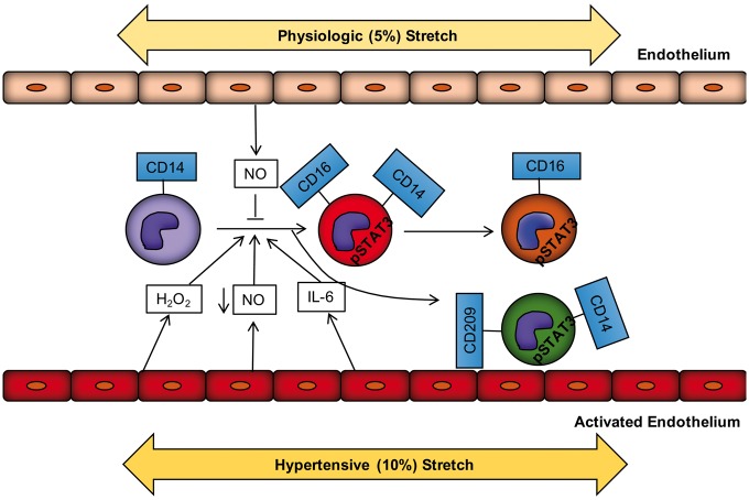
Conclusions: These findings provide insight into how monocytes are activated by the vascular endothelium during hypertension. This is likely in part due to a loss of NO signalling and increased release of IL-6 and hydrogen peroxide by the dysfunctional endothelium and a parallel increase in STAT activation in adjacent monocytes. Interventions to enhance bioavailable NO, reduce IL-6 or hydrogen peroxide production or to inhibit STAT3 may have anti-inflammatory roles in hypertension and related conditions.


Figures

Figure 1
Figure 1
Hypertensive mechanical stretch in human endothelial cells promotes monocyte activation and differentiation. Human CD14+ monocytes were isolated by magnetic sorting from PBMCs of normal human volunteers and cultured with HAECs exposed to cyclical stretch. (A) Schematic of the experimental design and (B) gating strategy for phenotyping human monocytes including classical (CD14++CD16), intermediate (CD14++CD16+) and non-classical monocytes (CD14lowCD16++). (C) Changes in numbers of cells for each subject are depicted by connected lines for CD14++CD16+ (n = 9) and (D) CD14++CD209+, (E) CD14lowCD16++, (F) macrophage population (CD14+CD163+), (G) CD14++CD16, (H) CD14CD83+ (n = 10). (I) Relative monocyte mRNA expression of IL-6, IL-1β, IL-23, TNFα, and CCL4 in adhered monocytes and in monocytes in suspension (5%, n = 15; 10%, n = 16). (J) Monocyte-HAECs cultures were stretched to either 5% or 10% for 48 h followed by sorting monocytes from HAECs using CD31+ isolation kit and FACS. Monocyte populations were cultured with CFSE-labelled T cells isolated from PBMCs of the same participants. Seven days later, we measured proliferation in the CD4+ and CD8+ T-cell populations by flow cytometry. Changes in number of proliferated CD4+ and CD8+ T cells after 7 days in culture for each subject are depicted by connected lines (n = 7). Comparisons were made using one-tail paired t-tests (*< 0.05, **< 0.01).
Figure 2
Figure 2
Effect of endothelial stretch on STAT3 activation in co-cultured monocytes. Human CD14+ monocytes were isolated from buffy coats of normal human volunteers and cultured with HAECs exposed to 5% or 10% cyclical stretch. (A) Representative flow cytometry plots are shown for intracellular staining of STAT3 phosphorylation in the tyrosine (Y) 705 and the serine (S) 727 and STAT1 phosphorylation in the Y701 in the CD14++CD16+ intermediate monocytes and (B) the CD14++CD209+ cells in the 5% stretch (blue), 10% stretch (red), and the dashed line represents FMO control. (CE) Changes in numbers of intermediate monocytes between 5% and 10% endothelial cell stretch expressing pSTAT3 (Y), pSTAT3 (S), and pSTAT1 are depicted by connected lines. (FH) Changes in numbers of CD14++CD209+ cells expressing pSTAT3 (Y), pSTAT3 (S), and pSTAT1 between 5% and 10% endothelial cell stretch. Comparisons were made using one-tail paired t-tests (n = 9, *< 0.05, **< 0.01).
Figure 3
Figure 3
STAT3 contributes to monocyte differentiation and activation during hypertensive mechanical stretch of endothelial cells. Human CD14+ monocytes were isolated from PBMCs of normal human volunteers and cultured with HAECs exposed to 10% or 10% stretch plus STAT3 inhibitor (5 µM), Stattic, for 48 h. (A) Flow cytometry gating examples are shown for the CD14++CD16+ intermediate monocyte population and (B) the CD14++CD209+ cells. (C) Individual data point for the effect of Stattic on the number intermediate monocytes and (D) CD14++CD209+ cells for each subject. (EG) Effect of Stattic on total number of cells expressing pSTAT3 (Y), pSTAT3 (S), and pSTAT1 within the intermediate monocyte population and (HJ) the CD14++CD209+ population. A total of n = 7 participants per group were used. (K) Relative monocyte mRNA expression of IL-6, IL-1β, IL-23 (10%, n= 11; 10% + Stattic, n = 9), TNFα (n = 7), and CCL4 (n =4) in monocytes. Comparisons were made using one-tail paired t-tests (*< 0.05, **< 0.01, ***< 0.001, ****< 0.0001).
Figure 4
Figure 4
IL-6 and hydrogen peroxide play a role in monocyte transformation and activation. Human CD14+ monocytes were isolated from PBMCs of normal human volunteers and cultured with HAECs exposed to 10% stretch, 10% plus anti-IL-6 neutralization antibody (10 µg/mL) or 10% plus PEG-Catalase (500 U/mL) for 48 h. (A) Schematic of methods and flow cytometry gating representatives are shown for the CD14++CD16+ intermediate monocyte population exposed to 10%, 10% + anti-IL-6, and 10% + PEG-Catalase. (B) Effect of anti-IL-6 on the total number of intermediate monocytes and (C) the CD14++CD209+ cells for each subject. (D) Effect of PEG-Catalase on total number of intermediate monocytes and (E) the CD14++CD209+ cells for each participant. (F) Effects of anti-IL-6 and (G) PEG-Catalase on number of cells expressing pSTAT3 (Y), pSTAT3 (S), and pSTAT1 within the intermediate monocyte population for each subject. Data with and without these interventions for each subject are shown by the connected lines. A total of n = 6 participants per group and per experimental treatment were used. Comparisons were made using one-tail paired t-tests (*< 0.05, **< 0.01, ***< 0.001).
Figure 5
Figure 5
Exposure of monocytes to NO donor inhibits human monocyte conversion and activation to its derived populations. Human CD14+ monocytes were isolated from buffy coats of normal human volunteers and cultured alone in untreated (UT) conditions, DETA-NONOate (DETA-NONO), an NO donor, at 300 µM or DETA-NONO at 1000 µM concentrations in static conditions for 48 h. (A) Flow cytometry representatives are shown for the CD14++CD16+ intermediate monocyte and (B) the CD14++CD209+ population. (C) Values for each subject without and with DETA-NONO are shown for the total number of cells from the CD14++CD16+ intermediate monocytes and for the total number of cells expressing (D) pSTAT3 (Y), (E) pSTAT3 (S), and (F) pSTAT1 within this population. (G) Effect of DETA-NONO on the number of CD14++CD209+ cells and the expression of (H) pSTAT3 (Y), (I) pSTAT3 (S), and (J) pSTAT1 for each subject. A total of n= 9 participants per group were used for these experiments. (K) Human CD14+ monocytes were cultured with HAECs exposed to 5% stretch or 5% plus NO synthase inhibitor, L-NAME (1000 µM) for 48 h. The number of CD14++CD16+ intermediate monocyte population expressing pSTAT3 (Y), pSTAT3 (S), and pSTAT1 for each subject are connected by lines. A total of n = 7 participants per group were used. For (CJ) the nonparametric Friedman’s test followed by Dunn’s multiple comparison tests was employed. For (K) a one-tailed paired t-tests was used (*< 0.05, **< 0.01, ***< 0.001).
Figure 6
Figure 6
Hypertension affects the distribution of circulating monocytes in humans. (A) A cohort of normotensive (n = 20), mildly hypertensive (systolic BP ∼130–140 mmHg, n = 52), and severely hypertensive (systolic BP >140 mmHg, n = 60) subjects were recruited and flow cytometry was used to analyse various monocyte populations. (A) Mean values are shown for the percent of positive CD14++CD16 classical, (B) CD14++CD16+ intermediate and (C) CD14lowCD16++ non-classical monocytes comparing normotensive, mild, and severely hypertensive subjects. Data were analysed using one-way ANOVA. (D) Representative flow cytometry dot plots showing classical, intermediate, and non-classical subset distribution are shown. (E) Histograms showing the CD14++CD16 classical, CD14++CD16+ intermediate, non-classical monocyte population comparing normotensive (black) and hypertensive (red) subjects. The dashed line represents the Mean fluorescent intensity (MFI) for the FMO control. (E) MFI for pSTAT3 (Y), pSTAT3 (S) (F), and pSTAT1 (G) in various monocyte subgroups in both normotensive (n = 15) and hypertensive (n = 12) subjects. Two-way ANOVA with Student Newman Keuls post hoc test was used (*< 0.05, **< 0.01, ***< 0.001, ****< 0.0001).
Figure 7
Figure 7
Angiotensin II-induced hypertension in wildtype C57Bl/6 mice promotes an increase in STAT3 phosphorylation in the immune cells from kidney and aorta. (A) Representative flow cytometry dot plots showing the gating strategy to identify macrophages, DCs and monocytes from C57Bl/6 wildtype mice infused with Ang II (490 ng/kg/min) or sham for 6 days. (BD) Mean values of absolute numbers of indicated cell types per thoracic aorta. (E) Mean values of pSTAT3 (Y) expression within the macrophage (Mφ), (F) DC and (G) monocyte populations per thoracic aorta. (HJ) Mean values of absolute numbers of indicated cell types per kidney. (K) Mean values of pSTAT3 (Y) expression within the macrophage, (L) DC and (G) monocyte populations per kidney. A total of n = 5, sham, and n =7, Ang II, treated mice per group were used. One-tail unpaired t-test was employed (*< 0.05, **< 0.01, ***< 0.001).
Figure 8
Figure 8
Increased endothelial stretch promotes monocyte transformation and activation. Under physiological stretch, the endothelium releases nitric oxide that prevents monocyte activation and transformation. In hypertension, increased vascular stretch promotes endothelial activation. The activated endothelium in turn releases ROS including hydrogen peroxide, cytokines including IL-6 and exhibits reduced NO bioavailability. These events initiate STAT3 activation in the monocytes and promote transformation of the classical monocyte into an intermediate and subsequently a non-classical monocyte as well as a cells bearing the CD209 DC marker.

Comment in

References

    1. Global Burden of Disease Risk Factors Collaborators. Global, regional, and national comparative risk assessment of 79 behavioural, environmental and occupational, and metabolic risks or clusters of risks, 1990–2015: a systematic analysis for the Global Burden of Disease Study 2015. Lancet 2016;388:1659–1724. - PMC - PubMed
    1. McMaster WG, Kirabo A, Madhur MS, Harrison DG.. Inflammation, immunity, and hypertensive end-organ damage. Circ Res 2015;116:1022–1033. - PMC - PubMed
    1. Wenzel P, Knorr M, Kossmann S, Stratmann J, Hausding M, Schuhmacher S, Karbach SH, Schwenk M, Yogev N, Schulz E, Oelze M, Grabbe S, Jonuleit H, Becker C, Daiber A, Waisman A, Munzel T.. Lysozyme M-positive monocytes mediate Angiotensin II-induced arterial hypertension and vascular dysfunction. Circulation 2011;124:1370–1381. - PubMed
    1. De Ciuceis C, Amiri F, Brassard P, Endemann DH, Touyz RM, Schiffrin EL.. Reduced vascular remodeling, endothelial dysfunction, and oxidative stress in resistance arteries of angiotensin II-infused macrophage colony-stimulating factor-deficient mice: evidence for a role in inflammation in angiotensin-induced vascular injury. Arterioscler Thromb Vasc Biol 2005;25:2106–2113. - PubMed
    1. Guzik TJ, Hoch NE, Brown KA, McCann LA, Rahman A, Dikalov S, Goronzy J, Weyand C, Harrison DG.. Role of the T cell in the genesis of angiotensin II induced hypertension and vascular dysfunction. J Exp Med 2007;204:2449–2460. - PMC - PubMed

Publication types

MeSH terms