. 2018 Aug;11(4):900-910.
doi: 10.1016/j.tranon.2018.04.008. Epub 2018 May 31.

Hydrogen Sulfide Demonstrates Promising Antitumor Efficacy in Gastric Carcinoma by Targeting MGAT5

Affiliations

Hydrogen Sulfide Demonstrates Promising Antitumor Efficacy in Gastric Carcinoma by Targeting MGAT5

Rui Wang et al. Transl Oncol. 2018 Aug.

Abstract

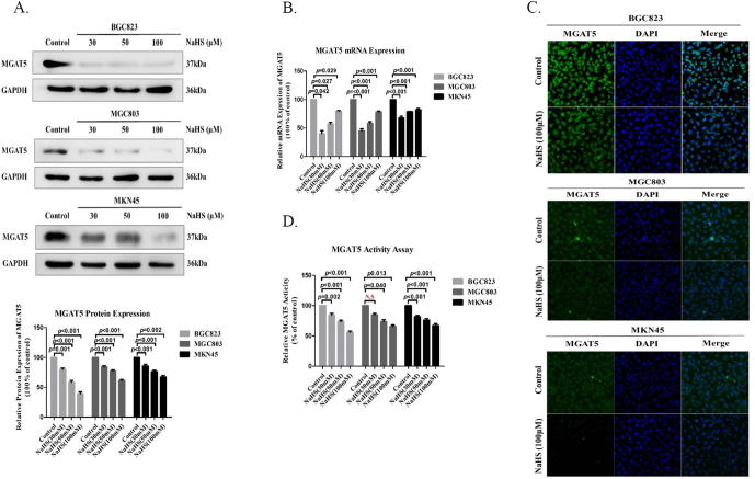
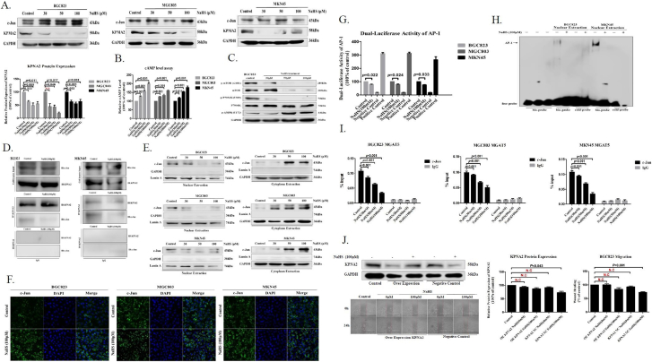
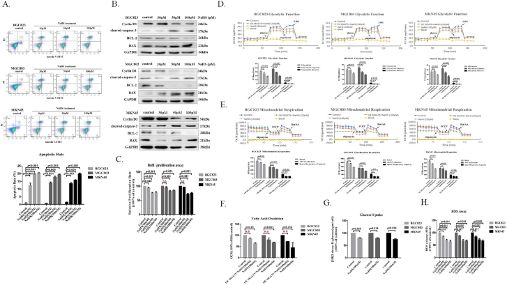
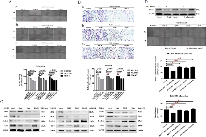
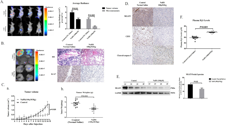
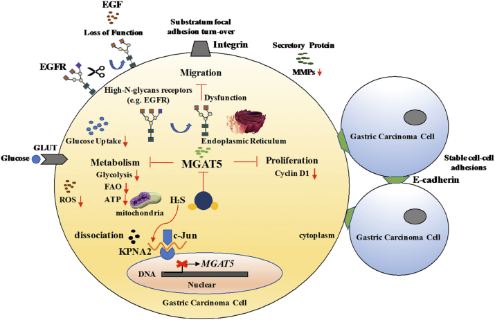
Mannosyl (alpha-1,6-)-Glycoprotein beta-1,6-N-acetyl-glucosaminyltransferase (MGAT5) is exclusively expressed in gastric carcinoma, and plays an essential role in cancer progression, but no targeted drug is available so far. The potential anti-cancer effect of Hydrogen Sulfide (H2S), has not been widely recognized. It intrigued broad interest to explore the clinical benefits of cancer therapy, with the current understanding of molecular mechanisms of H2S which remains very limited. In this study, we identify that H2S is an effective inhibitor of MGAT5, leading to reduce the expression of exclusively abnormal glycoprotein processes in gastric carcinoma. H2S specifically dissociation of karyopherin subunit alpha-2 (KPNA2) with Jun proto-oncogene (c-Jun) interaction, and blocking c-Jun nuclear translocation, and downregulation of MGAT5 expression at the level of gene and protein. Consequently, H2S impairs growth and metastasis in gastric carcinoma by targeting inhibits MGAT5 activity. In an animal tumor model study, H2S is well tolerated, inhibits gastric carcinoma growth and metastasis. Our preclinical work therefore supports that H2S acts as a novel inhibitor of MGAT5 that block tumorigenesis in gastric carcinoma.

Significance: This study shows that H2S can effective targeting inhibits MGAT5 activity, and demonstrates promising antitumor efficacy. These findings gain mechanistic insights into the anti-cancer capacity of H2S and may provide useful information for the clinical explorations of H2S in cancer treatment.


Figures

Figure 1
Figure 1
H2S downregulates the expression of MGAT5 and inhibits its activity. (A). After treatment with various concentrations of NaHS for 24 h, cell extracts were prepared and applied to immunoblotting with MGAT5. GAPDH was used as a loading control. (B). quantitative real-time PCR analysis the expression of MGAT5 mRNA in GC cells after 24 hours with treatment NaHS at various concentrations. (C). Immunofluorescence staining of MGAT5 in GC cells treated with NaHS at 100 μM after 24 hours. (D). The effect of H2S on MGAT5 activity inhibition in different cells. Various concentrations of NaHS were added to GC cells. The activity of MGAT5 was determined by the HPLC methods using pyridiylaminated GlcNAc2Man3GlcNAc2 as acceptor substrate in the absence of Mn2+. Each bar represents the means ± S.D. of three independent experiments.
Figure 2
Figure 2
H2S inhibits MGAT5 activity though specifically dissociation of KPNA2 with c-Jun interaction. (A). After treatment with various concentrations of NaHS for 24 h, cell extracts were prepared and applied to immunoblotting with c-jun and KPNA2. GAPDH was used as a loading control. (B). The cAMP activity was assayed by HTRF. Various concentrations of NaHS were added to GC cells. (C). Western blotting of protein on the mTOR signaling pathway in BGC823 cells after 24 hours with NaHS treatment indicated various concentrations. GAPDH was used as a loading control. (D). Co-Immunoprecipitation detection the relationship between KPNA2 and c-Jun with NaHS treatment after 24 hours in BGC823 and MKN45. (E). Western blotting assayed c-Jun from nuclear and cytoplasmic extracts of GC cells treated with various concentrations for 24 hours. Lamin A and GAPDH was used as a loading control. (F). Immunofluorescence staining of c-Jun in GC cells treated with NaHS at 100 μM after 24 hours. (G). Luciferase reporter assay measuring AP-1 activity in GC cells transiently co-transfected with pAP-1-Luc with NaHS treatment for 24 hours at 100 μM. (H). EMSA assay of DNA binding to AP-1 in the nuclei extracts from BGC823 and MKN45 cells after NaHS treatment for 24 hours at 100 μM. Results are repetitive from at least two independent experiments. (I). GC cells were incubated with NaHS and analyzed by a quantitative ChIP assay with anti-c-Jun antibody. (J). The inhibitory effect of H2S on migration of serum free stimulated stably KPNA2 over-expression in BGC823. Each bar represents the means ± S.D. of three independent experiments.
Figure 3
Figure 3
H2S suppresses MGAT5-promoted GC cells growth. (A). FACS analysis of apoptosis in GC cells after 24 hours with treatment NaHS at various concentrations. (B). After treatment with various concentrations of NaHS for 24 h, cell extracts were prepared and applied to immunoblotting with apoptosis relevant protein. GAPDH was used as a loading control. (C). BrdU proliferation assay measuring GC cells proliferation capacity with NaHS treatment on various concentrations. Seahorse XF24 Extracellular Flux Analyzer examined the inhibitory effect of H2S on cellular metabolism capacity of serum free stimulated stably MGAT5 over-expression in GC cells with NaHS treatment for 200 minutes at 100 μM. (D). Glycolytic Function. (E). Mitochondrial Respiration. (F). Fatty Acid Oxidation. (G). The effect of NaHS solution on 2-deoxtglucose uptake in GC cells. (H). Chemiluminescence analysis assayed the level of ROS in GC cells after 24 hours with treatment NaHS at various concentrations. Each bar represents the means ± S.D. of three independent experiments.
Figure 4
Figure 4
H2S inhibits MGAT5-promoted GC cells migration and invasion. (A). Wound healing assayed that the inhibitory effect of H2S to migration for serum free stimulated GC cells. Up panel: treatment of H2S, the migration capacity was detected. a. BGC823, b. MGC803, and c. MKN45. Down panel: quantification of the inhibition activity of H2S on migration. (B). Transwell assayed that the inhibitory effect of H2S to invasion for serum free stimulated GC cells. Up panel: treatment of H2S, the invasion capacity was detected. a. BGC823, b. MGC803, and c. MKN45. Down panel: quantification of the inhibition activity of H2S on invasion. (C). Western blotting assayed EMT relevant protein on GC cells after 24 hours of treatment with NaHS at various concentrations. GAPDH was used as a loading control. (D). The inhibitory effect of H2S on migration of serum free stimulated MGAT5 over-expression in BGC823. Each bar represents the means ± S.D. of three independent experiments.
Figure 5
Figure 5
H2S inhibits tumor growth and metastasis in gastric carcinoma Xenografts. (A). Effect of H2S on lung metastasis of human gastric carcinoma cell BGC823 in mice orthotopic Xenotransplantation model. Left. representative bioluminescence imaging of metastatic nodules on lungs. Right. The BGC823 colonies were measured. (n = 3 flanks and 4 mice in each group). (B). Effect of H2S on lung metastasis of human gastric carcinoma cell BGC823 in mice orthotopic Xenotransplantation model. Left. representative bioluminescence imaging of metastatic nodules on lungs. Right. HE staining and IHC staining of Ki67 representative photograph of metastatic nodules on lungs. (C). Tumor growth inhibition upon H2S treatment in BGC823 gastric carcinoma mice subcutaneous Xenografts tumor model. a. The curve of tumor growth after 15-days treatment of H2S. b. Experimental inhibitory effects of H2S on BGC823 Xenografts in nude mice. The percentage of relative tumor volume inhibition values was measured on the last day during the experiment. (D). Effect of H2S against primary tumor growth and angiogenesis. A typical photograph of IHC staining of MGAT5, CD31, and cleaved-caspase-3. (E). Inhibition of the expression of MGAT5 in BGC823 mice orthotopic Xenotransplantation model by H2S. Mice were humanely euthanized on the last day at 2 hours post-administration of H2S and the tumors were resected. Equal amounts of proteins of tumor tissues were evaluated for expression of MGAT5 levels. (F). The level of H2S in the plasma of mice orthotopic Xenotransplantation model. Data are shown as means ± S.D. (n = 3 flanks and 4 mice in each group).
Figure 6
Figure 6
The potential toxicity of H2S in vitro and in vivo. (A). Body weight of tumor-bearing mice measured at the indicated times. Apoptotic and metabolism assayed at the indicated concentration by FACS and Seahorse XF24 extracellular Flux Analyzer in human normal gastric cells GES-1. (B). Representative FACS plots and quantitative data of apoptotic rate. Treatment of H2S, (C). Glycolysis Function and (D). Mitochondrial Respiration rates were detected. Each bar represents the means ± S.D. of three independent experiments.
Figure 7
Figure 7
Hydrogen Sulfide demonstrates promising antitumor efficacy in gastric carcinoma by targeting MGAT5. Schematic illustration of our systems biology approach to identify H2S may be an important MGAT5 inhibitor and their corresponding targets. H2S specifically dissociation of KPNA2 with c-Jun interaction, and blocking c-Jun nuclear translocation, and downregulation of MGAT5 expression at the level of gene and protein. H2S inhibited MGAT5 activity lead to suppress metabolism, substratum focal adhesion turn-over, reduce the expression of exclusively abnormal glycoprotein processes, and disturb cell cyclin. It provides insights into a better understanding of the molecular mechanisms of H2S in anti-cancer effects which are required for further application in translational medicine.

Similar articles

Cited by

References

    1. Freddie B, Ahmedin J, Nathan G, Jacques F, David F. Global cancer transitions according to the Human Development Index (2008-2030): a population-based study. Lancet Oncol. 2012;13:790–801. - PubMed
    1. Huang B, Sun Z, Wang Z, Lu C, Zhao B, Xu H. Factors associated with peritoneal metastasis in non-serosa-invasive gastric cancer: a retrospective study of a prospectively-collected database. BMC Cancer. 2013;131:57. - PMC - PubMed
    1. Croci DO, Cerliani JP, Dalottomoreno T, Mendezhuergo SP, Mascanfroni ID, Dergandylon S, Rabinovich GA. Glycosylation-Dependent Lectin-Receptor Interactions Preserve Angiogenesis in Anti-VEGF Refractory Tumors. Cell. 2014;156:744–758. - PubMed
    1. Granovsky M, Fata JE, Pawling J, Muller WJ, Khokha R, Dennis JW. Suppression of tumor growth and metastasis in Mgat5-deficient mice. Nat Med. 2000;6:306–312. - PubMed
    1. Partridge EA, Roy CL, Guglielmo GM, Pawling J, Cheung P, Granovsky M, Dennis JW. Regulation of cytokine receptors by Golgi N-glycan processing and endocytosis. Science. 2004;306:120–124. - PubMed