. 2018 Apr 1;59(5):2167-2176.
doi: 10.1167/iovs.17-23304.

Importance of Considering the Middle Capillary Plexus on OCT Angiography in Diabetic Retinopathy

Affiliations

Importance of Considering the Middle Capillary Plexus on OCT Angiography in Diabetic Retinopathy

Alex C Onishi et al. Invest Ophthalmol Vis Sci. .

Abstract

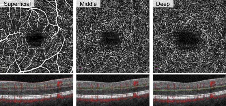
Purpose: To quantify microvasculature changes in the superficial (SCP), middle (MCP), and deep capillary plexuses (DCP) in diabetic retinopathy (DR).

Methods: Retrospective cross-sectional study at a tertiary academic referral center, in which 26 controls (44 eyes), 27 diabetic subjects without retinopathy (44 eyes), 32 subjects with nonproliferative retinopathy (52 eyes), and 27 subjects with proliferative retinopathy (40 eyes) were imaged with optical coherence tomography angiography (OCTA). Outcome measures included parafoveal vessel density (VD), percentage area of nonperfusion (PAN), and adjusted flow index (AFI) at the different plexuses.

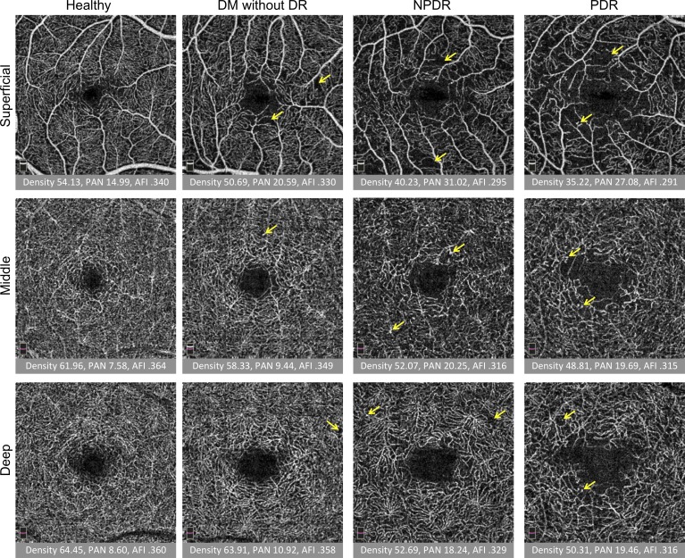
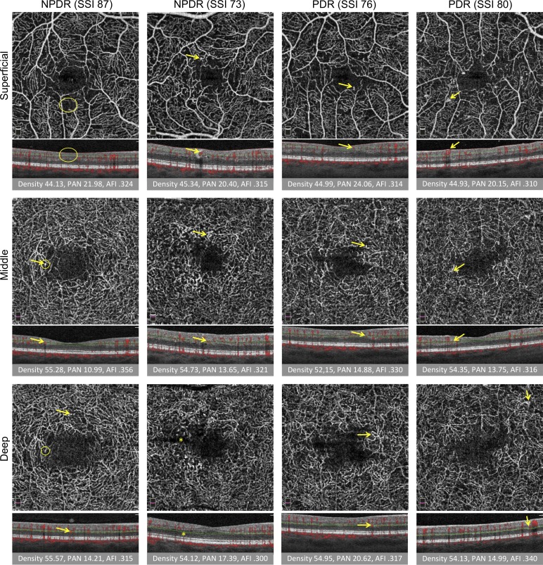
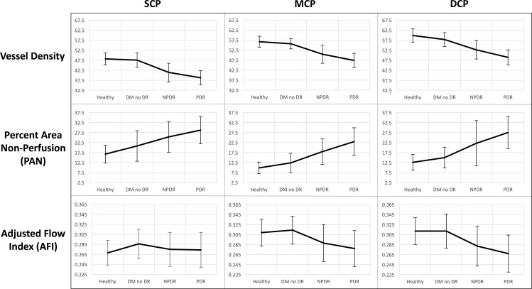
Results: MCP VD and MCP AFI decreased with worsening DR, while PAN increased, mirroring changes within the DCP. The fitted regression line for MCP and DCP AFI were significantly different than the SCP, while DCP PAN differed from SCP PAN with disease progression. Higher SCP AFI and PAN were different in eyes with diabetes without retinopathy compared with controls. Unexpectedly, sex was found to independently influence MCP VD and AFI with worsening disease.

Conclusions: OCTA parameters in the MCP and DCP displayed parallel changes with DR progression, different from the SCP, emphasizing the importance of physiologic considerations in the retinal capillaries. Thus, segmentation protocols that include the MCP within the SCP may be confounded. A difference in DCP PAN with worsening DR was unmasked relative to a prior study that included the MCP with SCP. We confirm that SCP AFI and PAN may serve as early indicators of microvascular changes in DR and identify an interaction between sex and the MCP deserving further study.


Figures

Figure 1
Figure 1
Segmentation of three capillary plexuses on OCTA. Left eye of patient with DM without DR. En face (top row) and cross-sectional (bottom row) OCTA of the superficial (left), middle (center), and deep (right) capillary plexuses. The red and green lines on cross-sectional OCTA show the segmentation boundaries for each layer.
Figure 2
Figure 2
Quantitative analysis of three capillary plexuses in a series of eyes with increasing disease severity. Columns from left to right: healthy subject, DM without DR, NPDR, and PDR. Rows from top to bottom: superficial, middle, and deep capillary plexus. Under each image, the vessel density (density), PAN, and AFI are reported. Overall, density and AFI decreased and PAN increased with severity. Projection artifact is seen as superficial vessels are cast onto the deeper layers, but was minimized by the exclusion of the hyperreflective plexiform layers in the segmentation scheme. Arrows represent vascular abnormalities, including microaneurysms, dilated vessels, and neovascularization.
Figure 3
Figure 3
Vascular abnormalities in three capillary plexuses in NPDR and PDR. First two columns are NPDR and last two columns are PDR. SSI is reported. Rows from top to bottom: SCP, MCP, and DCP. Under each image, the corresponding B-scan shows red flow overlay and red and green segmentation boundaries. Below B-scans, the vessel density (density), PAN, and AFI are reported. Arrows represent vascular abnormalities, including microaneurysms, dilated vessels, intraretinal microvascular abnormalities, and neovascularization. Large oval (SCP, left) represents nonperfusion, small circles (DCP, left) indicate one instance of projection artifact of a microaneurysm from the MCP onto the DCP. Ovals and arrows on the B-scan correspond to the en face to show the location of the abnormality within the retina. Projection artifact was minimized using our segmentation scheme, as discussed. Straight dark line in the deep plexus results from a failure in the segmentation algorithm (asterisk).
Figure 4
Figure 4
OCTA parameters across disease groups graphed continuously. Columns from left to right: SCP, MCP, and DCP. Rows from top to bottom: mean values for parafoveal vessel density, PAN, and AFI, respectively, graphically represented across healthy controls, eyes with DM without DR, NPDR, and PDR. Error bars: 1 SD.

References

    1. Nesper PL, Soetikno BT, Zhang HF, Fawzi AA. . OCT angiography and visible-light OCT in diabetic retinopathy. Vision Res. 2017; 139: 191– 203. - PMC - PubMed
    1. Miwa Y, Murakami T, Suzuma K,et al. . Relationship between functional and structural changes in diabetic vessels in optical coherence tomography angiography. Sci Rep. 2016; 6: 29064. - PMC - PubMed
    1. Couturier A, Mane V, Bonnin S,et al. . Capillary plexus anomalies in diabetic retinopathy on optical coherence tomography angiography. Retina. 2015; 35: 2384– 2391. - PubMed
    1. Mo S, Krawitz B, Efstathiadis E,et al. . Imaging foveal microvasculature: optical coherence tomography angiography versus adaptive optics scanning light ophthalmoscope fluorescein angiography. Invest Ophthalmol Vis Sci. 2016; 57: OCT130– OCT140. - PMC - PubMed
    1. Bhanushali D, Anegondi N, Gadde SG,et al. . Linking retinal microvasculature features with severity of diabetic retinopathy using optical coherence tomography angiography. Invest Ophthalmol Vis Sci. 2016; 57: OCT130– OCT140. - PubMed

Publication types