

Heat-stress-modulated induction of NF-κB leads to brucellacidal pro-inflammatory defense against Brucella abortus infection in murine macrophages and in a mouse model
- PMID: 29801438
- PMCID: PMC5970535
- DOI: 10.1186/s12866-018-1185-9
Heat-stress-modulated induction of NF-κB leads to brucellacidal pro-inflammatory defense against Brucella abortus infection in murine macrophages and in a mouse model
Abstract
Background: Brucella causes a chronic and debilitating infection that leads to great economic losses and a public health burden. In this study, we demonstrated the brucellacidal effect of heat shock mediated by the induction of pro-inflammatory cytokines, reactive oxygen species (ROS) accumulation and apoptosis in murine macrophages and in mice.
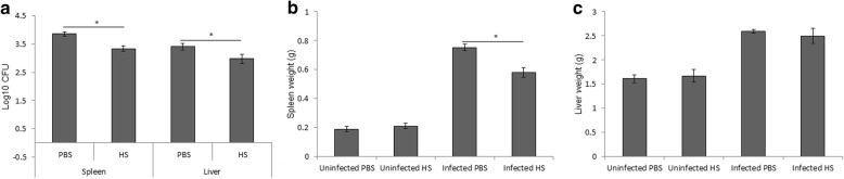
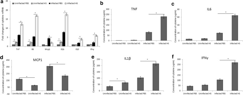
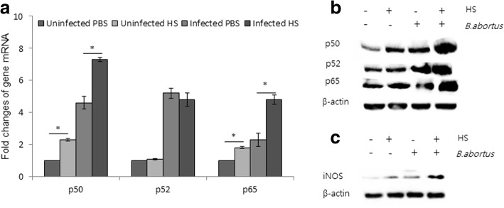
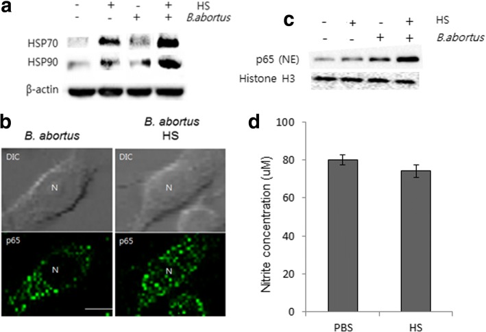
Results: RAW264.7 cells were incubated at 43 °C, and BALB/c mice were subjected to whole body hyperthermia. The data showed a reduction in bacterial survival in the mice after daily heat exposure. This was accompanied by increased levels of cytokines TNF, IL-6, IL-1β and IFN-γ in the sera of the mice. Gene expression of NF-κB and inducible nitric oxide production were also induced in the mouse splenic cells. In parallel with the bacterial reduction in the mouse model, an increased bactericidal effect was observed in RAW264.7 cells after exposure to heat stress. In addition, the heat stress increased both the nuclear translocation of NF-κB and the expression of the heat shock proteins HSP70 and HSP90 in murine macrophages. Furthermore, heat exposure induced the increase of pro-inflammatory cytokines, ROS accumulation and apoptosis but did not affect the production of nitric oxide (NO) in macrophages.
Conclusion: This study demonstrated the induction of innate immune responses by heat stress that significantly reduced the intracellular survival of B. abortus in vitro and in vivo. Transcriptional factor NF-κB, which is a master regulator, could be termed a key activator of heat-induced immunity against Brucella. The increase in the expression and activation of NF-κB in splenic cells and macrophages was followed by enhanced antimicrobial effectors, including cytokines, ROS and NO that may contribute to the reduction of bacterial survival.
Keywords: B. abortus; Heat stress; Macrophage; NF-κB; ROS.
Conflict of interest statement
Ethics approval and consent to participate
All of the procedures performed were reviewed and approved by the Animal Ethical Committee of Gyeongsang National University (Authorization Number GNU-120423-M0012).
Competing interests
The authors declare that they have no competing interests.
Publisher’s Note
Springer Nature remains neutral with regard to jurisdictional claims in published maps and institutional affiliations.
Figures
Similar articles
-
TLR2 and TLR4 signaling pathways are required for recombinant Brucella abortus BCSP31-induced cytokine production, functional upregulation of mouse macrophages, and the Th1 immune response in vivo and in vitro.Cell Mol Immunol. 2014 Sep;11(5):477-94. doi: 10.1038/cmi.2014.28. Epub 2014 Apr 28. Cell Mol Immunol. 2014. PMID: 24769793 Free PMC article.
-
Activation of NF-kB-Mediated TNF-Induced Antimicrobial Immunity Is Required for the Efficient Brucella abortus Clearance in RAW 264.7 Cells.Front Cell Infect Microbiol. 2017 Oct 9;7:437. doi: 10.3389/fcimb.2017.00437. eCollection 2017. Front Cell Infect Microbiol. 2017. PMID: 29062811 Free PMC article.
-
Inflammatory response of TLR4 deficient spleen macrophages (CRL 2471) to Brucella abortus S19 and an isogenic ΔmglA deletion mutant.Int J Med Microbiol. 2016 May;306(3):141-51. doi: 10.1016/j.ijmm.2016.02.006. Epub 2016 Feb 20. Int J Med Microbiol. 2016. PMID: 26946956
-
The role of innate immune signals in immunity to Brucella abortus.Front Cell Infect Microbiol. 2012 Oct 25;2:130. doi: 10.3389/fcimb.2012.00130. eCollection 2012. Front Cell Infect Microbiol. 2012. PMID: 23112959 Free PMC article. Review.
-
Macrophages and Brucella.Immunol Ser. 1994;60:363-80. Immunol Ser. 1994. PMID: 8251581 Review.
Cited by
-
Comprehensive transcriptome analysis based on RNA sequencing identifies critical genes for lipopolysaccharide-induced epididymitis in a rat model.Asian J Androl. 2019 Nov-Dec;21(6):605-611. doi: 10.4103/aja.aja_21_19. Asian J Androl. 2019. PMID: 31044753 Free PMC article.
-
NF‑κB/IκBα signaling pathways are essential for resistance to heat stress‑induced ROS production in pulmonary microvascular endothelial cells.Mol Med Rep. 2021 Nov;24(5):814. doi: 10.3892/mmr.2021.12454. Epub 2021 Sep 24. Mol Med Rep. 2021. PMID: 34558646 Free PMC article.
-
Intracellular Growth Inhibition and Host Immune Modulation of 3-Amino-1,2,4-triazole in Murine Brucellosis.Int J Mol Sci. 2023 Dec 11;24(24):17352. doi: 10.3390/ijms242417352. Int J Mol Sci. 2023. PMID: 38139181 Free PMC article.
-
The Role and Mechanism of S1PR5 in Colon Cancer.Cancer Manag Res. 2020 Jun 19;12:4759-4775. doi: 10.2147/CMAR.S239118. eCollection 2020. Cancer Manag Res. 2020. Retraction in: Cancer Manag Res. 2021 Jul 15;13:5723. doi: 10.2147/CMAR.S329184. PMID: 32606966 Free PMC article. Retracted.
-
Loci Associated With Antibody Response in Feral Swine (Sus scrofa) Infected With Brucella suis.Front Vet Sci. 2020 Nov 25;7:554674. doi: 10.3389/fvets.2020.554674. eCollection 2020. Front Vet Sci. 2020. PMID: 33324693 Free PMC article.
References
Publication types
MeSH terms
Substances
LinkOut - more resources
Full Text Sources
Other Literature Sources
