

Patient-derived multicellular tumor spheroids towards optimized treatment for patients with hepatocellular carcinoma
- PMID: 29801504
- PMCID: PMC5970513
- DOI: 10.1186/s13046-018-0752-0
Patient-derived multicellular tumor spheroids towards optimized treatment for patients with hepatocellular carcinoma
Abstract
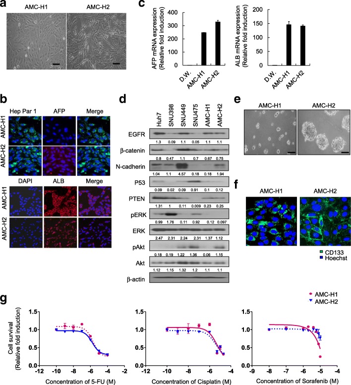
Background: Hepatocellular carcinoma (HCC) is one of the most common malignant tumors worldwide and has poor prognosis. Specially, patients with HCC usually have poor tolerance of systemic chemotherapy, because HCCs develop from chronically damaged tissue that contains considerable inflammation, fibrosis, and cirrhosis. Since HCC exhibits highly heterogeneous molecular characteristics, a proper in vitro system is required for the study of HCC pathogenesis. To this end, we have established two new hepatitis B virus (HBV) DNA-secreting HCC cell lines from infected patients.
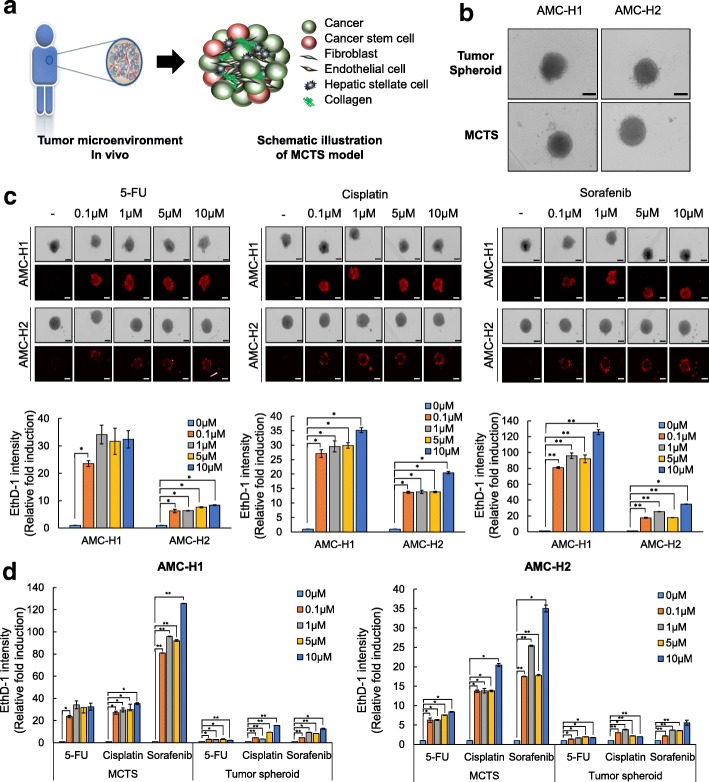
Methods: Based on these two new HCC cell lines, we have developed chemosensitivity assays for patient-derived multicellular tumor spheroids (MCTSs) in order to select optimized anti-cancer drugs to provide more informative data for clinical drug application. To monitor the effect of the interaction of cancer cells and stromal cells in MCTS, we used a 3D co-culture model with patient-derived HCC cells and stromal cells from human hepatic stellate cells, human fibroblasts, and human umbilical vein endothelial cells to facilitate screening for optimized cancer therapy.
Results: To validate our system, we performed a comparison of chemosensitivity of the three culture systems, which are monolayer culture system, tumor spheroids, and MCTSs of patient-derived cells, to sorafenib, 5-fluorouracil, and cisplatin, as these compounds are typically standard therapy for advanced HCC in South Korea.
Conclusion: In summary, these findings suggest that the MCTS culture system is the best methodology for screening for optimized treatment for each patients with HCC, because tumor spheroids not only mirror the 3D cellular context of the tumors but also exhibit therapeutically relevant pathophysiological gradients and heterogeneity of in vivo tumors.
Keywords: Hepatocellular carcinoma (HCC); MCTS-based chemosensitivity assays; Multicellular tumor spheroids (MCTS); Optimized treatment.
Conflict of interest statement
Ethics approval and consent to participate
The study was conducted in accordance with the Declaration of Helsinki principles. It was approved by the Human Research Ethics Committee of ASAN medical center. The institute review board in ASAN medical center complies with the related laws such as ICH, KGCP or bioethics and safety act. Written informed consent for the use of tissues for research was taken from patients at the time of procurement of tumor specimens.
Competing interests
The authors declare that they have no competing interests.
Publisher’s Note
Springer Nature remains neutral with regard to jurisdictional claims in published maps and institutional affiliations.
Figures

Similar articles
-
Activated hepatic stellate cells play pivotal roles in hepatocellular carcinoma cell chemoresistance and migration in multicellular tumor spheroids.Sci Rep. 2016 Nov 17;6:36750. doi: 10.1038/srep36750. Sci Rep. 2016. PMID: 27853186 Free PMC article.
-
Inhibitors of Na+/K+ ATPase exhibit antitumor effects on multicellular tumor spheroids of hepatocellular carcinoma.Sci Rep. 2020 Mar 24;10(1):5318. doi: 10.1038/s41598-020-62134-4. Sci Rep. 2020. PMID: 32210281 Free PMC article.
-
Identification of hepatic fibrosis inhibitors through morphometry analysis of a hepatic multicellular spheroids model.Sci Rep. 2021 May 25;11(1):10931. doi: 10.1038/s41598-021-90263-x. Sci Rep. 2021. PMID: 34035369 Free PMC article.
-
Applicability of tumor spheroids for in vitro chemosensitivity assays.Expert Opin Drug Metab Toxicol. 2019 Jan;15(1):15-23. doi: 10.1080/17425255.2019.1554055. Epub 2018 Dec 2. Expert Opin Drug Metab Toxicol. 2019. PMID: 30484335 Review.
-
Deciphering the underlying mechanism of liver diseases through utilization of multicellular hepatic spheroid models.BMB Rep. 2023 Apr;56(4):225-233. doi: 10.5483/BMBRep.2023-0010. BMB Rep. 2023. PMID: 36814078 Free PMC article. Review.
Cited by
-
Therapeutic Potential of CUDC-907 (Fimepinostat) for Hepatocarcinoma Treatment Revealed by Tumor Spheroids-Based Drug Screening.Front Pharmacol. 2021 Oct 29;12:658197. doi: 10.3389/fphar.2021.658197. eCollection 2021. Front Pharmacol. 2021. PMID: 34776939 Free PMC article.
-
Three-dimensional (3D) cell culture: a valuable step in advancing treatments for human hepatocellular carcinoma.Cancer Cell Int. 2022 Jul 30;22(1):243. doi: 10.1186/s12935-022-02662-3. Cancer Cell Int. 2022. PMID: 35908054 Free PMC article. Review.
-
Reconstructing the hepatocellular carcinoma microenvironment: the current status and challenges of 3D culture technology.Discov Oncol. 2025 Apr 10;16(1):506. doi: 10.1007/s12672-025-02290-z. Discov Oncol. 2025. PMID: 40208520 Free PMC article. Review.
-
Non-Destructive Tumor Aggregate Morphology and Viability Quantification at Cellular Resolution, During Development and in Response to Drug.Acta Biomater. 2020 Nov;117:322-334. doi: 10.1016/j.actbio.2020.09.042. Epub 2020 Sep 29. Acta Biomater. 2020. PMID: 33007490 Free PMC article.
-
LncRNA NEAT1 modulates sorafenib resistance in hepatocellular carcinoma through regulating the miR-149-5p/AKT1 axis.Saudi J Gastroenterol. 2020 May 26;26(4):194-203. doi: 10.4103/sjg.SJG_4_20. Online ahead of print. Saudi J Gastroenterol. 2020. PMID: 32461380 Free PMC article.
References
-
- Tovar V, Alsinet C, Villanueva A, Hoshida Y, Chiang DY, Sole M, Thung S, Moyano S, Toffanin S, Minguez B, et al. IGF activation in a molecular subclass of hepatocellular carcinoma and pre-clinical efficacy of IGF-1R blockage. J Hepatol. 2010;52(4):550–559. doi: 10.1016/j.jhep.2010.01.015. - DOI - PMC - PubMed
MeSH terms
Grants and funding
LinkOut - more resources
Full Text Sources
Other Literature Sources
Medical
