. 2018 Jun 19;48(6):1220-1232.e5.
doi: 10.1016/j.immuni.2018.04.009. Epub 2018 May 22.

Autoimmune Th17 Cells Induced Synovial Stromal and Innate Lymphoid Cell Secretion of the Cytokine GM-CSF to Initiate and Augment Autoimmune Arthritis

Affiliations

Autoimmune Th17 Cells Induced Synovial Stromal and Innate Lymphoid Cell Secretion of the Cytokine GM-CSF to Initiate and Augment Autoimmune Arthritis

Keiji Hirota et al. Immunity. .

Abstract

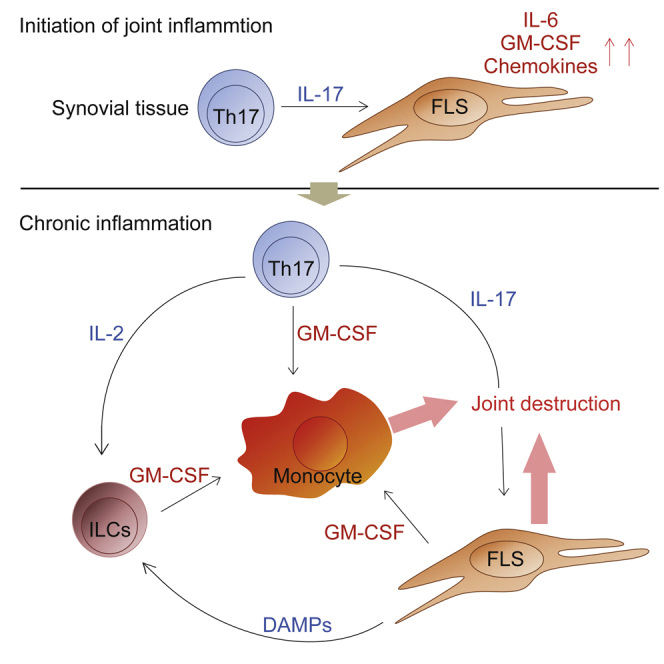
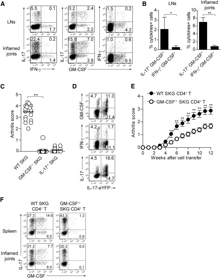
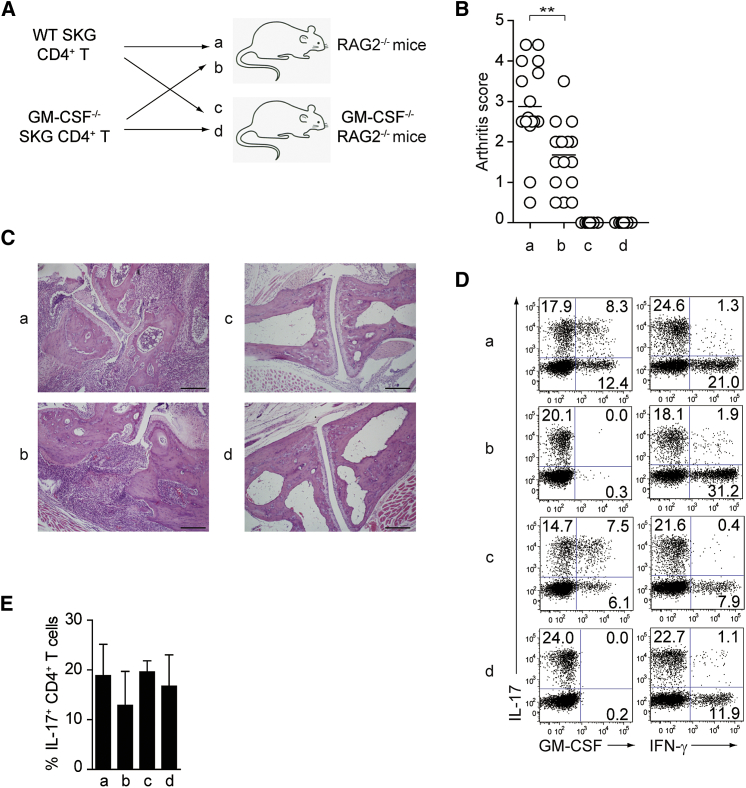
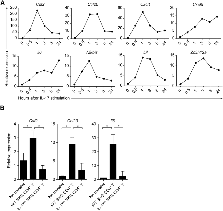
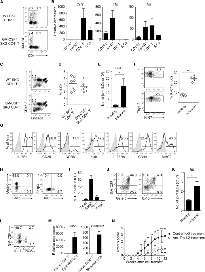
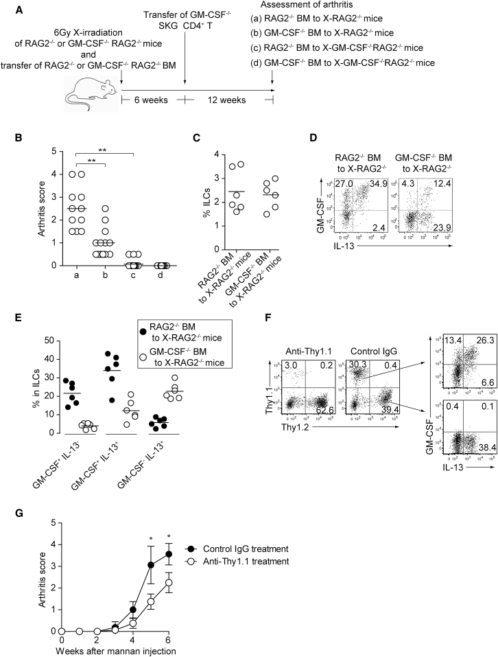
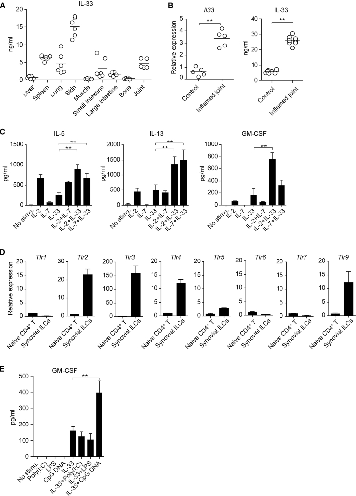
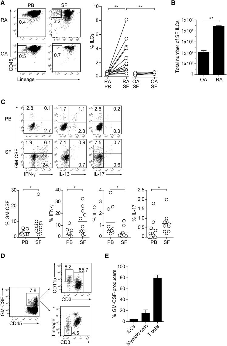
Despite the importance of Th17 cells in autoimmune diseases, it remains unclear how they control other inflammatory cells in autoimmune tissue damage. Using a model of spontaneous autoimmune arthritis, we showed that arthritogenic Th17 cells stimulated fibroblast-like synoviocytes via interleukin-17 (IL-17) to secrete the cytokine GM-CSF and also expanded synovial-resident innate lymphoid cells (ILCs) in inflamed joints. Activated synovial ILCs, which expressed CD25, IL-33Ra, and TLR9, produced abundant GM-CSF upon stimulation by IL-2, IL-33, or CpG DNA. Loss of GM-CSF production by either ILCs or radio-resistant stromal cells prevented Th17 cell-mediated arthritis. GM-CSF production by Th17 cells augmented chronic inflammation but was dispensable for the initiation of arthritis. We showed that GM-CSF-producing ILCs were present in inflamed joints of rheumatoid arthritis patients. Thus, a cellular cascade of autoimmune Th17 cells, ILCs, and stromal cells, via IL-17 and GM-CSF, mediates chronic joint inflammation and can be a target for therapeutic intervention.

Keywords: GM-CSF; IL-17; ILCs; SKG; Th17; arthritis; autoimmunity; innate lymphoid cells.


Figures

None
Graphical abstract
Figure 1
Figure 1
GM-CSF-Producing T Helper Cells Are Dispensable for GM-CSF-Dependent Autoimmune Arthritis Development (A) Intracellular IL-17, IFN-γ, and GM-CSF staining of CD4+ T cells from popliteal LNs or inflamed joints. (B) Proportion of cytokine-producing cells in CD4+ T cells from individual mice as shown in (A). Vertical bars mean SD (n = 3). (C) Arthritis scores assessed in individual SKG, Csf2−/− SKG, or Il17a−/− SKG mice (n = 20 each) 3 months after single i.p. injection of 20 mg mannan. (D) Intracellular IL-17, IFN-γ, and GM-CSF staining of CD4+ T cells from inflamed joints of Il17aCre R26ReYFP SKG mice. (E) Arthritis development after adoptive transfer of CD4+ T cells from WT or Csf2−/− SKG mice into Rag2−/− mice (n = 17 each, SEM). The severity of arthritis was monitored every week after transfer of 1 × 106 CD4+ T cells. (F) Intracellular IL-17 and GM-CSF staining of CD4+ T cells from spleens and inflamed joints of Rag2−/− mice with CD4+ T cells transfer as shown in (E). p < 0.05, ∗∗p < 0.01. Data are representative of three independent experiments (A, B, D, and F) or pooled from three experiments (C and E).
Figure 2
Figure 2
GM-CSF from Non-T Cells Is Crucial for the Initiation of Autoimmune Arthritis (A) Experimental design of adoptive transfer of CD4+ T cells from WT or Csf2−/− SKG mice into Rag2−/− or Csf2−/−Rag2−/− mice. Arthritis scores of four groups (a–d) of mice were assessed 3 months after transfer of 1 × 106 CD4+ T cells. (B) Arthritis scores of the four groups mice (n = 15 or 16 each) shown in (A). Horizontal bars indicate the means. (C) Representative joint histology of the groups shown in (A). Scale bars indicate 200 μm. (D) Flow cytometry of splenic CD4+ T cells stained for intracellular IL-17 and GM-CSF or IFN-γ. (E) Proportion of IL-17-producing CD4+ T cells from individual mice as shown in (D). Vertical bars mean SD (n = 3). ∗∗p < 0.01. Data are representative of three independent experiments (C–E) or pooled from three independent experiments (B).
Figure 3
Figure 3
Induction of GM-CSF in FLSs Stimulated with IL-17 (A) Quantitative RT-PCR analysis for the expression of designated genes in IL-17-stimulated FLSs. FLSs (2.5 × 104) were stimulated with 50 ng/mL rmIL-17 and harvested at the indicated time points. mRNA expression is presented relative to the expression of Hprt1. (B) Quantitative RT-PCR analysis for the expression of designated genes in synoviocytes from Rag2−/− mice with CD4+ T cell transfer. CD45Podoplanin+ synoviocytes (3 × 104) were sorted from inflamed joints of Rag2−/− mice 4 weeks after transfer of 1 × 106 SKG or Il17a−/− SKG CD4+ T cells. Vertical bars mean SD (n = 3). Data are representative of two independent experiments.
Figure 4
Figure 4
GM-CSF-Producing ILCs in Inflamed Joints (A) Flow cytometry analysis of GM-CSF expression by CD4+ T cells and CD4 cells among CD45+ joint infiltrating cells in Rag2−/− mice transferred with CD4+ T cells from WT or Csf2−/− SKG mice. (B) Quantitative RT-PCR analysis of Csf2, Il1b, and Tnf in CD11b+Ly-6G (CD11b+), CD11b+Ly-6G+ (Ly-6G+), CD4+ T cells, and ILCs sorted from arthritic joints of mannan-treated SKG mice (n = 3). mRNA expression is presented relative to the expression of Hprt1. (C) Flow cytometry of joint infiltrating cells in Rag2−/− mice transferred with CD4+ T cells from WT or Csf2−/− SKG mice. Cells were stained for CD45.2 and lineage markers (a cocktail of CD3, CD4, CD8, CD11b, CD11c, CD19, and DX-5). (D) Proportion of ILCs in Rag2−/− mice transferred with CD4+ T cells as shown in (C). Each symbol represents an individual mouse. Horizontal bars indicate the means. (E) Total cell number of ILCs from healthy or inflamed joints of SKG mice (n = 3). (F) Flow cytometry of synovial ILCs (CD45.2+ lineage markers-negative Thy1.2+ cells as shown in C) for Ki-67 expression. (G) Flow cytometry of synovial ILCs (CD45.2+ lineage markers-negative Thy1.2+ cells as shown in C) for cell surface expression of IL-7Ra, CD25, CCR6, c-kit, IL-33Ra, CD44, and MHC2. (H) Flow cytometry of synovial ILCs (as shown in C) for intranuclear expression of the transcription factor T-bet, Gata-3, Rorγt, and Foxp3. (I) Proportion of the transcription factor-expressing synovial ILCs (n = 3) as shown in (H). (J) Flow cytometry of synovial ILCs (as shown in C) for the expression of GM-CSF, Gata-3, and IL-13. (K) Total cell numbers of ILCs from healthy or inflamed joints of C57/BL6 (B6) mice with collagen antibody-induced arthritis (n = 3). Data are representative of two independent experiments. (L) Flow cytometry of synovial ILCs for the expression of GM-CSF and FP635 in arthritic Il17aCre R26RFP635 SKG mice. (M) Quantitative RT-PCR analysis of Csf2 and Bhlhe40 in splenic naive CD25CD44loCD4+ T cells (naive CD4+ T) and synovial ILCs (n = 3) as shown in (C). (N) The effects of ILC depletion on arthritis development. CD4+ T cells (1 × 106) from Thy1.1+ SKG mice were adoptively transferred into Thy1.2+Rag2−/− mice, which were i.v. injected with 500 μg anti-Thy1.2 mAb or control Rat IgG every week (n = 19 each). The severity of arthritis was monitored every week. p < 0.05, ∗∗p < 0.01. Data are representative of three independent experiments in (A)–(C), (E)–(J), (L), and (M) and pooled from more than two experiments in (D), (F), and (N). Vertical bars mean SD in (B), (E), (I), (K), (M), and (N).
Figure 5
Figure 5
GM-CSF from ILCs and Radio-Resistant Stromal Cells Is Crucial for Autoimmune Arthritis (A) Preparation of experimental groups for assessing arthritogenic effects of GM-CSF from ILCs or radio-resistant stromal cells. Rag2−/− or Csf2−/−Rag2−/− mice were x-irradiated (X-Rag2−/− mice) and transferred with BM cells from Csf2−/− or WT Rag2−/− mice. The resulting four groups of BM chimeras were transferred with CD4+ T cells from Csf2−/− SKG mice 6 weeks after BM reconstitution and assessed for arthritis development 12 weeks later. (B) Arthritis scores of four groups of mice shown in (A). (C) Proportion of total synovial ILCs from x-irradiated Rag2−/− mice reconstituted with Csf2−/− or WT Rag2−/− BM cells. (D) Flow cytometry of synovial ILCs for the expression of GM-CSF and IL-13 in arthritic joints of BM chimeras shown in (C). (E) Proportion of GM-CSF+IL-13, GM-CSF+IL-13+, and GM-CSFIL-13+ synovial ILCs (n = 6 each) as shown in (D). (F and G) The effects of ILC depletion on arthritis development. Thy1.1+Rag2−/− mice were x-irradiated and transferred with BM cells from Thy1.1+Rag2−/− and Thy1.2+Csf2−/− SKG mice. The resulting BM chimeras were i.p. injected with 20 mg mannan 6 weeks later, followed by i.v. injection with 500 μg anti-Thy1.1 mAb or control Rat IgG every week. Flow cytometry of synovial ILCs for GM-CSF and IL-13 expression (F). Arthritis scores in each group of mice monitored every week (G). Vertical bars mean SD (n = 8 each). p < 0.05, ∗∗p < 0.01. Data are representative of three independent experiments in (D) and (F), and pooled from three experiments in (B), (C), (E), and (G). Horizontal bars indicate the means in (B), (C), and (E).
Figure 6
Figure 6
Control of GM-CSF Production in Synovial ILCs by IL-2, IL-33, and TLR-9 Ligands (A) Quantity of the active form IL-33 assessed by ELISA (Biolegend, Mouse IL-33 ELISA kit) in indicated tissue homogenates. (B) Quantitative RT-PCR for Il33 expression and ELISA for IL-33 protein in arthritic or control joints. Symbols represent individual mice. (C) Production of cytokines by synovial ILCs. Synovial ILCs (5 × 103) from arthritic joints were purified and cultured for 24 hr with rhIL-2 (20 U/mL), rmIL-7 (20 ng/mL), and rmIL-33 (20 ng/mL) alone or in combination. The concentration of IL-5, IL-13, and GM-CSF in the supernatant was measured (n = 3). (D) Quantitative RT-PCR analysis of the expression of indicated TLR genes in naive CD4+ T cells and synovial ILCs as shown in Figure 4M (n = 3). (E) GM-CSF production by synovial ILCs. Synovial ILCs (5 × 103) were cultured for 24 hr with poly(I:C) (1 μg/mL), LPS (1 μg/mL), and CpG DNA (1 μM) alone or in combination with rmIL-33 (20 ng/mL). The concentration of GM-CSF in the supernatant was measured (n = 3). ∗∗p < 0.01. Data are representative of at least two independent experiments. Horizontal bars indicate the means in (A) and (B). Vertical bars indicate SD in (C)–(E).
Figure 7
Figure 7
GM-CSF-Producing ILCs in Synovial Fluid of RA Patients (A) The presence of ILCs (defined as CD45+CD3CD4CD8CD11bCD11cCD19CD56) from peripheral blood (PB) or synovial fluid (SF) of a patient with RA or OA (left). The percentages of ILCs in PB and SF from individual RA (n = 13) or OA (n = 6) patients. The lines indicate the sample pairs of the same patients (right). (B) Total numbers of ILCs in 1 mL of SF from OA and RA patients (n = 6). Vertical bars indicate SD. (C) Flow cytometry analysis of IFN-γ, IL-13, IL-17, and GM-CSF expression by ILCs (gated as in A) in PB or SF of a RA patient (top). The percentages of cytokine-producing ILCs from individual RA patients (n = 11) (bottom). (D) Gating strategies for GM-CSF+CD45+ lineage markers-negative (ILCs), GM-CSF+CD45+CD3CD11b+ (myeloid cells), and GM-CSF+CD45+CD11bCD3+ cells (T cells). (E) Proportion of GM-CSF-producing cells (n = 3). Vertical bars indicate SD. Symbols represent individual samples. Horizontal bars indicate the means. p < 0.05, ∗∗p < 0.01.

Comment in

Similar articles

Cited by

References

    1. Alvaro-Gracia J.M., Zvaifler N.J., Firestein G.S. Cytokines in chronic inflammatory arthritis. IV. Granulocyte/macrophage colony-stimulating factor-mediated induction of class II MHC antigen on human monocytes: a possible role in rheumatoid arthritis. J. Exp. Med. 1989;170:865–875. - PMC - PubMed
    1. Alvaro-Gracia J.M., Zvaifler N.J., Brown C.B., Kaushansky K., Firestein G.S. Cytokines in chronic inflammatory arthritis. VI. Analysis of the synovial cells involved in granulocyte-macrophage colony-stimulating factor production and gene expression in rheumatoid arthritis and its regulation by IL-1 and tumor necrosis factor-alpha. J. Immunol. 1991;146:3365–3371. - PubMed
    1. Bando J.K., Liang H.E., Locksley R.M. Identification and distribution of developing innate lymphoid cells in the fetal mouse intestine. Nat. Immunol. 2015;16:153–160. - PMC - PubMed
    1. Bartok B., Firestein G.S. Fibroblast-like synoviocytes: key effector cells in rheumatoid arthritis. Immunol. Rev. 2010;233:233–255. - PMC - PubMed
    1. Behrens F., Tak P.P., Østergaard M., Stoilov R., Wiland P., Huizinga T.W., Berenfus V.Y., Vladeva S., Rech J., Rubbert-Roth A. MOR103, a human monoclonal antibody to granulocyte-macrophage colony-stimulating factor, in the treatment of patients with moderate rheumatoid arthritis: results of a phase Ib/IIa randomised, double-blind, placebo-controlled, dose-escalation trial. Ann. Rheum. Dis. 2015;74:1058–1064. - PMC - PubMed

Publication types

MeSH terms

Substances