Review
. 2018 May 1;32(9-10):620-638.
doi: 10.1101/gad.314351.118.

The impact of mitotic errors on cell proliferation and tumorigenesis

Affiliations
Review

The impact of mitotic errors on cell proliferation and tumorigenesis

Michelle S Levine et al. Genes Dev. .

Abstract

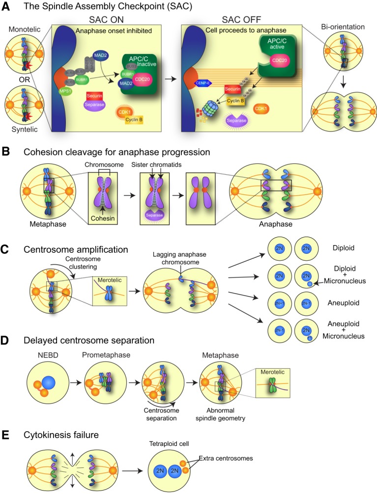
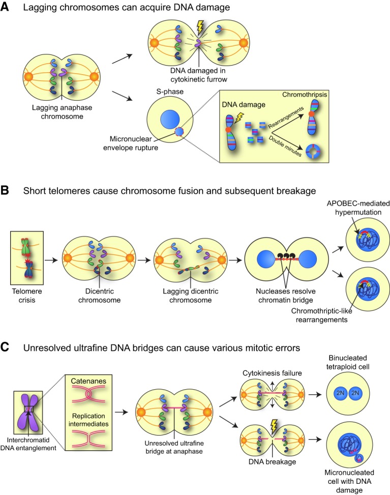
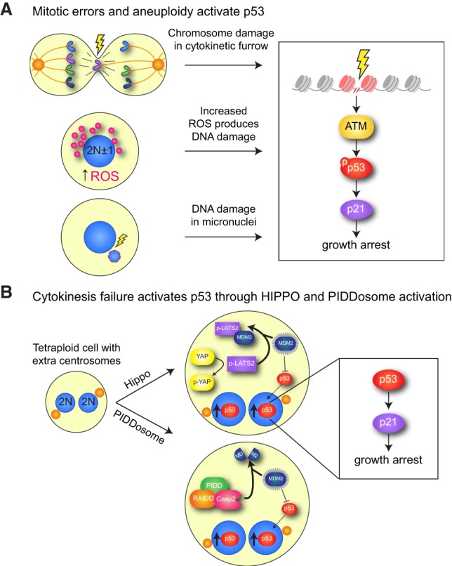
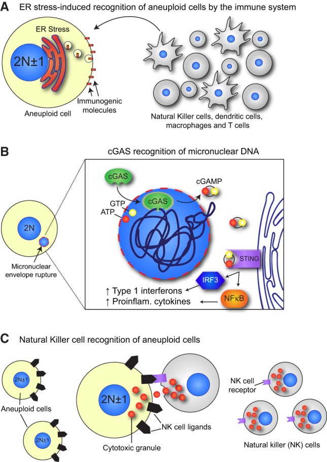
Mitosis is a delicate event that must be executed with high fidelity to ensure genomic stability. Recent work has provided insight into how mitotic errors shape cancer genomes by driving both numerical and structural alterations in chromosomes that contribute to tumor initiation and progression. Here, we review the sources of mitotic errors in human tumors and their effect on cell fitness and transformation. We discuss new findings that suggest that chromosome missegregation can produce a proinflammatory environment and impact tumor responsiveness to immunotherapy. Finally, we survey the vulnerabilities exposed by cell division errors and how they can be exploited therapeutically.

Keywords: aneuploidy; chromosomal instability; mitosis.


Figures

Figure 1.
Figure 1.
Chromosome segregation and sources of mitotic errors. (A) Unattached kinetochores activate an inhibitory SAC signal, which in turn blocks progression to anaphase. The target of the SAC is the APC/C, an E3 ubiquitin ligase that targets several proteins for degradation, including Cyclin B1 and Securin. When all kinetochores are correctly attached to MTs emerging from opposite poles of the cell (biorientation), the SAC is silenced, and APC/CCDC20 ubiquitinates and targets for degradation Cyclin B (to inactivate CDK1 and allow for mitotic exit) and Securin (to liberate the protease Separase and initiate the onset of anaphase). (B) Replicated sister chromatids are held together by the cohesin complex of proteins. Following silencing of the SAC, Securin is degraded, and the protease Separase is activated. Separase cleaves the cohesin complex to allow for sister chromatid separation and anaphase onset. (C) Extra centrosomes can generate a transient multipolar spindle, which, following centrosome clustering, leads to an increased rate of merotelic attachments, where one sister kinetochore is attached to MTs emerging from opposite poles. Merotelically attached chromosomes can lag in the middle of the spindle during anaphase and may subsequently be missegregated or incorporated into micronuclei. (D) After centrosome duplication, the two centrosomes are attached by a protein linker. This linker is disassembled prior to mitotic entry to allow the centrosomes to migrate apart and form opposite poles of the spindle. Delays in centrosome separation can lead to misattached chromosomes and/or abnormal spindle geometry that results in increased rates of chromosome missegregation. (E) Cleavage furrow regression leads to cytokinesis failure and the formation of a binucleate tetraploid cell with twice the normal centrosome content.
Figure 2.
Figure 2.
Mitotic errors can generate DNA damage. (A) Lagging chromosomes in anaphase can acquire DNA damage directly by being trapped in the spindle midzone during cytokinesis. In addition, lagging chromosomes that are partitioned into micronuclei can acquire DNA damage in interphase of the subsequent cell cycle. Extensive damage leads to chromosome shattering, a phenomenon known as chromothripsis, which results in the production of highly localized chromosome rearrangements and/or the production of double-minute chromosomes. (B) Extensive shortening of telomeres (telomere crisis) can result in the end-to-end fusion of two telomeres and the generation of a dicentric chromosome. Dicentric chromosomes can attach to opposite sides of the cell and be pulled apart during mitosis, resulting in a chromatin bridge that connects the two daughter nuclei. The nuclear membrane surrounding the bridging DNA ruptures in interphase, and the exposed DNA can be subject to cleavage by a cytoplasmic nuclease to resolve the bridge. The DNA exposed to the cytoplasm may undergo chromothriptic-like chromosome rearrangements and/or hypermutation generated by APOBEC cytidine deaminases. (C) DNA entanglements between sister chromatids can form at underreplicated regions or as a result of persistent DNA catenation. If these linkages are not resolved by topoisomerases and helicases, they can form ultrafine DNA bridges that connect the segregating sister chromatids in anaphase. Ultrafine bridges can lead to cytokinesis failure, resulting in a binucleated cell, or be broken during anaphase, creating DNA damage and micronuclei.
Figure 3.
Figure 3.
Mitotic errors can trigger activation of p53. (A) Lagging chromosomes that become damaged in the cleavage furrow or in micronuclei can elicit the canonical DNA damage repair pathway that activates p53. In addition, aneuploidy can also cause increased reactive oxidative species that lead to activation of DNA damage signaling. (B) Cytokinesis failure has been proposed to activate p53 through two distinct pathways. (1) Activation of the Hippo pathway. The Hippo pathway kinase LATS2 is activated in a tetraploid cell, leading to the phosphorylation and cytoplasmic sequestration of the transcription factor YAP. In addition, LATS2 binds and inactivates MDM2, a negative regulator of p53 stability. Inhibiting MDM2 allows for the increased accumulation of p53, which up-regulates p21 to elicit a growth arrest. (2) Activation of PIDDosome signaling. Tetraploid cells activate the PIDDosome, a multiprotein complex comprised of PIDD and RAIDD that in turn activates Caspase-2 (CASP2). Caspase 2 cleaves and inactivates MDM2, allowing p53 stabilization.
Figure 4.
Figure 4.
Mitotic errors activate the immune system. (A) Aneuploid cells exhibit a constitutive endoplasmic reticulum (ER) stress that leads to the increased surface exposure of immunogenic cell surface molecules, such as calreticulin. These are recognized by immune cells such as natural killer (NK) cells, dendritic cells, macrophages, and T cells that engulf or kill the aneuploid cell. (B) The micronuclear envelope is prone to rupture, leading to the exposure of the entrapped chromatin to cytoplasmic DNA-sensing molecules, such as cGAS. cGAS is activated by the exposed micronuclear DNA, allowing for conversion of ATP and GTP to the second messenger cGAMP. cGAMP activates STING, which causes IRF3- and NFκB-mediated expression of type 1 interferons and proinflammatory cytokines, respectively. (C) Aneuploid cells exhibit increased expression of NK cell-activating ligands, which allow recognition and killing of aneuploid cells by NK cells through their NKG2D and DNAM1 receptors.

References

    1. Ablasser A, Schmid-Burgk JL, Hemmerling I, Horvath GL, Schmidt T, Latz E, Hornung V. 2013. Cell intrinsic immunity spreads to bystander cells via the intercellular transfer of cGAMP. Nature 503: 530–534. - PMC - PubMed
    1. Acebes-Huerta A, Lorenzo-Herrero S, Folgueras AR, Huergo-Zapico L, Lopez-Larrea C, Lopez-Soto A, Gonzalez S. 2016. Drug-induced hyperploidy stimulates an antitumor NK cell response mediated by NKG2D and DNAM-1 receptors. Oncoimmunology 5: e1074378. - PMC - PubMed
    1. Andreassen PR, Lohez OD, Lacroix FB, Margolis RL. 2001. Tetraploid state induces p53-dependent arrest of nontransformed mammalian cells in G1. Mol Biol Cell 12: 1315–1328. - PMC - PubMed
    1. Artandi SE, Chang S, Lee SL, Alson S, Gottlieb GJ, Chin L, DePinho RA. 2000. Telomere dysfunction promotes non-reciprocal translocations and epithelial cancers in mice. Nature 406: 641–645. - PubMed
    1. Babu JR, Jeganathan KB, Baker DJ, Wu X, Kang-Decker N, van Deursen JM. 2003. Rae1 is an essential mitotic checkpoint regulator that cooperates with Bub3 to prevent chromosome missegregation. J Cell Biol 160: 341–353. - PMC - PubMed

Publication types