. 2018 May 25;8(1):8171.
doi: 10.1038/s41598-018-25837-3.

CD11c+ M1-like macrophages (MΦs) but not CD206+ M2-like MΦ are involved in folliculogenesis in mice ovary

Affiliations

CD11c+ M1-like macrophages (MΦs) but not CD206+ M2-like MΦ are involved in folliculogenesis in mice ovary

Yosuke Ono et al. Sci Rep. .

Abstract

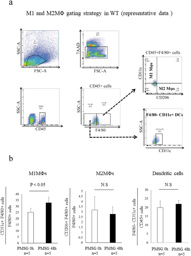
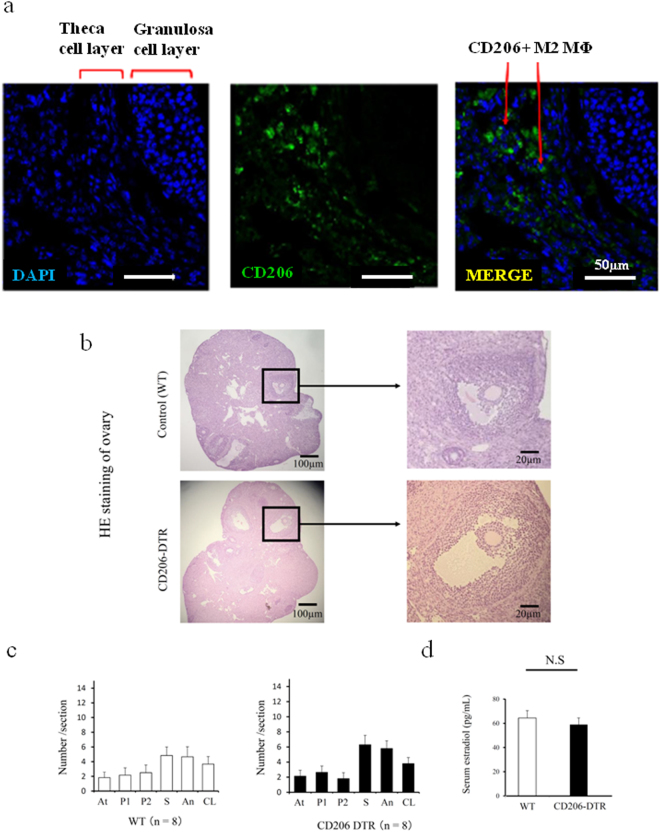
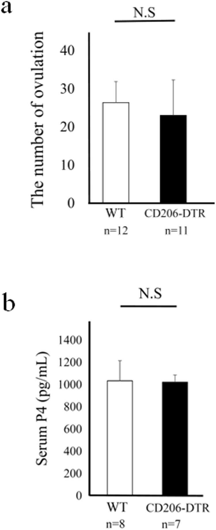
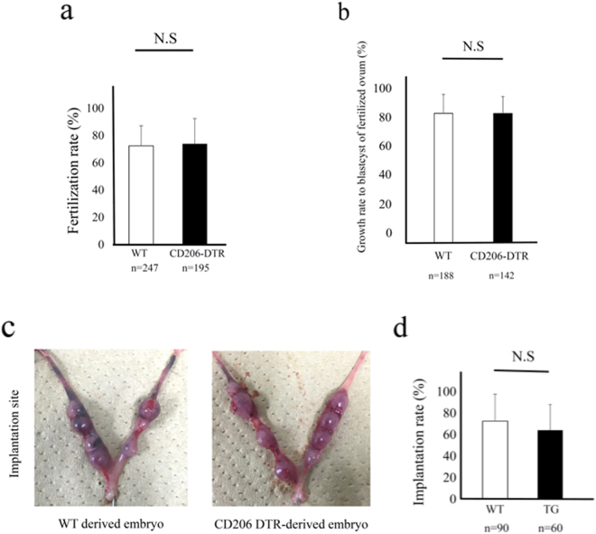
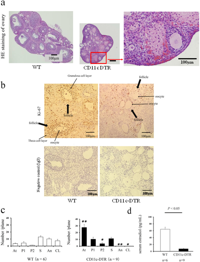
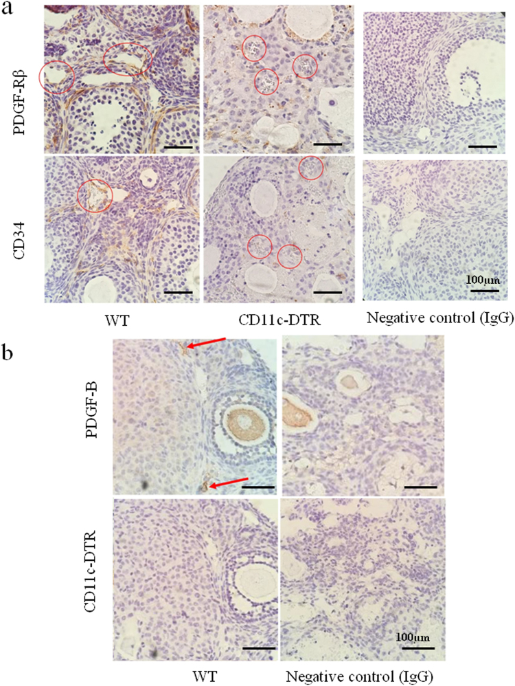
Macrophages (MΦs) are involved in folliculogenesis and ovulation. However, it is unknown which type of MΦ, M1 or M2, plays a more essential role in the ovary. CD206 or CD11c diphtheria toxin receptor transgenic (DTR) mice, which enable depletion of CD206+ M2 MΦs and CD11c+ MΦ or CD11c+ Dendritic cells (DCs), respectively, were used. Oocytes were used for in vitro fertilization and embryo transfer. In vitro fertilized embryos derived from M2 MΦ depleted oocytes were transferred to pseudo pregnant wild type mice. CD11c DTR mice were also used to investigate the role of CD11c cells, M1 MΦ and DCs in folliculogenesis. In WT mice, the proportion of CD206+ M2-like MΦs was not increased in follicular induction, while that of CD11c+ M1-like MΦs was increased. In CD206 DTR mice, folliculogenesis was normal and the ovulation number, fertilization rate, and implantation rate were similar to those in WT mice. In CD11c DTR mice, folliculogenesis was impaired with ovarian hemorrhage and the staining of platelet derived growth factor-receptor β (PDGF-Rβ), a marker of pericytes, and CD34, a marker of endothelial cells, was reduced. CD11c+ cells, M1 MΦs or DCs, may be involved in folliculogenesis, while M2 MΦs are not involved in folliculogenesis.


Conflict of interest statement

The authors declare no competing interests.

Figures

Figure 1
Figure 1
M1 and M2 macrophages (MΦs) in wild type (WT) mice ovary. (a) Representative flow cytometry analysis of M1 and M2 MΦs in the WT mice ovary. M1-like MΦ was defined as CD45+/F4/80+/CD11c+/CD206− cells, and M2-like MΦ was defined as CD45+/F4/80+/CD206+/CD11c− cells and Dendritic cells (DCs) was defined as F4/80− CD11c+ cells. (b) The proportion of CD11c+F4/80+ cells, M1-like MΦs (left panel) and CD206+ F4/80 cells, M2-like MΦs (middle panel) and F4/80− CD11c+ cells, DCs (right panel) in ovary. The proportion of M1-like MΦs significantly increased following follicular induction with PMSG 48 h, while that of M2-like MΦs and DCs was not increased. The data are shown as the means ± standard error of the mean (SEM). A P-value of < 0.05 was considered statistically significant by Mann-Whitney U test. N.S; not significant compared to WT mice. n; the number of mice.
Figure 2
Figure 2
The involvement of CD206+ F4/80+ M2-like macrophage (MΦ) in mouse ovary during folliculogenesis. (a) Localization of ovarian CD206+ M2-like MΦ in follicular induction with PMSG 48 h. (b) The Hematoxylin Eosin (HE) staining of ovaries in follicular induction with PMSG 48 h in wild type (WT) and CD206 Diphtheria toxin-receptor (DTR) mice. (c) The number of each follicle stage in follicular induction in WT and CD206 DTR mice. We counted the number of follicles at each stage, including atresia (At), primordial (P1), primary (P2), secondary (S), antral (An), and corpus luteum (CL) in CD206 DTR (right panel) and WT (left panel) mice, as previous described. (d) The serum estradiol levels after 48 h PMSG in WT and CD206 DTR mice.n; the number of mice. N.S; not significant compared to WT mice.
Figure 3
Figure 3
The involvement of CD206+ F4/80+ M2-like macrophage (MΦ) in mouse ovary during ovulation and luteinization. (a) The number of ovulations in wild type (WT) and CD206 diphtheria toxin-receptor (DTR) mice treated with superovulation. (b) Serum Progesterone (P4) levels in WT and CD206 DTR mice in ovulatory induction. n; the number of mice. N.S; not significant compared to WT mice.
Figure 4
Figure 4
The involvement of CD206+ M2-like MΦs in fertilization and implantation. (a) In vitro fertilization rate of oocytes derived from wild type (WT) and CD206 diphtheria toxin receptor (DTR) mice. n; the number of oocytes. (b) The embryo growth rate to blastocyst of fertilized ovum derived from WT and CD206 DTR mice. (c) The photographs of implantation site after embryo transfer derived from WT mice (left panel) and CD206 DTR mice (right panel). (d) The implantation rate of ovum derived from CD206 DTR mice compared to WT mice. n; the number of embryo. N.S; significant compared to WT mice.
Figure 5
Figure 5
The involvement of CD11c+ cells, M1 macrophages (MΦs) and dendritic cells (DCs) in folliculogenesis. (a) The Hematoxylin Eosin (HE) staining of ovaries in follicular induction with PMSG 48 h in Wild Type (WT) (left Panel) and CD11c diphtheria toxin receptor (DTR) mice (middle). (b) The Ki-67 staining, a cell proliferation marker, in follicular induced ovary with PMSG 48 h in WT and CD11c DTR mice. Negative control data are also shown. (c) The number of each follicle stage in follicular induction in WT and CD11c DTR mice. We counted the number of follicles at each stage, such as atresia (At), primordial (P1), primary (P2), secondary (S), antral (An), and corpus luteum (CL) in CD206 DTR (right panel) and WT (left panel) mice, as previous described. (d) The serum estradiol levels after 48 h PMSG in WT and CD11c DTR mice. The data are shown as the means ± SEM. A P-value of < 0.05 was considered statistically significant by Mann–Whitney U test. n; the number of mice. N.S: not significant compared to WT.
Figure 6
Figure 6
(a) Localization of PDGF-Rβ+ pericytes, and CD34+ endothelial cells, around follicles in WT and CD11c DTR mice. (b) Localization of PDGF-B around follicles in WT and CD11c DTR mice. Negative control data are shown.

References

    1. Fernando O, et al. The M1 and M2 paradigm of macrophage activation: time for reassessment. F1000 Prime Reports. 2014;6:13. - PMC - PubMed
    1. Wu R, et al. Macrophage contributions to ovarian function. Hum Reprod Update. 2004;10(No2):pp.119–133. doi: 10.1093/humupd/dmh011. - DOI - PubMed
    1. Hume DA, et al. The mononuclear phagocyte system of the mouse defined by immunohistochemical localisation of antigen F4/80: macrophages of endocrine organs. Proc. Natl. Acad. Sci. 1984;81:174–4177. doi: 10.1073/pnas.81.13.4174. - DOI - PMC - PubMed
    1. Best CL, et al. Localisation and characterisation of white blood cell populations within the human ovary throughout the menstrual cycle and menopause. Hum Reprod. 1996;11:790–797. doi: 10.1093/oxfordjournals.humrep.a019256. - DOI - PubMed
    1. Petrovska M, et al. Quantitative changes in macrophage distribution in normal mouse ovary over the course of the estrous cycle examined with an image analysis system. Am J Reprod Immunol. 1996;36:175–183. doi: 10.1111/j.1600-0897.1996.tb00159.x. - DOI - PubMed

Publication types