. 2018 May 25;8(1):8094.
doi: 10.1038/s41598-018-26549-4.

An in vitro method to keep human aortic tissue sections functionally and structurally intact

Affiliations

An in vitro method to keep human aortic tissue sections functionally and structurally intact

Jorn P Meekel et al. Sci Rep. .

Abstract

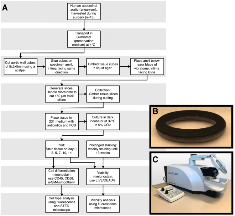
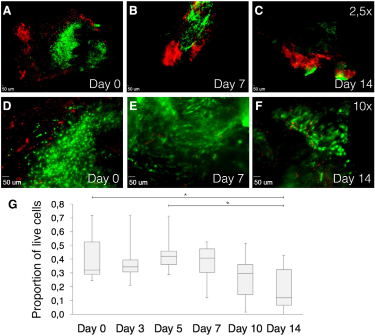
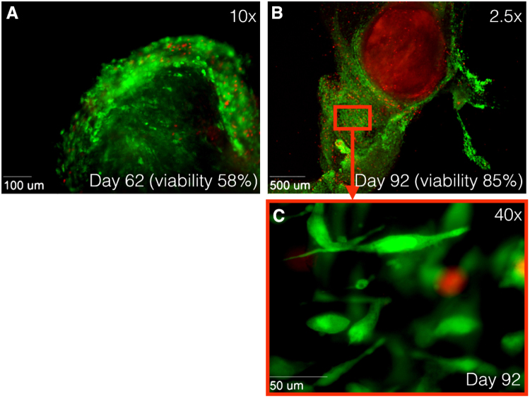
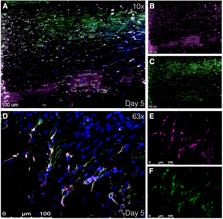
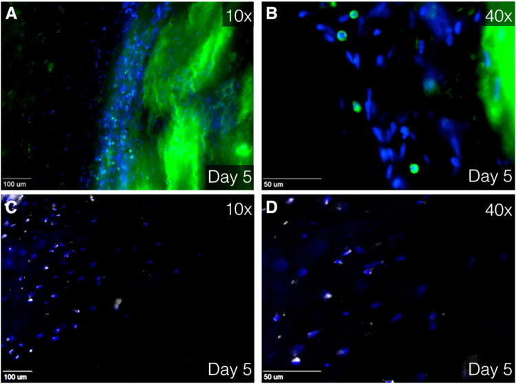
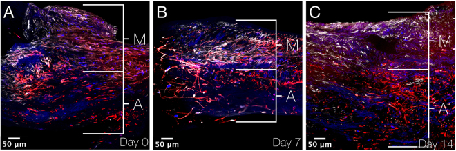
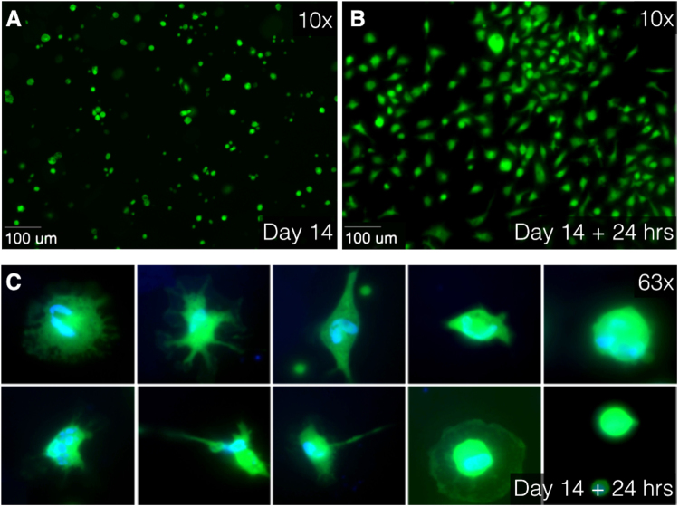
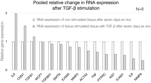
The pathophysiology of aortic aneurysms (AA) is far from being understood. One reason for this lack of understanding is basic research being constrained to fixated cells or isolated cell cultures, by which cell-to-cell and cell-to-matrix communications are missed. We present a new, in vitro method for extended preservation of aortic wall sections to study pathophysiological processes. Intraoperatively harvested, live aortic specimens were cut into 150 μm sections and cultured. Viability was quantified up to 92 days using immunofluorescence. Cell types were characterized using immunostaining. After 14 days, individual cells of enzymatically digested tissues were examined for cell type and viability. Analysis of AA sections (N = 8) showed a viability of 40% at 7 days and smooth muscle cells, leukocytes, and macrophages were observed. Protocol optimization (N = 4) showed higher stable viability at day 62 and proliferation of new cells at day 92. Digested tissues showed different cell types and a viability up to 75% at day 14. Aortic tissue viability can be preserved until at least 62 days after harvesting. Cultured tissues can be digested into viable single cells for additional techniques. Present protocol provides an appropriate ex vivo setting to discover and study pathways and mechanisms in cultured human aneurysmal aortic tissue.


Conflict of interest statement

The authors declare no competing interests.

Figures

Figure 1
Figure 1
Overview of experimental setup. (A) Flowchart of protocol: vascular tissue cubes were cut into sections using a vibrating blade microtome (Leica VT1200S). Tissue sections were cultured in supplemented culture medium in 24-well plates in a dark humidified atmosphere at 37 °C in 5% CO2. (B) Photograph of self-designed 3D-printed mould in which tissue cubes are fixed in agarose. (C) Photograph of vibratome Leica VT1200S, used for tissue sectioning. α-SMA indicates alpha smooth muscle actin; FCS, fetal calf serum and STED, stimulated emission depletion.
Figure 2
Figure 2
Immunofluorescence images using Zeiss Axiovert 200 M Marianas™ Microscope. Cells stained with LIVE/DEAD® Viability/Cytotoxicity Kit. (AB) 2.5× magnification of cultured tissues at day 0, 7 and 14, respectively. (DF) 10× magnification of cultured tissues at day 0, 7 and 14, respectively. Green fluorescence shows live cells, while red fluorescence indicates dead nuclei. Live cells are mainly located central to tissue. (G) Quantification of live human cells in harvested vascular tissues of distinct patients (N = 9). Box plots show proportions of square micron of green fluorescence divided by the sum of green and red fluorescence. *P < 0.05 compared with other time points using ANOVA with Bonferroni test.
Figure 3
Figure 3
Immunofluorescence images at 10× magnification using Zeiss Axiovert 200 M Marianas™ Microscope. Human tissue stained with LIVE/DEAD® Viability/Cytotoxicity Kit. Green fluorescence shows live cells, while red fluorescence indicates dead nuclei. (A) Alive tissue at day 62 after harvesting. Tissue viability of 58%. (B) Alive tissue at day 62 after harvesting at 2.5× magnification and (C) at 40× magnification. Outgrowth of new cells is observed after 92 days, while original tissue shows only staining of EthD-1 (dead cells). Tissue viability of 85%. EthD-1 indicates ethdium homodimer-1.
Figure 4
Figure 4
Immunofluorescence images showing smooth muscle cells. (AC) Immunofluorescence images at 40× magnification using using Zeiss Axiovert 200 M Marianas™ Microscope at day 5 after harvesting. (DF) Immunofluorescence image at 63× magnification using super resolution Confocal Laser Scanning Microscope Leica TCS SP8. Human tissue stained with α-SMA and smoothelin (smooth muscle cell markers) at day 5 after harvesting. (A,D) Merged image of α-SMA (purple), smoothelin (green) and DAPI (blue). (B,E) Isolated α-SMA staining. (C,F) Isolated smoothelin staining. α-SMA indicates alpha smooth muscle actin and DAPI, 4′,6-diamidino-2-phenylindole.
Figure 5
Figure 5
Immunofluorescence image at 10× (A,C) and 40× (B,D) magnification using Zeiss Axiovert 200 M Marianas™ Microscope. Human tissue stained with DAPI (blue), CD45 (green, A,B) and CD68 (white, C,D) Green staining at the right side (A,B) is due to unintentional fluorescence of elastin. CD indicates cluster of differentiation and DAPI, 4′,6-diamidino-2-phenylindole.
Figure 6
Figure 6
Immunofluorescence images showing aneurysmal aortic wall sections. (A–C) XY stitched images at 40× magnification using Nikon A1R confocal microscope on day 0, 7 and 14 after culturing. Smooth muscle cells are immunostained by smoothelin (white), actin (found in practically all eukaryotic cells) by phalloidin (red) and cell nuclei by DAPI (blue). Smooth muscle cells are found in the media and other cells (mainly fibroblasts) in the adventitia. M indicates media; A, adventitia and DAPI, 4′,6-diamidino-2-phenylindole.
Figure 7
Figure 7
Immunofluorescence images at 10× (A,B) and 63× oil (C) magnification using Zeiss Axiovert 200 M Marianas™ Microscope. (A) After 14 days of culturing, tissues were enzymatically digested using collagenase. Live separate cells, stained with Calcein AM (green), float as round cells in culture medium. (B) After 24 hours of additional culturing, the cells were attached to the slide. (C) Different cell type characteristics and differences in cell nuclei are observed in attached cells. Live cytoplasm is stained with Calcein AM (green) and cell nuclei are stained with DAPI (blue). AM indicates acetoxymethyl and DAPI, 4′,6-diamidino-2-phenylindole.
Figure 8
Figure 8
Quantification of gene expression of pooled non-stimulated (white) and TGF-β-stimulated (grey) AAA patient tissue sections after seven days ex vivo. Relative expression is shown as ratio based on non-stimulated tissue sections. IL6 indicates interleukin 6; CNN1, calponin 1; TGFB1, transforming growth factor beta 1; MCP1, monocyte chemotactic protein 1; TFBR1, transforming growth factor beta receptor 1; SMTN, smoothelin; ICAM3, intercellular adhesion molecule 3; MMP2, matrix metalloproteinase-2; ACTA2, alpha smooth muscle actin 2; TNF, tumor necrosis factor; PTPRC, protein tyrosine phosphatase, receptor type, C; Ki67, antigen KI-67; ICAM1, intercellular adhesion molecule 1; IL8 (or CXCL8), interleukin 8 and MMP9, matrix metalloproteinase-9.

References

    1. Dua A, Kuy S, Lee CJ, Upchurch GR, Desai SS. Epidemiology of aortic aneurysm repair in the United States from 2000 to 2010. J. Vasc. Surg. 2014;59:1512–1517. doi: 10.1016/j.jvs.2014.01.007. - DOI - PubMed
    1. Sidloff D, et al. Aneurysm global epidemiology study public health measures can further reduce abdominal aortic aneurysm mortality. Circulation. 2014;129:747–753. doi: 10.1161/CIRCULATIONAHA.113.005457. - DOI - PubMed
    1. Lozano R, et al. Global and regional mortality from 235 causes of death for 20 age groups in 1990 and 2010: A systematic analysis for the Global Burden of Disease Study 2010. Lancet. 2012;380:2095–2128. doi: 10.1016/S0140-6736(12)61728-0. - DOI - PMC - PubMed
    1. Davis FM, Rateri DL, Daugherty A. Mechanisms of aortic aneurysm formation: translating preclinical studies into clinical therapies. Heart (British Cardiac Society) 2014;100:1498–1505. - PubMed
    1. Hellenthal FAMVI, Buurman WA, Wodzig WKWH, Schurink GWH. Biomarkers of AAA progression. Part 1: Extracellular matrix degeneration. Nature Reviews Cardiology. 2009;6:464–474. doi: 10.1038/nrcardio.2009.80. - DOI - PubMed

Publication types

LinkOut - more resources