. 2018 May 25;9(1):2061.
doi: 10.1038/s41467-018-04523-y.

Zinc ion flux during mammalian sperm capacitation

Affiliations

Zinc ion flux during mammalian sperm capacitation

Karl Kerns et al. Nat Commun. .

Abstract

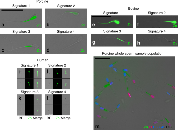
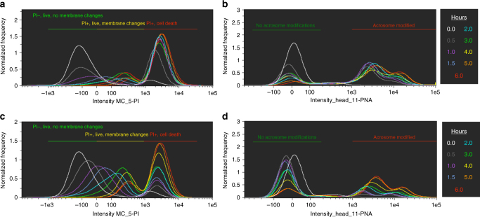
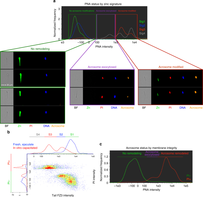
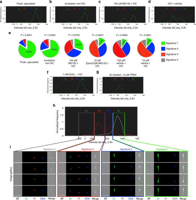
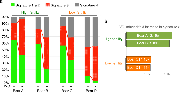
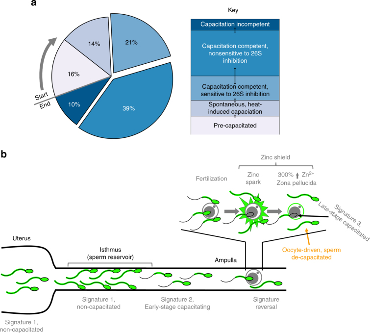
Sperm capacitation, the ultimate maturation event preparing mammalian spermatozoa for fertilization, was first described in 1951, yet its regulatory mechanisms remain poorly understood. The capacitation process encompasses an influx of bicarbonate and calcium ions, removal of decapacitating factors, changes of pH and sperm proteasomal activities, and the increased protein tyrosine phosphorylation. Here, we document a novel biological phenomenon of a unique zinc (Zn2+) ion redistribution associated with mammalian sperm in vitro capacitation (IVC). Using image-based flow cytometry (IBFC), we identified four distinct types of sperm zinc ion distribution patterns (further zinc signature) and their changes during IVC. The zinc signature was altered after sperm capacitation, reduced by proteasomal inhibitors, removed by zinc chelators, and maintained with addition of external ZnCl2. These findings represent a fundamental shift in the understanding of mammalian fertilization, paving the way for improved semen analysis, in vitro fertilization (IVF), and artificial insemination (AI).


Conflict of interest statement

The authors declare no competing interests.

Figures

Fig. 1
Fig. 1
Mammalian sperm zinc signature. ad Epifluorescence microscopy of boar sperm zinc signature (green). eh Epifluorescence microscopy of bull sperm zinc signature. il FlowSight image gallery of human sperm zinc signature (scale bar: 20 μm). m Boar spermatozoa after 72 h of storage in Beltsville thaw solution (BTS semen extender) show varied zinc signatures. Imprecise fluorescent to bright-field overlay illustrates motile status (all scale bars: 25 μm, unless noted)
Fig. 2
Fig. 2
Zinc signature time course with high vs. low bicarbonate IVC. Time-lapse recordings of zinc signature during IVC in high, 15 mM sodium bicarbonate media: (a) plasma membrane changes as identified by PI status, distinguishing between IVC-induced PI+ subpopulations (PI+ live with plasma membrane changes vs. PI+ cell death). b Acrosomal modifications. Time course of zinc signature modification in low, 2 mM sodium bicarbonate IVC media: (c) plasma membrane changes, and d acrosomal modifications (corresponding histogram color code for time points in figure legend)
Fig. 3
Fig. 3
Acrosomal status and membrane integrity of zinc signature. a Most spermatozoa in zinc signature 1 and 2 states had no capacitation-like acrosomal remodeling compared to zinc signature 3 and 4 (P < 0.0001; see Table 1). Capacitation-like acrosomal remodeling was most prevalent with zinc signatures 3 and 4 compared to zinc signatures 1 and 2 (P < 0.0001; 4 biological replicates; 10,000 spermatozoa analyzed per treatment). Acrosomal exocytosis occurred within the subpopulation of spermatozoa with zinc signature 4 and was greater than zinc signatures 1, 2, and 3 (P < 0.001; scale bar: 20 μm). b Zinc signature status corresponds with PI plasma membrane integrity in fresh (blue) and IVC spermatozoa (red). c As sperm plasma membrane integrity decreased, acrosomal remodeling and exocytosis occurred. P-values determined by the general linear model procedure in SAS 9.4
Fig. 4
Fig. 4
Modulation of internal and external Zn2+ during IVC. a Fresh, ejaculated spermatozoa have mostly signature 1. b After 4 h under non-IVC conditions, few spermatozoa underwent spontaneous early-stage capacitation. c Proteasomal inhibitor MG132 (100 µM) prevented some of the IVC-induced zinc signature changes compared to d (IVC with MG132-vehicle); see Supplementary Fig. 2g for no vehicle IVC control. e Ten-micrometer Zn-chelator TPEN altered the zinc signature; see Supplementary Fig. 2h for TPEN vehicle treatment (P-value by treatment across replicates). f Addition of 1 mM ZnCl2 + IVC prevented IVC-induced zinc signature changes. g Pie chart illustration of select IVC treatments (green: signature 1; blue: signature 2; red: signature 3; gray: signature 4). Treatment P-values are shown in Table 2 as determined by the general linear model procedure in SAS 9.4. 10,000 sperm per sample analyzed. h Histograms of non-capacitated (non-IVC, white) and capacitated (IVC, red) sperm populations from IBFC analysis i Images from IBFC gallery representing individual zinc signatures (scale bar: 20 μm). Each spermatozoon analyzed has the following images acquired: bright field (BF); Zn2+ reporting probe FZ3 (Zn); sperm viability/plasma membrane integrity probe propidium iodide (PI); live DNA stain Hoechst 33342 (DNA); and side scatter (not displayed), with a merger of the four images (Merge)
Fig. 5
Fig. 5
Zinc signature associated with varied fertility in AI boars. a Zinc signature in four boars of known high or low fertility, before and after IVC. b High-fertility boars had double the amount of spermatozoa with signature 3 after IVC compared to minimal increase in low-fertility boars
Fig. 6
Fig. 6
Proposed zinc signature population interpretation. a Interpretation of zinc signature meaning and population segregation: 16% of fresh, ejaculated spermatozoa had undergone early-stage capacitation upon semen collection (lightest blue working to darkest); 14% of spermatozoa spontaneously undergo early-stage capacitation during incubation without IVC inducers; 60% of spermatozoa remained capacitation competent with IVC inducers, with 21% sensitive to proteasomal inhibition; remaining 10% of sperm were capacitation incompetent under IVC conditions (darkest blue) (s.e. bars included). b Proposed zinc signature changes throughout female reproductive tract and oocyte zinc spark interference with sperm zinc signature as a combined polyspermy defense mechanism, the zinc shield

References

    1. Que EL, et al. Quantitative mapping of zinc fluxes in the mammalian egg reveals the origin of fertilization-induced zinc sparks. Nat. Chem. 2015;7:130–139. doi: 10.1038/nchem.2133. - DOI - PMC - PubMed
    1. Zhang N, Duncan FE, Que EL, O’Halloran TV, Woodruff TK. The fertilization-induced zinc spark is a novel biomarker of mouse embryo quality and early development. Sci. Rep. 2016;6:22772. doi: 10.1038/srep22772. - DOI - PMC - PubMed
    1. Lishko PV, Botchkina IL, Fedorenko A, Kirichok Y. Acid extrusion from human spermatozoa is mediated by flagellar voltage-gated proton channel. Cell. 2010;140:327–337. doi: 10.1016/j.cell.2009.12.053. - DOI - PubMed
    1. Tardif S, Dube C, Bailey JL. Porcine sperm capacitation and tyrosine kinase activity are dependent on bicarbonate and calcium but protein tyrosine phosphorylation is only associated with calcium. Biol. Reprod. 2003;68:207–213. doi: 10.1095/biolreprod.102.005082. - DOI - PubMed
    1. Boerke A, Tsai PS, Garcia-Gil N, Brewis IA, Gadella BM. Capacitation-dependent reorganization of microdomains in the apical sperm head plasma membrane: functional relationship with zona binding and the zona-induced acrosome reaction. Theriogenology. 2008;70:1188–1196. doi: 10.1016/j.theriogenology.2008.06.021. - DOI - PubMed

Publication types

LinkOut - more resources