Review
. 2019 Jan 1:688:14-25.
doi: 10.1016/j.neulet.2018.05.032. Epub 2018 May 23.

What cerebellar malformations tell us about cerebellar development

Affiliations
Review

What cerebellar malformations tell us about cerebellar development

Parthiv Haldipur et al. Neurosci Lett. .

Abstract

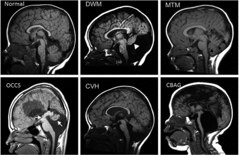
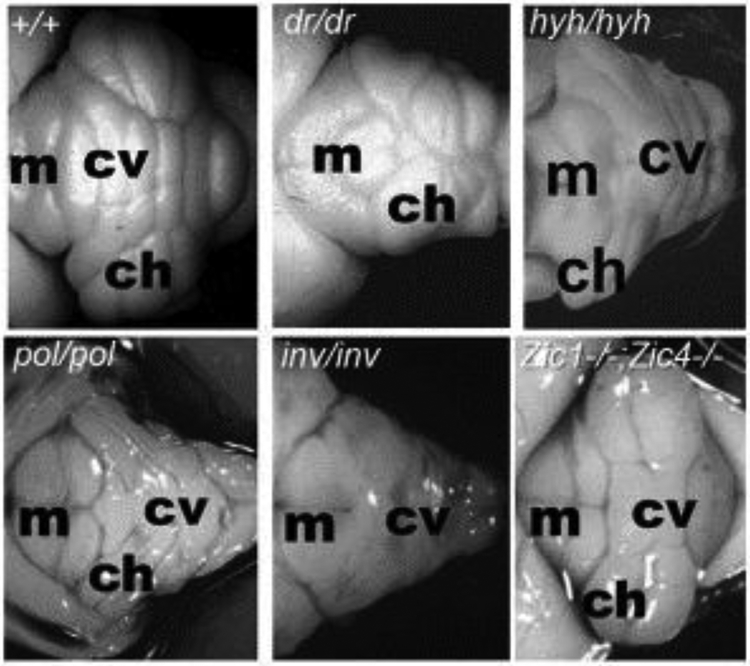
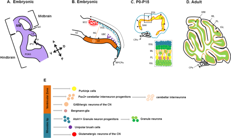
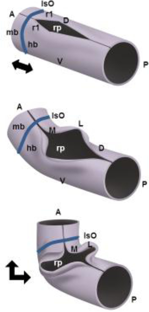
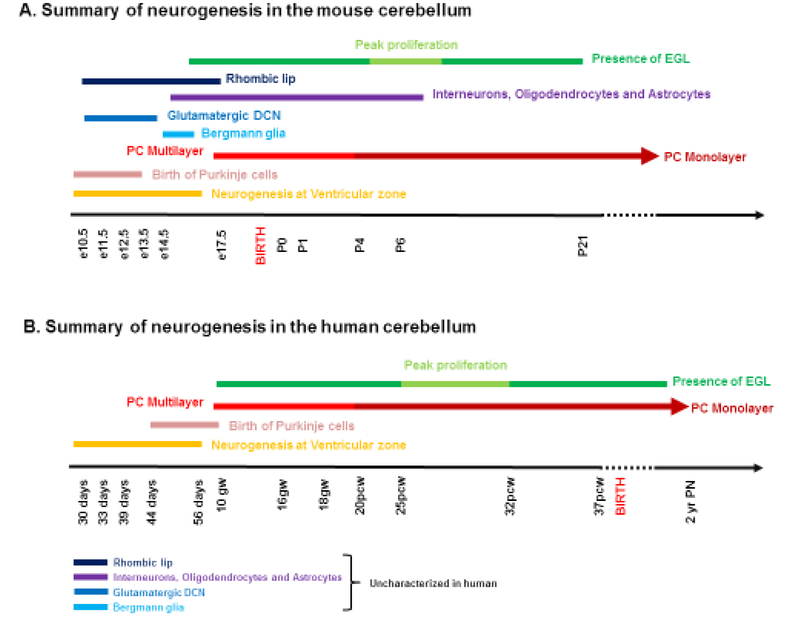
Structural birth defects of the cerebellum, or cerebellar malformations, in humans, have long been recognized. However, until recently there has been little progress in elucidating their developmental pathogenesis. Innovations in brain imaging and human genetic technologies over the last 2 decades have led to better classifications of these disorders and identification of several causative genes. In contrast, cerebellar malformations in model organisms, particularly mice, have been the focus of intense study for more than 70 years. As a result, many of the molecular, genetic and cellular programs that drive formation of the cerebellum have been delineated in mice. In this review, we overview the basic epochs and key molecular regulators of the developmental programs that build the structure of the mouse cerebellum. This mouse-centric approach has been a useful to interpret the developmental pathogenesis of human cerebellar malformations. However, it is becoming apparent that we actually know very little regarding the specifics of human cerebellar development beyond what is inferred from mice. A better understanding of human cerebellar development will not only facilitate improved diagnosis of human cerebellar malformations, but also lead to the development of treatment paradigms for these important neurodevelopmental disorders.

Keywords: Cerebellar malformation; Development; Human; Model organism; Mouse; Neurogenetics; Pathogenesis.


Figures

Figure 1:
Figure 1:. Examples of Human cerebellar malformations.
Mid sagittal MRI views of human cerebellar malformations with an unaffected individual shown for comparison. Dandy-Walker malformation (DWM) with white arrowhead highlighting the small cerebellar vermis rotated away from the brainstem in an enlarged posterior fossa encompassing a very large fourth ventricle. Molar Tooth Malformation (MTM) with black arrowheads marking the edge of the small vermis with cerebellar hemispheres occupying the residual space in a normally sized posterior fossa. Occulocerebrocutaneous syndrome (OCCS) with left white arrow indicating the third ventricle and black arrow highlighting a massively enlarged tectum; right white arrow points to rudimentary cerebellum. Also note lack of corpus callosum. Cerebellar vermis hypoplasia (CVH). Cerebellar agenesis (CBAG); note reduced size of pontine nucleus of the small brain stem.
Figure 2
Figure 2. Examples of mouse cerebellar malformations.
Dorsal whole mount views of cerebellar malformations in 4 spontaneous mutants and 1 engineered mouse strain. Wild-type (+/+) cerebellum with cerebellar vermis (cv) and cerebellar hemispheres (ch) indicated showing stereotypical foliation pattern. Disruption of this patterning is obvious in many mouse mutant strains. For example, in dreher (dr) homygous mutants, a reduced cerebellar vermis causes juxtaposition of the cerebellar hemispheres. In hydrocephalus with hop gait (hyh) homozygous mutants, the vermis is more prominent than the hemispheres. Although these mice are not models for any specific human malformation, investigation of the underlying pathogenesis has provided insights into the role of the roof plate in cerebellar development and vermis formation. Polaris (pol) and inversus (inv) homozygous mutants have severely disrupted cerebellar morphology and are models for cilia related MTM human cerebellar malformations. Zic1/4 double homozygous mouse mutants model human DWM and display simplified vermis foliation. Anterior is to the left, indicated by the presence of midbrain colliculi (m). Photos are optimized to show pattering differences and are not all at the same magnification.
Figure 3 -
Figure 3 -
(A) Schematic representation of an embryonic mouse cerebellum between e12.5-e18.5 (CB) sectioned along the sagittal plane. The cerebellum is derived from the dorsal region of rhombomere 1 (rh1) under the influence of signaling factors from the Isthmic organizer (IsO) and roof plate (RP). (B) A composite of embryonic developmental processes during embryogenesis. The developing cerebellum has two zones of neurogenesis, the ventricular zone (VZ) and the rhombic lip (RL). The cerebellar ventricular zone consists of a lining of radial glia (RG) and gives rise to all cerebellar GABAergic neurons and interneurons. GABAergic cerebellar nuclei neurons are produced first, followed by Purkinje cells and PAX2-expressing cerebellar interneuron progenitors. Bergmann Glia are also derived from the cerebellar ventricular zone. The rhombic lip on the other hand gives rise to the three major glutamatergic neuronal subtypes that populate the cerebellum. Firstly, cerebellar nuclei projection neurons migrate from the rhombic lip into the Nuclear Transitory Zone (NTZ) over the anlage as the rostral migratory stream. As embryonic development proceeds, granule neuron progenitors (GNPs) next migrate out of the rhombic lip between embryonic day 12.5 and 16. These cell progenitors migrate tangentially under the pial surface to establish the EGL of the developing cerebellum in an anterior to posterior manner. The RL also gives rise to unipolar brush cells (UBC) later in development, that migrate into the cerebellar anlage, (C) The EGL is a secondary germinal zone, or transit amplifying center. The EGL is composed of 2 sublayers – a proliferating external zone and an inner differentiating zone. Proliferation of GNPs takes place during postnatal days P0–P14. This proliferation is largely driven by the mitogen sonic hedgehog (SHH) secreted from Purkinje cells which have formed the Purkinje layer (PL) under the EGL. (D) Proliferation of GNPs in the EGL is responsible for the dramatic size increase of the post-natal mouse cerebellum. As granule neurons exit the cell cycle, they migrate tangentially within the inner EGL and then exit the EGL migrating radially inward to settle below the developing Purkinje cell layer to form the internal granule layer (IGL), resulting in the final laminar arrangement of the mature cerebellum. (E) Schematic representation of the multiple cell types that arise in the cerebellar ventricular zone and rhombic lip. Reference : Haldipur P., Dang D. and Millen K.J., (In press) Embryology. In: M. Manto and T.A.G.M. Huisman (Eds.) The cerebellum in children and adults. Elsevier, Amsterdam.
Figure 4 -
Figure 4 -
The cerebellum is a derivative of the anterior-most dorsal hindbrain, or dorsal (D) rhombomere 1 (r1). The establishment mid-hindbrain (mb-hb) boundary results in formation of a transient signaling center called the Isthmic Organizer (IsO), which secretes Fibroblast Growth Family 8 (FGF8) and WNT1 which are required for cell survival and pattern the adjacent tissue from e8–11.5 in mice. The developing cerebellar anlage undergoes a series of morphogenetic events between mouse embryonic (e) days 9 to e12.5 that rotate its anterior posterior (AP) axis by 90 degrees and convert it to the medio-lateral axis (ML) of the bilateral cerebellar wings. This reorientation is in large part driven by pontine flexure which converts the horizontal alignment of mid/hindbrain to nearly right angles (indicated by black double headed arrows). The roof plate (rp) is the single layer thick roof of the dorsal midline of the early neural tube which acts as another transient signaling center, expressing BMP and WNT secreted factors. The roof plate will eventually differentiate into the choroid plexus epithelium of the fourth ventricle. In rhombomere 1, roof plate derived Wnt expression is required to drive early cerebellar anlage ventricular zone proliferation, while secreted Bone morphogenetic protein (BMP) gene expression is required to induce the cerebellar rhombic lip and correctly pattern expression of Pancreatic transcription factor (Ptf1a) in the ventricular zone of the nascent cerebellar anlage.
Figure 5 -
Figure 5 -
Summary of neurogenesis in the developing (A) mouse and (B) human cerebellum. Human cerebellar development is highly protracted compared to mice. In mice, the cerebellum develops over a period of 30–35 days with peak EGL expansion, foliation and IGL formation and Purkinje cell maturation occurring during the first two postnatal weeks. In striking contrast, human cerebellar development extends from the early first trimester to final circuit maturity which is achieved by the end of the second postnatal year. Also, a significant portion of human cerebellar development occurs in utero, including peak proliferation of GNPs and folia formation during the last trimester. Reference: Haldipur P., Dang D., and Millen K.J., (In press) Embryology. In: M. Manto and T.A.G.M. Huisman (Eds.) The cerebellum in children and adults. Elsevier, Amsterdam
Figure 6 :
Figure 6 :
MRI views of human Joubert syndrome, a syndromic malformation of the brainstem involving a distinctive elongation of the cerebellar peduncles in addition to cerebellar vermis hypoplasia (C,D) and rhombencephalosynapsis (E, F) with an unaffected individual (A,B) shown for comparison. A,C and E are midsagittal views while B,D and F are along the horizontal plane. JS is characterized by distinct molar tooth sign (D, arrow), while RCS is characterized by missing cerebellar vermis with apparent fusion of the cerebellar hemispheres (F).

References

    1. Aldinger KA and Doherty D (2016) ‘The genetics of cerebellar malformations’, Seminars in fetal & neonatal medicine 21(5): 321–32. - PMC - PubMed
    1. Aldinger KA, Lehmann OJ, Hudgins L, Chizhikov VV, Bassuk AG, Ades LC, Krantz ID, Dobyns WB and Millen KJ (2009) ‘FOXC1 is required for normal cerebellar development and is a major contributor to chromosome 6p25.3 Dandy-Walker malformation’, Nature genetics 41(9): 1037–42. - PMC - PubMed
    1. Artzt K (2012) ‘Mammalian developmental genetics in the twentieth century’, Genetics 192(4): 1151–63. - PMC - PubMed
    1. Baptista CA, Hatten ME, Blazeski R and Mason CA (1994) ‘Cell-cell interactions influence survival and differentiation of purified Purkinje cells in vitro’, Neuron 12(2): 243–60. - PubMed
    1. Barkovich AJ, Guerrini R, Kuzniecky RI, Jackson GD and Dobyns WB (2012) ‘A developmental and genetic classification for malformations of cortical development: update 2012’, Brain : a journal of neurology 135(Pt 5): 1348–69. - PMC - PubMed

Publication types