. 2018 May 26;17(1):93.
doi: 10.1186/s12943-018-0841-x.

The long non-coding RNA PTTG3P promotes cell growth and metastasis via up-regulating PTTG1 and activating PI3K/AKT signaling in hepatocellular carcinoma

Affiliations

The long non-coding RNA PTTG3P promotes cell growth and metastasis via up-regulating PTTG1 and activating PI3K/AKT signaling in hepatocellular carcinoma

Jin-Lan Huang et al. Mol Cancer. .

Abstract

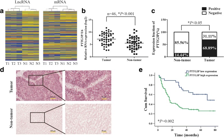
Background: Dysfunctions of long non-coding RNA (lncRNAs) have been associated with the initiation and progression of hepatocellular carcinoma (HCC), but the clinicopathologic significance and potential role of lncRNA PTTG3P (pituitary tumor-transforming 3, pseudogene) in HCC remains largely unknown.

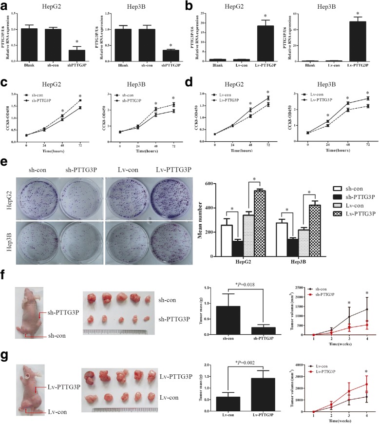
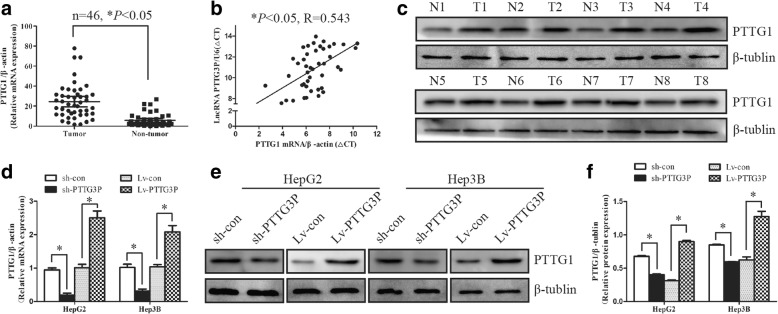
Methods: We compared the expression profiles of lncRNAs in 3 HCC tumor tissues and adjacent non-tumor tissues by microarrays. In situ hybridization (ISH) and quantitative real-time polymerase chain reaction (qRT-PCR) were applied to assess the level of PTTG3P and prognostic values of PTTG3P were assayed in two HCC cohorts (n = 46 and 90). Artificial modulation of PTTG3P (down- and over-expression) was performed to explore the role of PTTG3P in tumor growth and metastasis in vitro and in vivo. Involvement of PTTG1 (pituitary tumor-transforming 1), PI3K/AKT signaling and its downstream signals were validated by qRT-PCR and western blot.

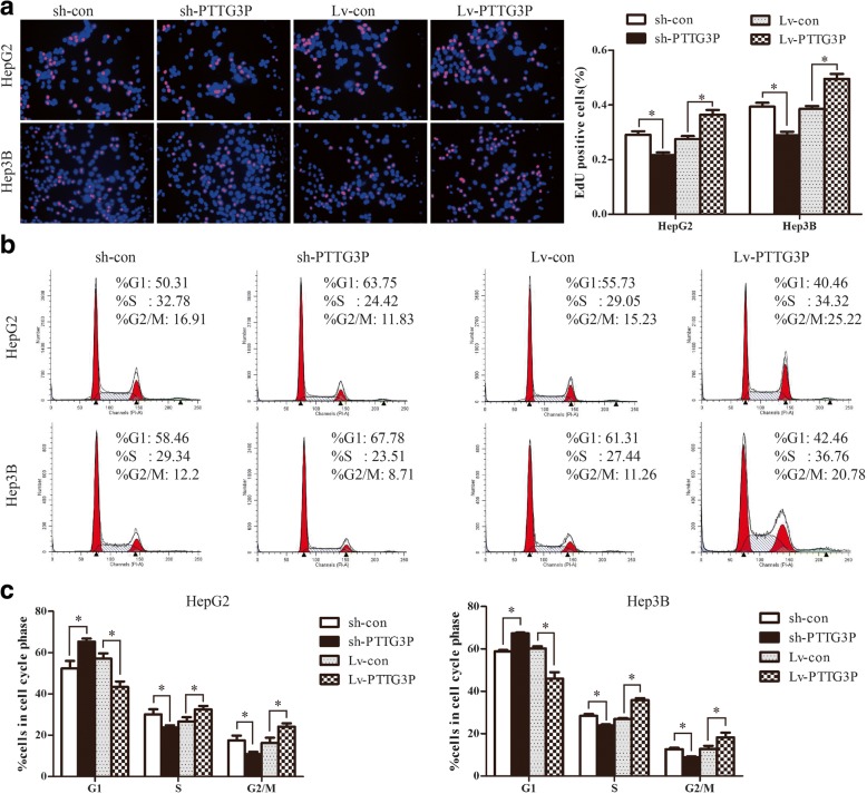
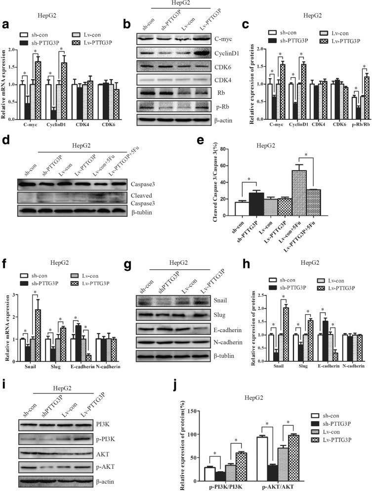
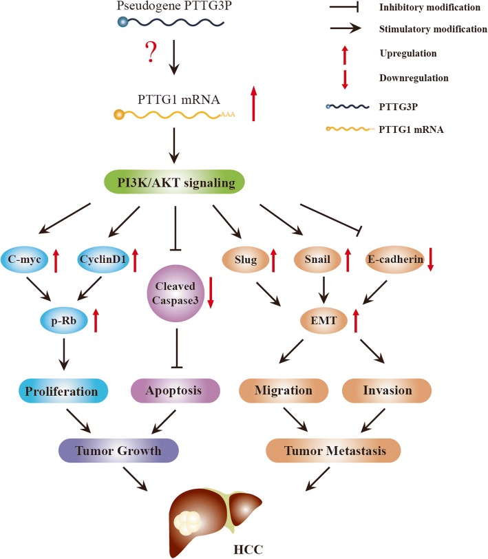
Results: We found that PTTG3P was frequently up-regulated in HCC and its level was positively correlated to tumor size, TNM stage and poor survival of patients with HCC. Enforced expression of PTTG3P significantly promoted cell proliferation, migration, and invasion in vitro, as well as tumorigenesis and metastasis in vivo. Conversely, PTTG3P knockdown had opposite effects. Mechanistically, over-expression of PTTG3P up-regulated PTTG1, activated PI3K/AKT signaling and its downstream signals including cell cycle progression, cell apoptosis and epithelial-mesenchymal transition (EMT)-associated genes.

Conclusions: Our findings suggest that PTTG3P, a valuable marker of HCC prognosis, promotes tumor growth and metastasis via up-regulating PTTG1 and activating PI3K/AKT signaling in HCC and might represent a potential target for gene-based therapy.

Keywords: Hepatocellular Carcinoma; Long non-coding RNA; PI3K/AKT signaling; PTTG1; PTTG3P.


Conflict of interest statement

Ethics approval and consent to participate

Written informed consent for the biological studies was obtained from each patient involved in the study, and the study was approved by the Ethics Committee of Nanfang Hospital. All animal studies were approved by the Animal Experimental Committee of Nanfang Hospital.

Consent for publication

Written consent for publication was obtained from all the patients involved in our study.

Competing interests

The authors declare no conflict of interest.

Publisher’s Note

Springer Nature remains neutral with regard to jurisdictional claims in published maps and institutional affiliations.

Figures

Fig. 1
Fig. 1
LncRNA PTTG3P is frequently up-regulated in HCC. (a) Hierarchical clustering analysis of 884 differentially expressed lncRNAs and 979 differentially expressed mRNAs between 3 HCC tumor tissues and paired non-tumor tissues. Up-regulation is shown in yellow and down-regulation is in blue. T, tumor tissues; N, non-tumor tissues. (b) The level of lncRNA PTTG3P was detected in 46 HCC tumor tissues and paired non-tumor tissues by qRT-PCR.U6 was used as a housekeeping gene. (c) The levels of PTTG3P in HCC tissues and adjacent non-tumor tissues was evaluated by ISH assays (cohort 2, n = 90). (d) Representative images of lncRNA PTTG3P expression from HCC tumor tissues and non-tumor tissues by ISH assays. (e) Kaplan-Meier survival analysis of overall survival in 90 patients with HCC (cohort 2) according to PTTG3P expression. Accumulation expression of PTTG3P was unfavorable for HCC prognosis. The log-rank test was used to calculate P values.*P < 0.05
Fig. 2
Fig. 2
Over-expression of PTTG3P accelerates HCC cell growth in vitro and in vivo. (a) Knockdown of endogenous PTTG3P in specific shRNA transduced HepG2 and Hep3B cells. U6 was used as a housekeeping gene for qRT-PCR. (b) HepG2 and Hep3B cells were infected with lentivirus carrying the PTTG3P gene. The level of PTTG3P was significantly increased in HepG2 and Hep3B cells over-expressing PTTG3P when compared with control cells. U6 was used as a housekeeping gene for qRT-PCR. (c) After knockdown of PTTG3P in HepG2 and Hep3B cells, the cell viability was assessed by CCK-8 assays daily for 3 days. (d) Ectopic expression of PTTG3P promotes cell growth as determined by CCK-8 assays. (e) The effects of PTTG3P on cellular survival were assessed by colony formation assays. Colonies are shown in purple post staining with crystal violet (left). (f) Effects of PTTG3P over-expression on tumorigenesis in vivo. Representative images of tumors formed in nude mice injected subcutaneously with PTTG3P–silencing HepG2 cells were shown. The tumor mass were measured. The tumor volume was periodically tested for each mouse and tumor growth curve was plotted. (g) Effects of blocked PTTG3P expression on tumorigenesis in vivo. Red arrows indicate HCC tumors. Error bars represent mean ± SD from 3 independent experiments. *, P < 0.05
Fig. 3
Fig. 3
LncRNAPTTG3P induces cell-cycle progression in HCC cells. (a) HepG2 and Hep3B cells with enhanced or silenced PTTG3P expression were seeded on 96-well plates, and cell proliferation was examined by EdU immunofluorescence staining. The bar graph on the right shows the percentage of EdU-positive nuclei. (b) FACS analysis of HepG2 and He3B cells with elevated or blocked PTTG3P expression. (c) Proportion of cells in various phases of the cell cycle in HepG2 and Hep3B cells. Data are presented as mean ± SD for at least three independent experiments.*, P < 0.05
Fig. 4
Fig. 4
LncRNAPTTG3P inhibits cellular apoptosis of HCC cells. (a) HepG2 and Hep3B cells with enhanced or silenced PTTG3P expression were stained with a combination of annexin V and 7-AAD and analyzed by FACS. Cells positive for annexin V staining were counted as apoptotic cells. (b) The HepG2 and Hep3B cells over-expressing PTTG3P were treated with 5-fluorouracil for 48 h, stained with a combination of annexin-V and 7-AAD, and analyzed by flow cytometry. (c) The bar graph showed the percentage of apoptotic cells. The results are means of 3 independent experiments ± SD from 3 independent experiments. *, P < 0.05
Fig. 5
Fig. 5
LncRNA PTTG3P promotes HCC cell metastasis in vitro and in vivo. (a) Transwell chamber assays showed that knockdown of PTTG3Preduced the migration ability while over-expression of PTTG3P enhanced the migration ability of HepG2 and Hep3B cells. (b) Boyden chamber assays revealed that stably suppressed PTTG3P expression inhibited invasiveness of HepG2 and Hep3B cells in vitro, whereas elevated PTTG3P expression had the opposite effects. (c) Representative images of hepatic metastasis tumors were obtained 40 days after spleen injection with sh-con, sh-PTTG3P, Lv-con or Lv-PTTG3P HepG2 cells, respectively(upper panel). H&E staining of metastastic tumor tissues were shown (middle panel). The boundary between tumor and normal tissue was indicated by black line. The level of PTTG3P in metastastic tumor tissues was assessed by ISH assays (lower panel). All values shown are mean ± SD of triplicate measurements and have been repeated 3 times with similar results. Independent t test was used to determine the differences between two groups.*, P < 0.05
Fig. 6
Fig. 6
LncRNA PTTG3P regulates the level of multiple genes involved in cell cycle, cell apoptosis and EMT through PI3K/AKT pathway in HCC cells. (a-c) The relative levels of cell cycle associated genes, including C-myc, CyclinD1, CDK6, CDK4, Rb and phosphorylated Rb (p-Rb), were detected in HepG2 cells overexpressing PTTG3P and HepG2 cells with stably suppressed PTTG3P expression by qRT-PCR (a) and western blot with quantitative analysis (b and c). β-actin was used as a housekeeping gene for qRT-PCR and an internal control for western blotting analysis. (d-e)Western blot analysis of the levels of Caspase3 and cleaved Caspase3 in HepG2 cells with silenced or enhanced PTTG3P. β-tublin was used as an internal control. (f-h) Knockdown of endogenous PTTG3P in HepG2 cells reduced the mRNA (f) and protein levels (g and h) of several EMT-marker genes including Snail and Slug but enhanced E-cadherin expression. In contrast, the level of Snail and Slug increased and the mRNA (f) and protein (g and h) level of E-cadherin decreased in HepG2 cells with elevated PTTG3P expression. β-actin was used as a housekeeping gene for qRT-PCR. β-tublin was used as an internal control for western blot analysis. (i-j) The levels of PI3K, phosphorylated PI3K (p-PI3K), AKT, phosphorylated AKT (p-AKT) were examined by western blot analysis in HepG2 cells with silenced or enhanced PTTG3P. β-actin was used as an internal control. The experiments were performed in triplicate; the data are expressed as the mean ± SD. *, P < 0.05
Fig. 7
Fig. 7
LncRNA PTTG3P up-regulates PTTG1 expression in HCC. (a) The level of PTTG1 was detected in 46 HCC tumor tissues and paired non-tumor tissues by qRT-PCR. β-actin was used as a housekeeping gene. (b) The correlation between lncRNA PTTG3P and PTTG1 mRNA in 46 HCC tissues. The ΔCt values were subjected to spearman correlation analysis. (c) The protein level of PTTG1 was assessed in 8 HCC tissues and paired non-tumor tissues by western blot. β-tublin was used as a loading control. T, tumor tissues; N, non-tumor tissues. (d-f) Overexpression of PTTG3P induced the expression of PTTG1 mRNA (d) and protein (e, f) in HepG2 and Hep3B cells. Knockdown of PTTG3P blocked PTTG1 expression (d-f). β-tublin was used as a loading control. The experiments were performed in triplicate, *P < 0.05
Fig. 8
Fig. 8
A schematic model of lncRNA PTTG3P functions during tumor growth and metastasis cascade in HCC. LncRNA PTTG3P up-regulates PTTG1 and then activates PI3K/AKT signaling. Activation of PI3K/AKT signaling elevates the expression of C-myc and CyclinD1, then increasing the level of phosphorylated Rb, and finally promoting cell proliferation in HCC. On the other hand, activation of PI3K/AKT inactivates caspase3 and thus inhibits cell apoptosis, thereby accelerating tumor growth. In addition, PTTG3P promotes HCC cell migration and invasion by activating PI3K/AKT signaling, up-regulating Snail and Slug, down-regulating E-cadherin, then inducing EMT

Similar articles

Cited by

References

    1. Torre LA, Siegel RL, Ward EM, Jemal A. Global Cancer Incidence and Mortality Rates and Trends--An Update. Cancer Epidemiol Biomark Prev. 2016;25:16–27. doi: 10.1158/1055-9965.EPI-15-0578. - DOI - PubMed
    1. Buendia MA, Neuveut C. Hepatocellular carcinoma. Cold Spring Harb Perspect Med. 2015;5:a021444. doi: 10.1101/cshperspect.a021444. - DOI - PMC - PubMed
    1. Dutta R, Mahato RI. Recent advances in hepatocellular carcinoma therapy. Pharmacol Ther. 2017;173:106–117. doi: 10.1016/j.pharmthera.2017.02.010. - DOI - PMC - PubMed
    1. Yang X, Xie X, Xiao Y-F, Xie R, Hu C-J, Tang B, et al. The emergence of long non-coding RNAs in the tumorigenesis of hepatocellular carcinoma. Cancer Lett. 2015;360:119–124. doi: 10.1016/j.canlet.2015.02.035. - DOI - PubMed
    1. Bruix J, Gores GJ, Mazzaferro V. Hepatocellular carcinoma: clinical frontiers and perspectives. Gut. 2014;63:844–855. doi: 10.1136/gutjnl-2013-306627. - DOI - PMC - PubMed

Publication types

MeSH terms

Substances