. 2018 Aug 1:123:125-137.
doi: 10.1016/j.freeradbiomed.2018.05.073. Epub 2018 May 24.

Administration of nicotinamide riboside prevents oxidative stress and organ injury in sepsis

Affiliations

Administration of nicotinamide riboside prevents oxidative stress and organ injury in sepsis

Guangliang Hong et al. Free Radic Biol Med. .

Abstract

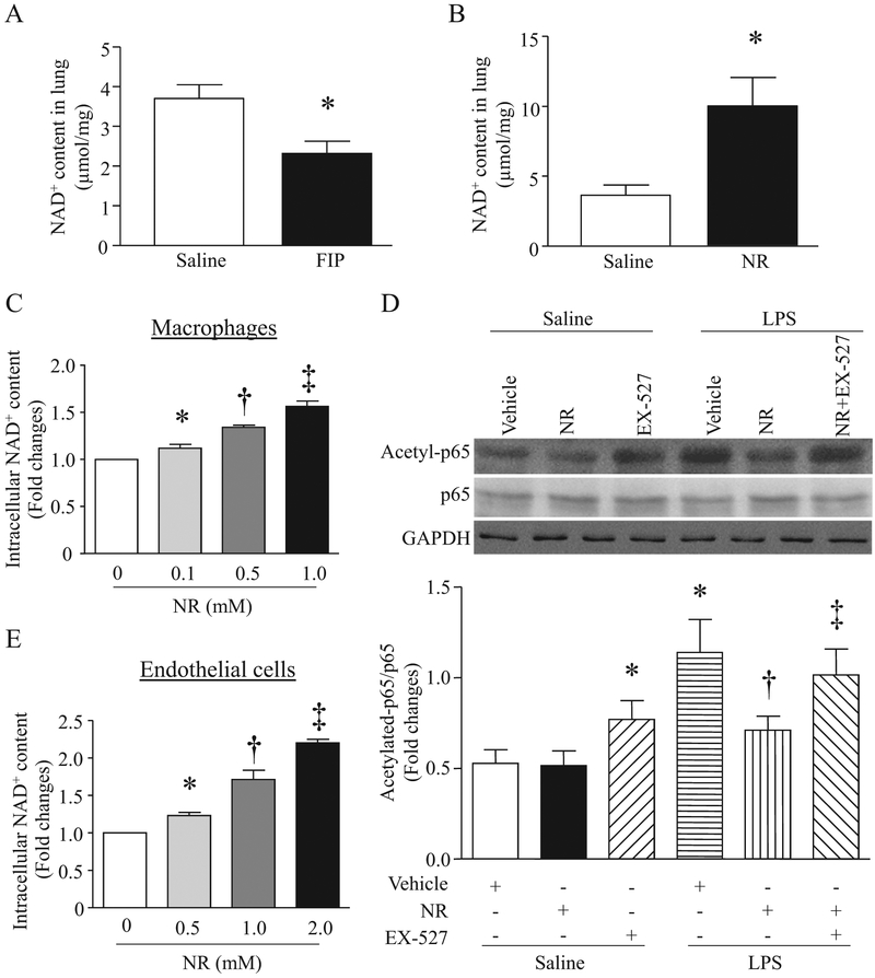
Aims: Sepsis-caused multiple organ failure remains the major cause of morbidity and mortality in intensive care units. Nicotinamide riboside (NR) is a precursor of nicotinamide adenine dinucleotide (NAD+), which is important in regulating oxidative stress. This study investigated whether administration of NR prevented oxidative stress and organ injury in sepsis.

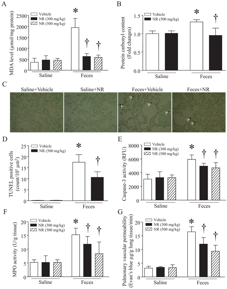
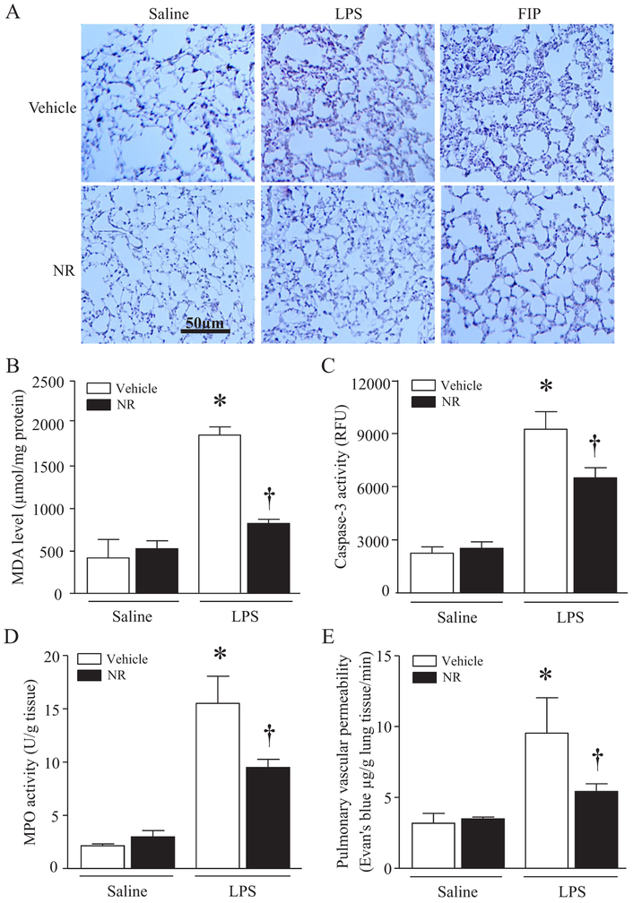
Methods: Mouse sepsis models were induced by injection of lipopolysaccharides (LPS) or feces-injection-in-peritoneum. NR was given before sepsis onset. Cultured macrophages and endothelial cells were incubated with various agents.

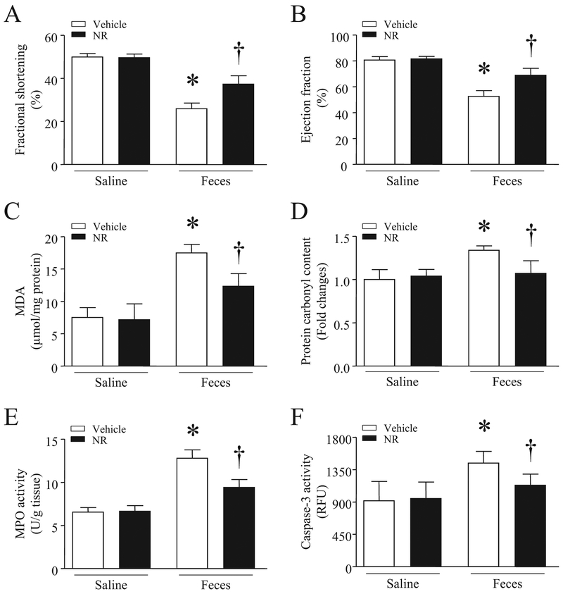
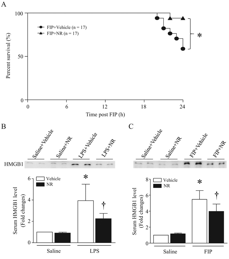
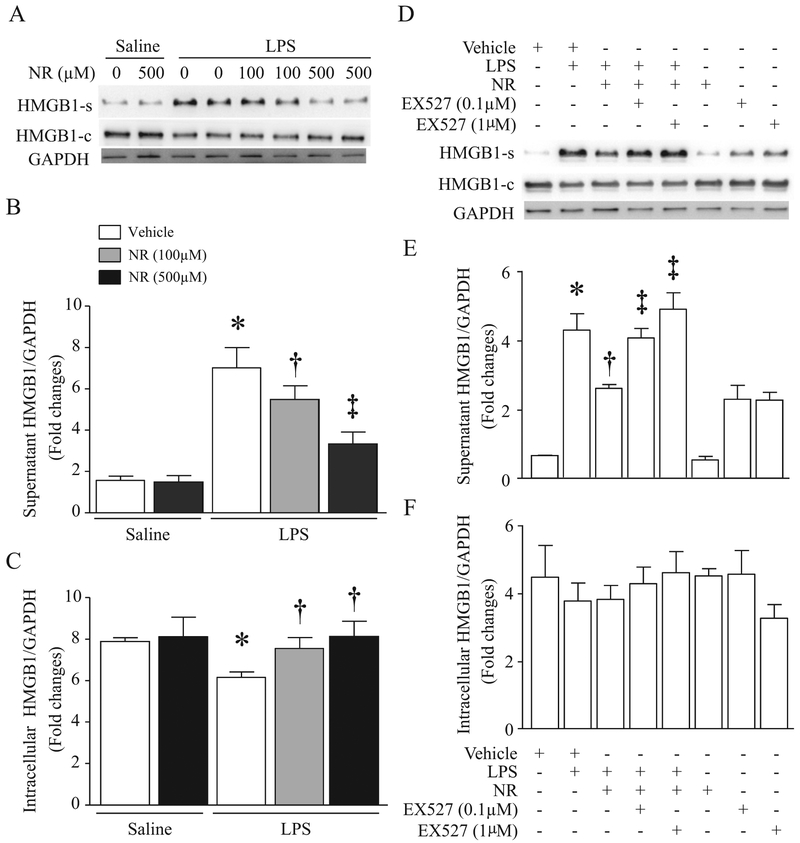
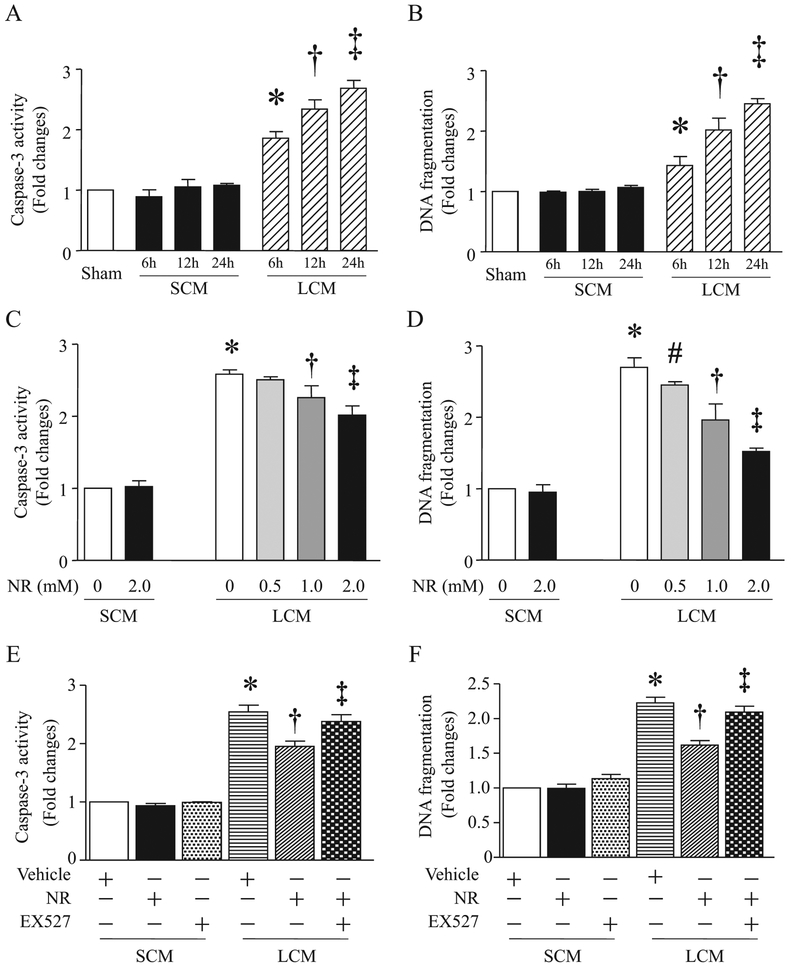
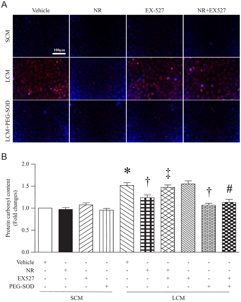
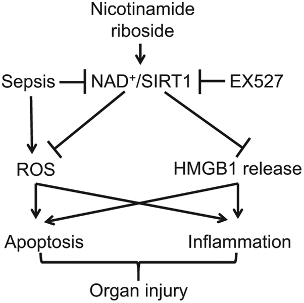
Results: Administration of NR elevated the NAD+ levels, and elicited a reduction of oxidative stress, inflammation and caspase-3 activity in lung and heart tissues, which correlated with attenuation of pulmonary microvascular permeability and myocardial dysfunction, leading to less mortality in sepsis models. These protective effects of NR were associated with decreased levels of plasma high mobility group box-1 (HMGB1) in septic mice. Consistently, pre-treatment of macrophages with NR increased NAD+ content and reduced HMGB1 release upon LPS stimulation. NR also prevented reactive oxygen species (ROS) production and apoptosis in endothelial cells induced by a conditioned-medium collected from LPS-treated macrophages. Furthermore, inhibition of SIRT1 by EX527 offset the negative effects of NR on HMGB1 release in macrophages, and ROS and apoptosis in endothelial cells.

Conclusions: Administration of NR prevents lung and heart injury, and improves the survival in sepsis, likely by inhibiting HMGB1 release and oxidative stress via the NAD+/SIRT1 signaling. Given NR has been used as a health supplement, it may be a useful agent to prevent organ injury in sepsis.

Keywords: HMGB1; Nicotinamide riboside; Oxidative stress; SIRT1; Sepsis.


Figures

Fig. 1.
Fig. 1.
Effects of NR on NAD+ levels and Sirt1 activation. (A) Mice were intraperitoneally injected with feces (3.75 g/kg, n = 6) or saline (n = 5). Six hours later, NAD+ level in lungs were determined. (B) Mice were intraperitoneally administrated with NR (300 mg/kg, n = 5) or saline (n = 5). NAD+ levels in lungs were assessed at 6 h after NR injection. (C) Macrophages (RAW264.7) were incubated with a normal culture medium containing various doses of NR for 36 h and then collected for NAD+ assay. Data are mean ± SD (n = 3). *P<0.05 vs saline, P<0.05 vs NR (0.1 mM), P<0.05 NR (0.5 mM). (D) Macrophages were pretreated with NR (1 mM) or EX527 (1 μM) alone or in combination, or vehicle, and then incubated with LPS (0.1 μg/ml) or saline. Western Blot analysis was performed to determine acetylated p65, total p65 and GAPDH. Upper panel is representative western blots from 3 independent experiments, and lower panel is the quantification of acetylated p65/p65 ratio. Data are mean ± SD (n = 3). *P<0.05 vs saline + vehicle, P<0.05 vs LPS + vehicle, P<0.05 vs LPS+NR. (E) Mouse cardiac microvascular endothelial cells were incubated with a normal culture medium containing NR for 12 h, and collected for NAD+ assay. Data are mean ± SD (n = 3). *P<0.05 vs saline, P<0.05 vs NR (0.5 mM), P<0.05 vs NR (1.0 mM).
Fig. 2.
Fig. 2.
Protective effects of NR on acute lung injury in septic mice. After receiving an intraperitoneal injection of NR (300 or 500 mg/kg) or vehicle, mice were challenged with feces (3.75 g/kg, i.p.) or saline. Six hours later, MDA level (A), carbonyl protein content (B), TUNEL staining (C, representative micro-pictures with white arrows indicating positive staining; D, quantification of TUNEL positive cells per μm2), caspase-3 activity (E) and MPO activity (F) were determined in lung tissues, and pulmonary microvascular albumin leak was measured (G). Data are mean ± SD (n = 5–7 mice in each group). *P<0.05 vs sham or vehicle, P<0.05 vs vehicle + feces.
Fig. 3.
Fig. 3.
Protective effects of NR on acute lung injury in endotoxemic mice. After receiving an intraperitoneal injection of NR (300 mg/kg) or vehicle, mice were challenged with LPS (4 mg/kg), FIP or saline. (A) Lung tissues were collected, fixed in formalin and embedded in wax. After sectioning and processing, H&E staining was performed. A representative H&E staining for histological changes is presented from 5 different hearts. (B–D) Four hours later after LPS injection, MDA level (B), caspase-3 activity (C) and MPO (D) were analyzed in lung tissues, and pulmonary microvascular albumin leak was determined (E). Data are mean ± SD (n = 5 mice in each group). *P<0.05 vs saline + vehicle, P<0.05 vs vehicle + LPS.
Fig. 4.
Fig. 4.
Effects of NR on myocardial injury in septic mice. After receiving an intraperitoneal injection of NR (500 mg/kg) or vehicle, mice were challenged with feces (3.75 g/kg, i.p.) or saline. Six hours later, myocardial function (A and B), MDA level (C), carbonyl protein content (D), MPO activity (E) and caspase-3 activity (F) were measured in heart tissues. Data are mean ± SD (n = 7 mice in each group). *P<0.05 vs saline + vehicle, P<0.05 vs vehicle + feces.
Fig. 5.
Fig. 5.
Effect of NR on survival in septic mice and serum HMGB1 in endotoxemic and septic mice. (A) After receiving an intraperitoneal injection of NR (300 mg/kg) or vehicle, mice were challenged with feces. Survival was monitored for 24 h in septic mice. *P < 0.05 (n = 17 in each group). (B and C) Mice received NR (300 mg/kg) or vehicle and then LPS, FIP or saline (i.p.). After four hours of LPS injection (B) or six hours of FIP (C), the protein levels of HMGB1 in serum were determined by western blot analysis. Data are mean ± SD (n = 5 mice in each group). *P<0.05 vs sham + vehicle, P<0.05 vs vehicle + LPS or vehicle + feces.
Fig. 6.
Fig. 6.
Effects of NR on LPS-induced HMGB1 release in Macrophages. (A–C) Cultured RAW264.7 cells were pretreated with NR (100 μM, 500 μM) or saline for 36 h and then challenged by LPS (0.1 μg/ml). After 24 h of LPS treatment, HMGB1 in supernatants (A, B) and cells (A, C) were determined by western blot analysis. Data are mean ± SD (n = 3). *P<0.05 vs saline + vehicle, P<0.05 vs vehicle + LPS, and P<0.05 vs vehicle + LPS + NR (100 μM). (D–F) Cultured RAW264.7 cells were treated with NR (500 μM) and/or EX527 (0.1 μM, 1 μM) for 36 h and then challenged with or without LPS (100 ng/ml). Twenty-four hours later, HMGB1 in supernatants (D, E) and cells (D, F) were measured by western blot analysis. Data are mean ± SD (n = 3 independent experiments). *P<0.05 vs vehicle, P<0.05 vs vehicle + LPS, and P<0.05 vs LPS + NR.
Fig. 7.
Fig. 7.
Effects of NR on conditioned medium-induced apoptosis in endothelial cells. (A and B) Mouse cardiac microvascular endothelial cells (MCECs) were challenged by a conditioned medium (LCM) from RAW264.7 cells stimulated with LPS or control medium (SCM) from cultured RAW264.7 cells treated with saline for different times. Caspase-3 (A) and DNA fragmentation (B) were analyzed. Data are mean ± SD (n = 3 independent experiments). *P<0.05 vs 0 h, P<0.05 vs 6 h, P <0.05 vs 12 h. (C and D) MCECs were treated with NR (0.5 mM, 1 mM, 2 mM) or saline for 12 h, and then challenged by LCM or SCM from RAW264.7 cells. Twelve hours after addition of LCM and SCM, caspase-3 activity (C) and DNA fragmentation (D) were measured. Data are mean ± SD (n = 3). *P<0.05 vs 0 + SCM, #P < 0.05 vs 0 + LCM, P<0.05 vs 0.5 + LCM, P<0.05 vs 1.0 + CM. (E and F) MCECs were pretreated with NR (2 mM) or saline for 12 h, and then stimulated with LCM or SCM in presence of EX527 (1 μM) or vehicle. Twelve hours later, caspase-3 activity (E) and DNA fragmentation (F) were assessed. Data are mean ± SD (n = 3 independent experiments). *P<0.05 vs vehicle + SCM, P<0.05 vs vehicle + LCM, P<0.05 vs NR + LCM.
Fig. 8.
Fig. 8.
Effects of NR on LPS-conditioned medium (LCM)-induced oxidant stress in endothelial cells. After 12 h of treatment with NR (2 mM), EX527 (1 μM), PEGSOD (100 units/ml) or vehicle alone or in combinations, MCECs were challenged by LCM or saline-conditioned medium (SCM) for 12 h. Intracellular ROS production was measured in living cells by DHE staining. (A) A representative fluorescent microscopic picture for DHE staining (red color) and nuclear staining (blue color) from 3 independent experiments. (B) Carbonyl protein content was determined in cell lysates. Data are mean ± SD (n = 3). *P<0.05 vs vehicle + SCM, P<0.05 vs vehicle + LCM, P<0.05 vs LCM + NR, #P < 0.05 vs EX527 + LCM.
Fig. 9.
Fig. 9.
Schematic of nicotinamide riboside/NAD/SIRT1 pathway in sepsis. Sepsis induces ROS production and inhibits NAD+/SIRT1 signaling, leading to HMGB1 release, apoptosis and inflammation, which contribute to organ injury. Nicotinamide riboside is converted to NAD+ and subsequently activates SIRT1 signaling. Activation of SIRT1 inhibits ROS production and HMGB1 release thereby attenuating apoptosis and inflammation, and finally protecting against organ injury in sepsis. EX527 inhibits SIRT1 activation and reverses the protective effects of nicotinamide riboside on septic organ injury.

References

    1. Martin CM, Priestap F, Fisher H, Fowler RA, Heyland DK, Keenan SP, Longo CJ, Morrison T, Bentley D, Antman N, A prospective, observational registry of patients with severe sepsis: the Canadian sepsis treatment and response registry, Crit. Care Med 37 (1) (2009) 81–88. - PubMed
    1. Singer M, Deutschman CS, Seymour CW, Shankar-Hari M, Annane D, Bauer M, Bellomo R, Bernard GR, Chiche JD, Coopersmith CM, Hotchkiss RS, Levy MM, Marshall JC, Martin GS, Opal SM, Rubenfeld GD, van der Poll T, Vincent JL, Angus DC, The third international consensus definitions for sepsis and septic shock (Sepsis-3), JAMA 315 (8) (2016) 801–810. - PMC - PubMed
    1. Howell MD, Davis AM, Management of sepsis and septic shock, JAMA 317 (8) (2017) 847–848. - PubMed
    1. Wheeler AP, Bernard GR, Acute lung injury and the acute respiratory distress syndrome: a clinical review, Lancet 369 (9572) (2007) 1553–1564. - PubMed
    1. Micek ST, McEvoy C, McKenzie M, Hampton N, Doherty JA, Kollef MH, Fluid balance and cardiac function in septic shock as predictors of hospital mortality, Crit. Care 17 (5) (2013) R246. - PMC - PubMed

Publication types

MeSH terms