. 2018 Oct;69(4):886-895.
doi: 10.1016/j.jhep.2018.05.021. Epub 2018 May 25.

Intestinal HIF-1α deletion exacerbates alcoholic liver disease by inducing intestinal dysbiosis and barrier dysfunction

Affiliations

Intestinal HIF-1α deletion exacerbates alcoholic liver disease by inducing intestinal dysbiosis and barrier dysfunction

Tuo Shao et al. J Hepatol. 2018 Oct.

Abstract

Background & aims: Alcoholic liver disease (ALD) is characterized by gut dysbiosis and increased gut permeability. Hypoxia inducible factor 1α (HIF-1α) has been implicated in transcriptional regulation of intestinal barrier integrity and inflammation. We aimed to test the hypothesis that HIF-1α plays a critical role in gut microbiota homeostasis and the maintenance of intestinal barrier integrity in a mouse model of ALD.

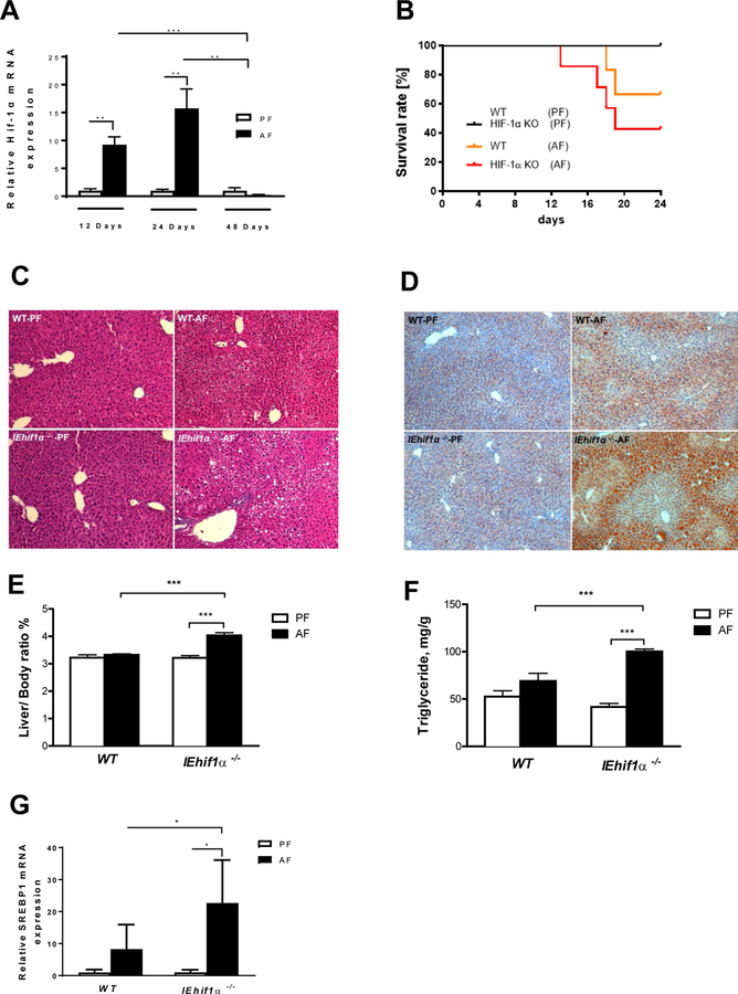
Methods: Wild-type (WT) and intestinal epithelial-specific Hif1a knockout mice (IEhif1α-/-) were pair-fed modified Lieber-DeCarli liquid diet containing 5% (w/v) alcohol or isocaloric maltose dextrin for 24 days. Serum levels of alanine aminotransferase and endotoxin were determined. Fecal microbiota were assessed. Liver steatosis and injury, and intestinal barrier integrity were evaluated.

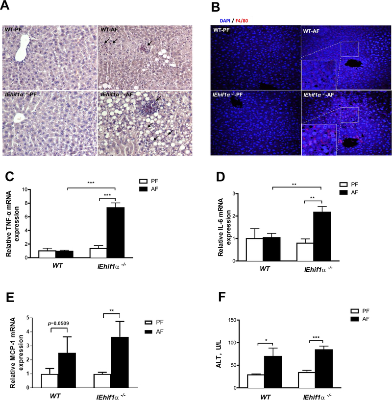
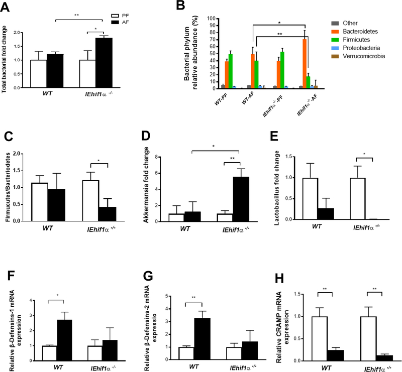
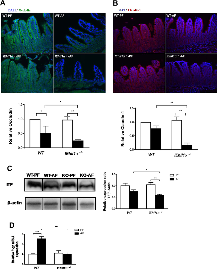
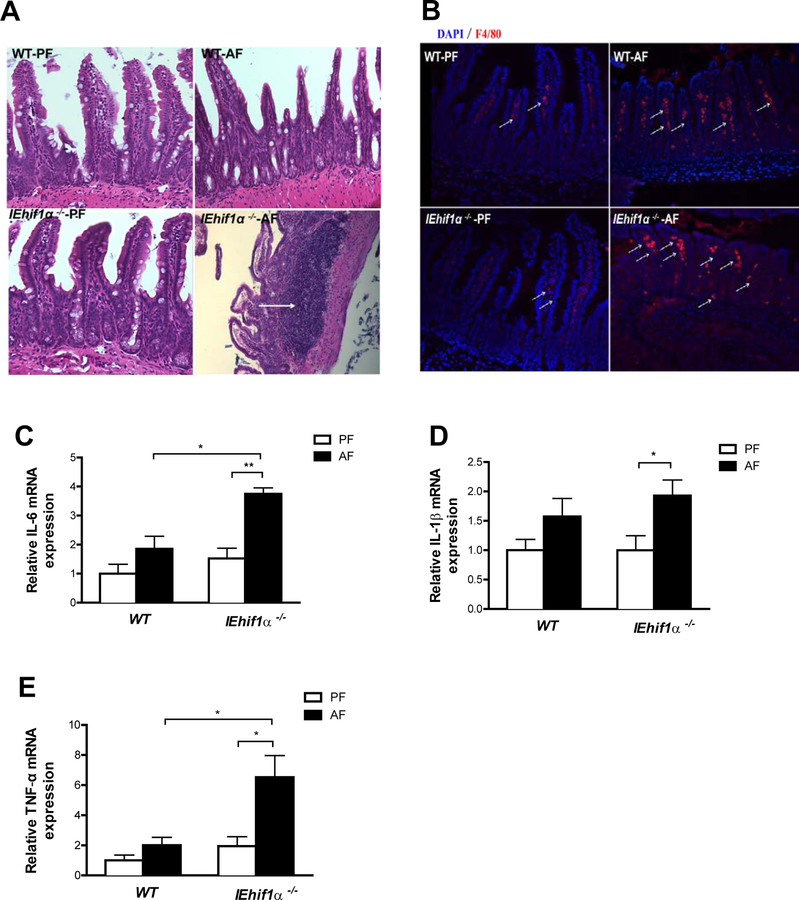
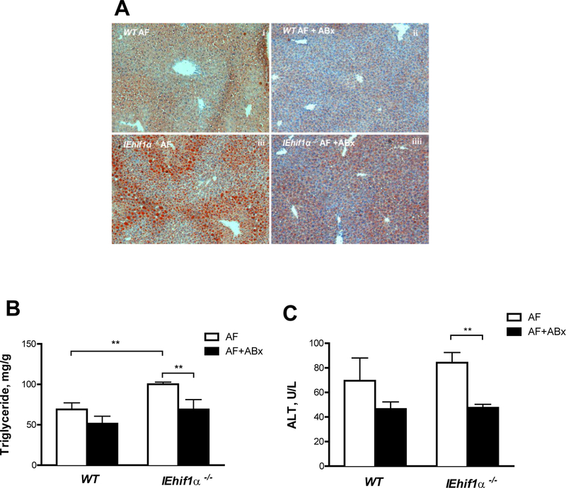
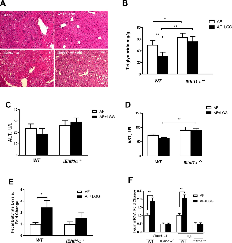
Results: Alcohol feeding increased serum levels of alanine aminotransferase and lipopolysaccharide, hepatic triglyceride concentration, and liver injury in the WT mice. These deleterious effects were exaggerated in IEhif1α-/- mice. Alcohol exposure resulted in greater reduction of the expression of intestinal epithelial tight junction proteins, claudin-1 and occludin, in IEhif1α-/- mice. In addition, cathelicidin-related antimicrobial peptide and intestinal trefoil factor were further decreased by alcohol in IEhif1α-/- mice. Metagenomic analysis showed increased gut dysbiosis and significantly decreased Firmicutes/Bacteroidetes ratio in IEhif1α-/- mice compared to the WT mice exposed to alcohol. An increased abundance of Akkermansia and a decreased level of Lactobacillus in IEhif1α-/- mice were also observed. Non-absorbable antibiotic treatment reversed the liver steatosis in both WT and IEhif1α-/- mice.

Conclusion: Intestinal HIF-1α is essential for the adaptative response to alcohol-induced changes in intestinal microbiota and barrier function associated with elevated endotoxemia and hepatic steatosis and injury.

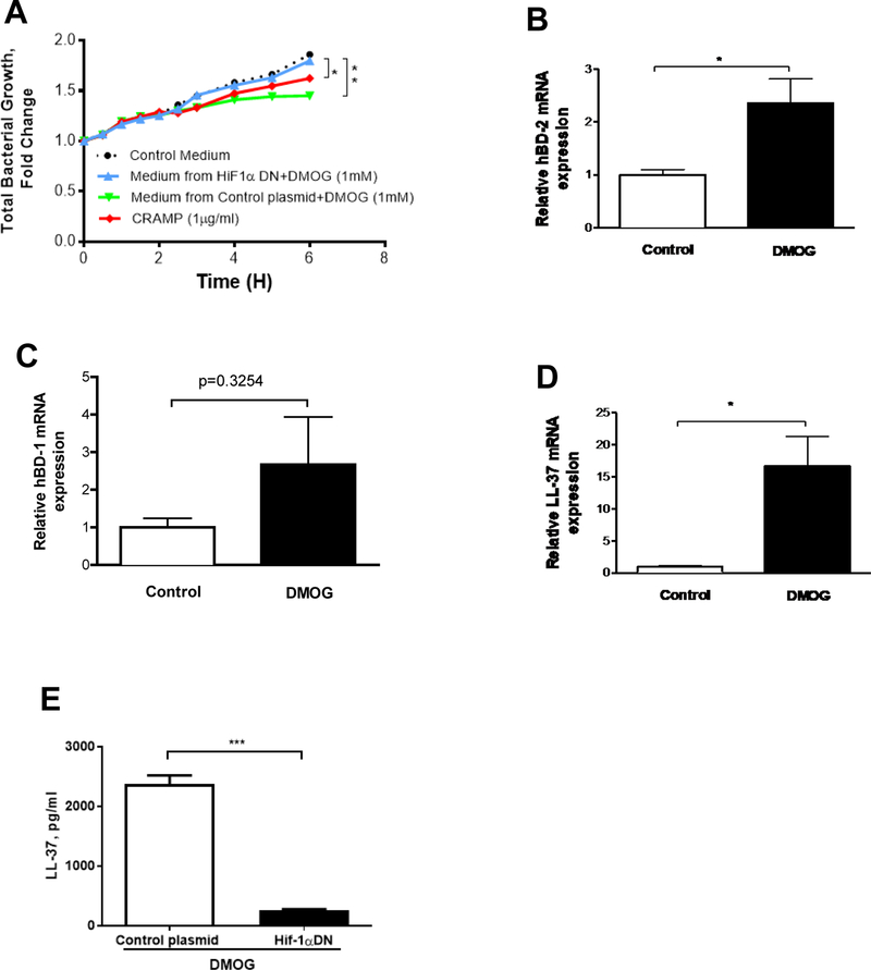
Lay summary: Alcohol consumption alters gut microbiota and multiple intestinal barrier protecting factors that are regulated by intestinal hypoxia-inducible factor 1α (HIF-1α). Absence of intestinal HIF-1α exacerbates gut leakiness leading to an increased translocation of bacteria and bacterial products to the liver, consequently causing alcoholic liver disease. Intestinal specific upregulation of HIF-1α could be developed as a novel approach for the treatment of alcoholic liver disease.

Keywords: Alcoholic Liver Disease; Gut Microbiota; Gut-Liver axis; Hypoxia-Inducible Factor-1α; Intestinal Barrier.


Conflict of interest statement

Conflicts of Interest: The authors declare no conflict of interest.

Figures

Figure 1
Figure 1
Intestinal Hif1α KO (IEhif1α−/−) mice display exacerbated chronic alcoholic liver injury and steatosis. WT and IEhif1α−/− mice were fed alcohol-containing Lieber DeCarli diet as described in the Material and Methods. (A) Hif-1α gene expression in WT mice fed 5% alcohol-containing Lieber DeCarli diet for 12 (n=4 for PF group; n=6 for AF group), 24 (n=4 for PF group; n=5 for AF group), and 48 (n=4 for PF group; n=5 for AF group) days, respectively. (B) Comparison of survival rate between WT and KO mice groups with or without EtOH (n=9 for each group). (C) Hematoxylin and eosin staining of the liver sections of the mice after PF or AF treatment for 24 days. Original magnification, ×10. (D) Oil red O staining of the liver neutral lipids in mice described as in C. (E) Liver weight to body weight ratio of mice described as in C. (F) Quantification of hepatic triglyceride content in mice described as in C. (G) Gene expression of SREBP-1 in liver tissues of mice described as in C. Data are expressed as mean ± SEM. AF, alcohol-fed; PF, pair-fed.
Figure 2
Figure 2
Inflammatory responses in EtOH-induced liver injury. WT and IEhif1α−/− mice were pair- or alcohol-fed for 24 days. (A) Chloroacetate esterase (CAE) localization (cell-localized fuchsia staining) in livers. Original magnification, ×10. Arrows indicate neutrophil infiltration. (B) F4/80 staining of the liver tissues. DAPI (blue) was used to counterstain nuclei. Original magnification, ×10. (C, D, E) Hepatic gene expression of proinflammatory cytokines TNF-α, IL-6 and chemokine MCP-1, respectively. (F) Serum alanine aminotransferase (ALT) activities. Data are expressed as mean ± SEM. AF, alcohol-fed; PF, pair-fed.
Figure 3
Figure 3
Effect of Hif1α on alcohol-induced dysbiosis. Mice were treated as described in Fig 2. (A) The total bacterial load in the cecum of mice. (B) Phylum levels of microbiota in fecal samples. (C) The ratio of Firmucutes/bacteriodetes level. (D, E) Akkermansia (Verrucomicrobia phylum) and Lactobacillus (Firmicutes phylum) levels. (F, G, H) β-Defensins (β-Defensins-1, 2) and cathelicidin-related antimicrobial peptide (CRAMP) gene expression in the ileal tissues. Data are expressed as mean ± SEM. AF, alcohol-fed; PF, pair-fed.
Figure 4
Figure 4
Effect of HIF activation in T84 cells on the bacterial growth and antimicrobial peptide gene expression. (A) Mouse fecal bacterial growth in response to control plasmid+DMOG, HIF-1αDN+DMOG, and synthetic CRAMP peptide. (B-D) Gene expression of LL-37 and human β-Defensins (hBD-1, 2). (E) The medium concentration of LL-37 by ELISA. Data are expressed as mean ± SEM of a minimum of three independent experiments.
Figure 5
Figure 5
Effect of Hif-1α on intestinal barrier integrity. Mice were treated as described in Fig 2. (A, B) Immunoflorescent staining of occludin (green) and claudin-1 (blue) in the ileal tissues. DAPI (blue) was used to counterstain of nuclei. Original magnification, ×10. (C) ITF protein Western blotting and quantification of ileal tissues normalized by β-actin. The bands are composite images of selected bands;and that the corresponding β-actin controls are shown. (D) P-gp gene expression in ileal tissues. Data are expressed as mean ± SEM. (AF, alcohol-fed; PF, pair-fed).
Figure 6
Figure 6
Effect of Hif-1α on intestinal inflammation. Mice were treated as described in in Fig 2. (A) HE staining of ileal tissues. Original magnification, ×10. (B) Immunofluorescent staining of F4/80 (red) showed increased macrophages in ileum of AF IEhif1α−/− mice (arrows). DAPI (blue) was used to visualize nuclei. Original magnification: ×10. (C, D, E) Ileum IL-6, IL-1β and TNF-α mRNA levels. Data are expressed as mean ± SEM. AF, alcohol-fed; PF, pair-fed.
Figure 7
Figure 7
Effects of non-absorbable broad-spectrum antibiotics (ABx) on EtOH-induced liver steatosis and injury. WT and IEhif1α−/− mice were fed 5% alcohol liquid diets for 24 days (n=4–6). Antibiotics treatment was started at day 12 after alcohol feeding, and mice were gavaged daily until harvesting. (A) Oil red O staining of hepatic tissues. Original magnification, ×10. (B) Quantification of hepatic triglyceride content. (C) Serum ALT levels. Data are mean ± SEM. AF, alcohol-fed; PF, pair-fed.
Figure 8
Figure 8
Effects of Hif-1α on Lactobacillus rhamnosus GG treatment in ALD. Mice were fed alcohol following the NIAAA model (n=5 for each group). (A) HE staining revealed LGG administration reduced steatosis by alcohol exposure in WT mice. However, LGG protective effects were diminished in IEhif1α−/− mice. (B) Quantification of hepatic triglyceride content. (C,D) Serum ALT and AST levels. (E) Fecal butyrate concentration. (F) Ileum Claudin-1 and P-gp mRNA levels. Data are expressed as mean ± SEM. AF, alcohol-fed; PF, pair-fed.

Comment in

References

    1. Gao B, Bataller R. Alcoholic Liver Disease: Pathogenesis and New Therapeutic Targets. Gastroenterology 2011;141:1572–1585. - PMC - PubMed
    1. Adachi Y, Moore LE, Bradford BU, Gao W, Thurman RG. Antibiotics prevent liver injury in rats following long-term exposure to ethanol. Gastroenterology 1995;108:218–224. - PubMed
    1. Parlesak A, Schafer C, Schutz T, Bode JC, Bode C. Increased intestinal permeability to macromolecules and endotoxemia in patients with chronic alcohol abuse in different stages of alcohol-induced liver disease. J Hepatol 2000;32:742–747. - PubMed
    1. Chen P, Miyamoto Y, Mazagova M, Lee KC, Eckmann L, Schnabl B. Microbiota Protects Mice Against Acute Alcohol-Induced Liver Injury. Alcohol Clin Exp Res 2015;39:2313–2323. - PMC - PubMed
    1. Szabo G, Bala S. Alcoholic liver disease and the gut-liver axis. World J Gastroenterol 2010;16:1321–1329. - PMC - PubMed

Substances

LinkOut - more resources