. 2018:144:33-74.
doi: 10.1016/bs.mcb.2018.03.040.

Dissecting the role of the tubulin code in mitosis

Affiliations

Dissecting the role of the tubulin code in mitosis

Luísa T Ferreira et al. Methods Cell Biol. 2018.

Abstract

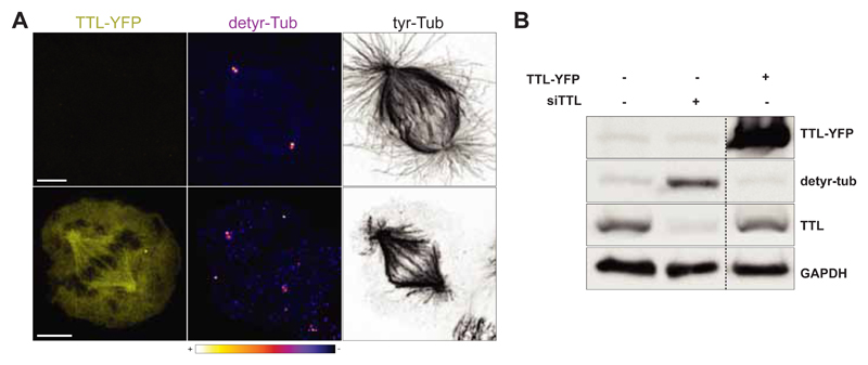
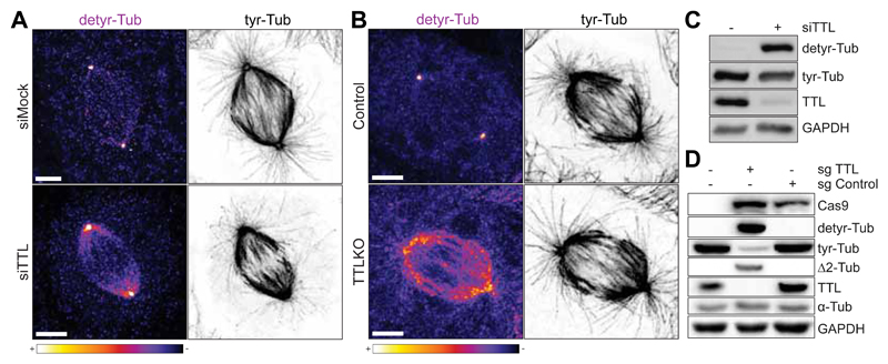
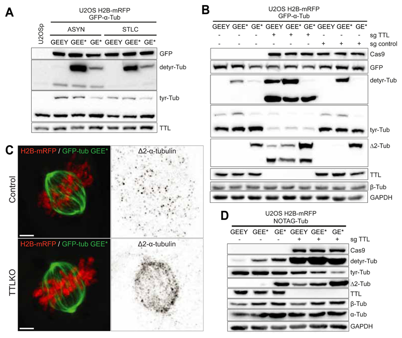
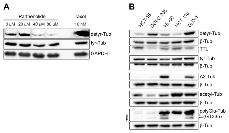
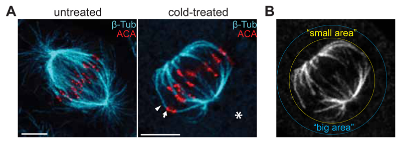
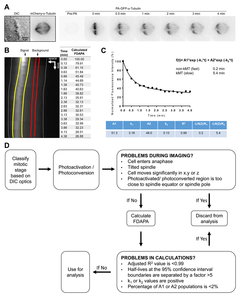
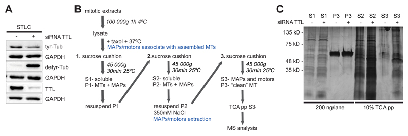
Mitosis is an essential process that takes place in all eukaryotes and involves the equal division of genetic material from a parental cell into two identical daughter cells. During mitosis, chromosome movement and segregation are orchestrated by a specialized structure known as the mitotic spindle, composed of a bipolar array of microtubules. The fundamental structure of microtubules comprises of α/β-tubulin heterodimers that associate head-to-tail and laterally to form hollow filaments. In vivo, microtubules are modified by abundant and evolutionarily conserved tubulin posttranslational modifications (PTMs), giving these filaments the potential for a wide chemical diversity. In recent years, the concept of a "tubulin code" has emerged as an extralayer of regulation governing microtubule function. A range of tubulin isoforms, each with a diverse set of PTMs, provides a readable code for microtubule motors and other microtubule-associated proteins. This chapter focuses on the complexity of tubulin PTMs with an emphasis on detyrosination and summarizes the methods currently used in our laboratory to experimentally manipulate these modifications and study their impact in mitosis.

Keywords: Detyrosination; Microtubules; Mitosis; Mitotic spindle; Tubulin code; Tubulin posttranslational modifications; Tyrosination.


Figures

Fig. 1
Fig. 1
A. Confocal image of a representative U2OS cell transiently overexpressing TTL-YFP (bottom panel). A non-transfected U2OS cell (upper panel) is represented as control. Scale bar, 5 μm. B. Western-blot analysis of U2OS cell lysate transiently overexpressing TTL-YFP.
Fig. 2
Fig. 2
A. Confocal image of U2OS cells transfected with non-targeting siRNA (siMock) or with siRNA for TTL (siTTL). B. Confocal image of U2OS control and TTL KO cells. Scale bar, 5 μm. C. Western-blot analysis of U2OS cell lysates before and after siRNA-mediated depletion of TTL. D. Western-blot analysis of U2OS cell lysates in control (sgcontrol) and TTL KO cells (sgTTL).
Fig. 3
Fig. 3
Western-blot analysis of U2OS cell lysates. A. Transiently overexpressing VASH1-GFP, VASH2-FLAG and SVBP-FLAG in control and TTL KO cells; B. Stably co-expressing VASH1-GFP and VASH2-FLAG or C. transfected with siRNAs for endogenous tubulins or TTL.
Fig. 4
Fig. 4
A. Western-blot analysis of U2OS cell lysates stably expressing H2B-mRFP and GFP-α-Tub (GEEY, GEE* or GE*) in asynchronous and mitotic populations (cells treated with STLC for 14h). B. Western-blot analysis of U2OS cell lysates stably expressing H2B-mRFP and GFP-α-Tub (GEEY, GEE* or GE*) in control (sgcontrol) and TTL KO cells (sgTTL). C. Deconvolved immunofluorescence showing the cellular distribution of ?2-tubulin in U2OS cells stably expressing H2B-mRFP and GFP-GEE* in the presence or absence of TTL. Scale bar, 5 μm. D. Western-blot analysis of U2OS cells lysates stably expressing H2B-mRFP and NOTAG-α-Tub (GEEY, GEE* or GE*), in control and TTLKO (sgTTL) cells.
Fig. 5
Fig. 5
A. Western-blot analysis of U2OS cells treated with the indicated concentrations of Parthenolide for 1h. Treatment with 10nM taxol for 1h was used as a control. B. Analysis of the expression profile of tubulin PTMs in the indicated cell lines by Western blot. β-tubulin was used as loading control.
Fig. 6
Fig. 6
A. Immunofluorescence of U2OS cells before and after cold-induced MT destabilization. B. Sum projection of the tubulin channel showing the mitotic spindle circumscribed by two ROIs (small area and big area) used to quantify the fluorescence intensity at the spindle region.
Fig. 7
Fig. 7
Calculating MT turnover through photoactivation. A. DIC and time-lapse fluorescent images of a representative metaphase U2OS cell expressing PA-GFP-α-tubulin and mCherry-α-tubulin. The mitotic spindle is visualized by mCherry fluorescence. Fluorescent images are inverted for better visualization of the photoactivated GFP molecules. B. Sum-projected, whole-spindle kymograph generated to quantify the Fluorescence Dissipation After Photo-Activation (FDAPA). Dashed white lines indicate the spindle poles; yellow lines indicate the boundaries used to quantify the signal generated from PA; red lines indicate the boundaries used for determining background levels. C. Normalized fluorescence intensity. Once fitted as a double exponential curve, the values obtained allow for the calculation of the dynamics of fast and slow MT populations. D. Photoactivation troubleshooting flowchart used to determine the exclusion criteria for calculating MT turnover using PA.
Fig. 8
Fig. 8
A. Western blot analysis of mitotic HeLa cells lysates after treatment with siRNA for TTL. B. Schematic diagram of the protocol used for the purification of MAPs and motors from mitotic extracts. Protocol adapted from (Sloboda, 2015). C. Silver staining analysis of fractions obtained following protocol B.

Similar articles

Cited by

References

    1. Aiken J, Sept D, Costanzo M, Boone C, Cooper JA, Moore JK. Genome-wide analysis reveals novel and discrete functions for tubulin carboxy-terminal tails. Curr Biol. 2014;24(12):1295–1303. doi: 10.1016/j.cub.2014.03.078. - DOI - PMC - PubMed
    1. Aillaud C, Bosc C, Peris L, Bosson A, Heemeryck P, Van Dijk J, et al. Moutin MJ. Vasohibins/SVBP are tubulin carboxypeptidases (TCPs) that regulate neuron differentiation. Science. 2017;358(6369):1448–1453. doi: 10.1126/science.aao4165. - DOI - PubMed
    1. Aillaud C, Bosc C, Saoudi Y, Denarier E, Peris L, Sago L, et al. Moutin MJ. Evidence for new C-terminally truncated variants of alpha- and beta-tubulins. Mol Biol Cell. 2016;27(4):640–653. doi: 10.1091/mbc.E15-03-0137. - DOI - PMC - PubMed
    1. Akella JS, Wloga D, Kim J, Starostina NG, Lyons-Abbott S, Morrissette NS, et al. Gaertig J. MEC-17 is an alpha-tubulin acetyltransferase. Nature. 2010;467(7312):218–222. doi: 10.1038/nature09324. - DOI - PMC - PubMed
    1. Akera T, Chmatal L, Trimm E, Yang K, Aonbangkhen C, Chenoweth DM, et al. Lampson MA. Spindle asymmetry drives non-Mendelian chromosome segregation. Science. 2017;358(6363):668–672. doi: 10.1126/science.aan0092. - DOI - PMC - PubMed

Publication types