. 2018 Jun 4;28(11):1818-1824.e2.
doi: 10.1016/j.cub.2018.04.063. Epub 2018 May 24.

Stereotyped Synaptic Connectivity Is Restored during Circuit Repair in the Adult Mammalian Retina

Affiliations

Stereotyped Synaptic Connectivity Is Restored during Circuit Repair in the Adult Mammalian Retina

Corinne Beier et al. Curr Biol. .

Abstract

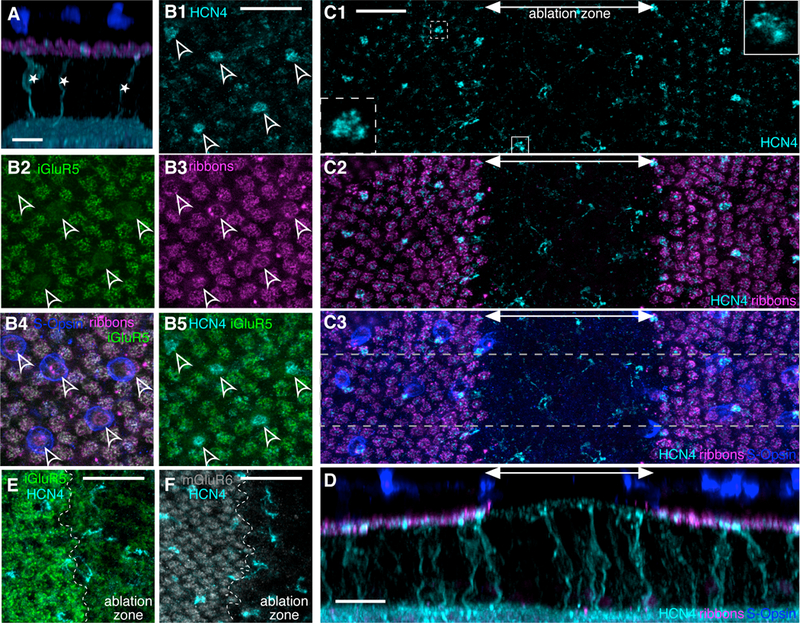
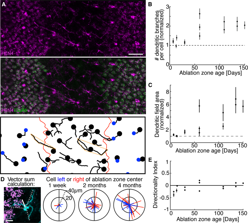
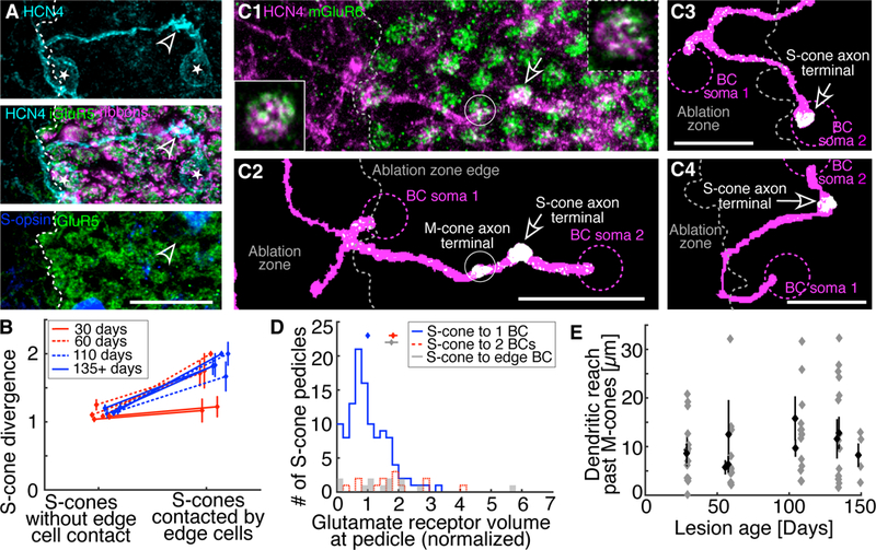
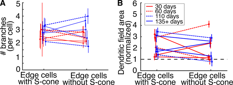
Proper function of the central nervous system (CNS) depends on the specificity of synaptic connections between cells of various types. Cellular and molecular mechanisms responsible for the establishment and refinement of these connections during development are the subject of an active area of research [1-6]. However, it is unknown if the adult mammalian CNS can form new type-selective synapses following neural injury or disease. Here, we assess whether selective synaptic connections can be reestablished after circuit disruption in the adult mammalian retina. The stereotyped circuitry at the first synapse in the retina, as well as the relatively short distances new neurites must travel compared to other areas of the CNS, make the retina well suited to probing for synaptic specificity during circuit reassembly. Selective connections between short-wavelength sensitive cone photoreceptors (S-cones) and S-cone bipolar cells provides the foundation of the primordial blue-yellow vision, common to all mammals [7-18]. We take advantage of the ground squirrel retina, which has a one-to-one S-cone-to-S-cone-bipolar-cell connection, to test if this connectivity can be reestablished following local photoreceptor loss [8, 19]. We find that after in vivo selective photoreceptor ablation, deafferented S-cone bipolar cells expand their dendritic trees. The new dendrites randomly explore the proper synaptic layer, bypass medium-wavelength sensitive cone photoreceptors (M-cones), and selectively synapse with S-cones. However, non-connected dendrites are not pruned back to resemble unperturbed S-cone bipolar cells. We show, for the first time, that circuit repair in the adult mammalian retina can recreate stereotypic selective wiring.


Conflict of interest statement

DECLARATION OF INTEREST

The authors declare no competing interests.

Figures

Figure 1
Figure 1. S-cone bipolar cells are completely deafferented by local photoreceptor ablation.
A. Confocal reconstruction of the healthy retina cross section. S-cone bipolar cells are immunostained for HCN4 (cyan). S-cone bipolar cell bodies (*) sit in the inner nuclear layer and send a single dendrite towards the photoreceptor ribbons (CtBP2, magenta) in the outer plexiform layer (OPL) to synapse with a single S-cone photoreceptor (outer segments stained with S-opsin, blue). B1. Maximal Z-projection in the OPL of healthy retina. Bright HCN4 puncta define locations of the S-cone bipolar cells’ dendritic tips (arrowheads). B2. In the same field of view as in (B1), iGluR5 immunostaining marks the location of M-cone axon terminal synapses, but not S-cone axon terminals. B3. Photoreceptor ribbons (CtBP2) mark both M- and S-cone axon terminal locations. B4. S-opsin staining from the outer nuclear layer (ONL) is overlaid with projections from the OPL, (B2) and (B3). S-cone cell bodies, though slightly offset from their axon terminals, align with the marked S-cone axon terminal locations. B5. Overlay of (B1) and (B2). S-cone bipolar cell dendritic tips (HCN4) terminate at S-cone axon terminals (lack of iGluR5). C1. Maximal Z-projection of the OPL of a 1-week-old ablation zone. S-cone bipolar cell dendritic tips (HCN4, cyan) are visible inside and outside the ablation zone. A double headed arrow marks the approximate width of the ablation zone area. Insets: Dendritic tip of an S-cone bipolar cell outside (dashed outline) and inside (solid outline) ablation zone. Inset images show a 5×5µm area of a single 0.3µm thick optical plane. C2. The ablation zone (marked by double headed arrow) is identified by the lack of photoreceptor ribbons (CtBP2, magenta). C3. S-opsin immunostaining from the ONL overlaid with (C2) marks S-cone locations. S-opsin staining is absent in the 1-week-old ablation zone. D. Cross section of the retinal region outlined in gray dashed lines in (C3). S-cone bipolar cell dendritic stalks extend vertically towards the OPL inside and outside the ablation zone. E and F show a portion of the OPL at the edge of a 1-week-old ablation zone. The ablation zone is to the right of the ablation zone edge (edge marked by dashed white line). Both iGluR5 (E) and mGluR6 (F) labeling is decreased inside the lesion. Scale bars are 20µm in all panels.
Figure 2
Figure 2. S-cone bipolar cells expand their dendritic trees in response to deafferentation.
A. Numerous dendritic branches of S-cone bipolar cells (HCN4, cyan) visible in a 60- day-old ablation zone (area lacking iGluR5, green). Bottom panel: S-cone bipolar cell dendrites (traced in 3D). Bipolar cell soma locations and S-cone axon terminals are marked by the black dots and blue dots, respectively. Red lines mark ablation zone border. Dashed orange polygons outline the dendritic fields of example cells. Scale bar 20µm. B. The average number of dendritic branches per cell inside the ablation zone normalized to the average dendritic branch number per cell outside the ablation zone (data from 14 ablation zones are shown as mean ± SEM, n ≥ 30 cells for each data point). S-cone bipolar cells inside the ablation zone have more dendritic branches than S-cone bipolar cells outside the ablation zone (binomial probability, p < 1e-6 for all ablation zones 60 days and older). Dendritic branches per cell increase with lesion age (correlation coefficient = 0.74, p < 2e-3). C. Dendritic field area (see dashed orange polygons in A for examples) dependence on ablation zone age for the same 14 ablation zones as in B. Dendritic field areas of cells inside each ablation zone are normalized by dendritic field areas in the surrounding retina. Dendritic fields inside ablation zones are larger than dendritic fields outside of the ablation zone (KS test, p < 0.001 for all lesions 60 days and older, n ≥ 30 cells for each ablation zone). Dendritic field areas increase with lesion age (correlation coefficient = 0.85, p < 2e-4). D. Left panel: Dendritic branch reach and angle (dashed lines) of an S-cone bipolar cells (cyan) were summed as vectors to estimate the dendritic directionality of the cell (white line). Three right panels: Vectors representing individual cells were colored red/blue if the cell was to the right/left of the ablation zone center, respectively. The ablation zone has been oriented vertically (black line) in each plot. Scale bar 5µm. E. Average directionality index of S-cone bipolar cells calculated in the 14 ablation zones at different times after photoreceptor ablation (mean ± SEM, n ≥ 10 cells for each ablation zone). Deafferented cell dendrites do not show consistent directional bias.
Figure 3
Figure 3. Deafferented S-cone bipolar cell dendrites bypass M-cones to synapse with new S-cones.
A. Two S-cone bipolar cells (somas marked by *) contact the same S-cone axon terminal (arrowhead). The dendritic tree of the leftmost bipolar cell begins within the ablation zone (area lacking iGluR5, green and CtBP2, magenta). The S-cone bipolar cell dendrite travels parallel to the OPL, through M-cone territory (iGluR5, green), to contact the S-cone. Scale bar 20µm. B. S-cones contacted by edge cells are significantly more likely to diverge to more than 1 S-cone bipolar cell than S-cones that are not contacted by edge cells (binomial probability, p ≤ 1e-7 for ablation zones 60 days and older, n ≥ 14 cells with and without contact in each age group: 30 days, 60 days, 110 days and 135–150 days). C1. S-cone bipolar cell dendrites (HCN4, magenta) and mGluR6 (green) located at cone axon terminals visible in a maximal Z-projection of the OPL. Insets: HCN4 and mGluR6 overlap in a single 0.3µm thick optical plane at S-cone bipolar cell dendritic tips. Inset with solid outline: S-cone terminal contacted by a restructured S-cone bipolar cell (same synapse as indicated by arrow). Inset with dashed outline: S-cone terminal contacted by a non-restructured S-cone bipolar cell, far from the ablation zone edge not shown in the image. Insets are 5×5µm. C2: Z-projection of C1 after conversion into binary images. mGluR6 staining not overlapping with HCN4 dendritic traces was removed, leaving only colocalized mGluR6 (white). See Methods for details. Additional examples in C3 and C4. In all C panels, two S-cone bipolar cells (dashed magenta circles indicate somas) synapse with a single S-cone (arrow). In each case, one S-cone bipolar cell’s dendritic tree begins within the ablation zone and travels out to make the synapse, where mGluR6 is present. We rarely (n = 4/23 edge cells) found evidence for an M-cone synapse. See C2 for the rare example. Scale bars 20µm. D. The volume overlap between HCN4 and mGluR6 at S-cone axon terminals (mean ± SEM above histograms). mGluR6 volume at individual S-cone terminals is normalized to the average mGluR6 volume at all S-cone terminals. S-cones connected to 1 S-cone bipolar cell have less mGluR6 volume than S-cones diverging to 2 S-cone bipolar cells (Student’s t-test, p = 8e-5), including S-cones that diverge to edge cells (p = 0.0011). S-cones in healthy retina diverging to 2 S-cone bipolar cells have the same mGluR6 volume as S-cones that diverge to edge cells (p = 0.92). E. The dendritic reach through M-cone territory of individual edge bipolar cells that contact an S-cone (grey diamonds). Black diamonds show mean ± SEM of the 10 ablation zones that are 30 days and older.
Figure 4
Figure 4. Dendritic tree simplification does not occur after new S-cone synapses are established.
A. The number of dendritic branches per edge cell with or without S-cones. Mean ± SEM. S-cone contact does not affect the number of dendritic branches per cell (binomial probability, p > 0.06, n ≥ 8 cells with and without S-cones in each age group). B. The dendritic field area of edge cells with and without S-cones. The dendritic fields of edge cells have been normalized to the dendritic fields of cells within the ablation zone. Mean ± SEM. Dendritic field areas do not change with S-cone contact (KS test, p > 0.8, n ≥ 8 cells with and without S-cones in each age group).

Comment in

Similar articles

Cited by

References

    1. Sanes JR, and Yamagata M (2009). Many Paths to Synaptic Specificity. Annu. Rev. Cell Dev. Biol 25, 161–195. - PubMed
    1. Margeta MA, and Shen K (2010). Cellular and Molecular Mechanisms of Synaptic Specificity. Mol. Cell. Neurosci 43, 261–267. - PMC - PubMed
    1. de Wit J, and Ghosh A (2015). Specification of synaptic connectivity by cell surface interactions. Nat. Rev. Neurosci 17, 4–4. - PubMed
    1. Luo J, McQueen PG, Shi B, Lee C-H, and Ting C-Y (2016). Wiring dendrites in layers and columns. J. Neurogenet 30, 69–79. - PMC - PubMed
    1. Zhang C, Kolodkin AL, Wong RO, and James RE (2017). Establishing Wiring Specificity in Visual System Circuits: From the Retina to the Brain. Annu. Rev. Neurosci 40, annurev-neuro-072116-031607. - PubMed

Publication types