. 2018 Jul 3;26(7):960-971.e4.
doi: 10.1016/j.str.2018.04.012. Epub 2018 May 24.

Mis16 Switches Function from a Histone H4 Chaperone to a CENP-ACnp1-Specific Assembly Factor through Eic1 Interaction

Affiliations

Mis16 Switches Function from a Histone H4 Chaperone to a CENP-ACnp1-Specific Assembly Factor through Eic1 Interaction

Sojin An et al. Structure. .

Abstract

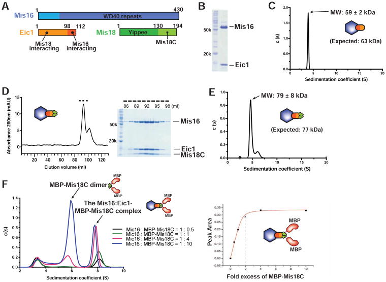
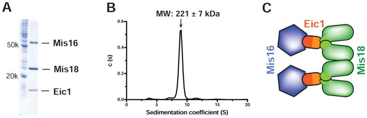
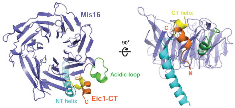
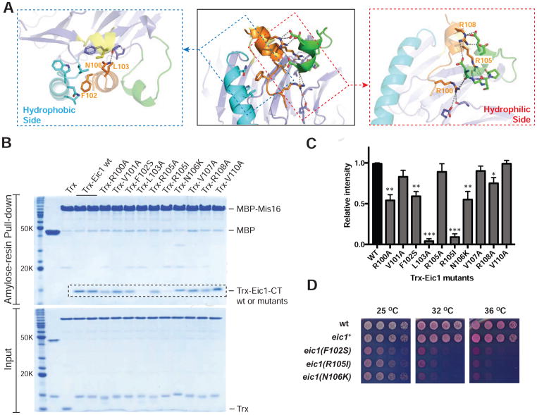
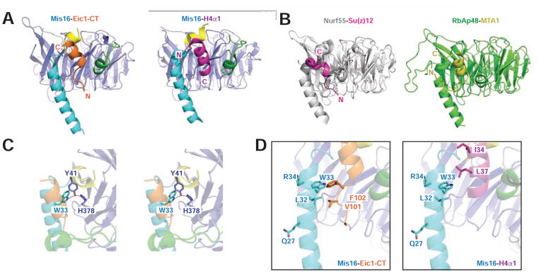
The Mis18 complex, composed of Mis16, Eic1, and Mis18 in fission yeast, selectively deposits the centromere-specific histone H3 variant, CENP-ACnp1, at centromeres. How the intact Mis18 holo-complex oligomerizes and how Mis16, a well-known ubiquitous histone H4 chaperone, plays a centromere-specific role in the Mis18 holo-complex, remain unclear. Here, we report the stoichiometry of the intact Mis18 holo-complex as (Mis16)2:(Eic1)2:(Mis18)4 using analytical ultracentrifugation. We further determine the crystal structure of Schizosaccharomyces pombe Mis16 in complex with the C-terminal portion of Eic1 (Eic1-CT). Notably, Mis16 accommodates Eic1-CT through the binding pocket normally occupied by histone H4, indicating that Eic1 and H4 compete for the same binding site, providing a mechanism for Mis16 to switch its binding partner from histone H4 to Eic1. Thus, our analyses not only determine the stoichiometry of the intact Mis18 holo-complex but also uncover the molecular mechanism by which Mis16 plays a centromere-specific role through Eic1 association.

Keywords: CENP-A; Eic1; Mis16; X-ray crystallography; analytical ultracentrifugation; centromere; fission yeast; histone chaperone; kinetochore; the Mis18 holo-complex.


Conflict of interest statement

DECLARATION OF INTERESTS

The authors declare no competing financial interests.

Figures

Figure 1
Figure 1. Stoichiometry of the S. pombe Mis18 sub-Complexes
(A) Domain organization of the S. pombe Mis18 holo-complex components. (B) SDS-PAGE analysis of the purified S. pombe Mis16-Eic1 subcomplex expressed in baculovirus insect cells. (C) SV-AUC analysis of the purified Mis16-Eic1 complex. The calculated MW corresponds to a heterodimer of Mis16 and Eic1. (D) SEC profile of the Mis16-Eic1-Mis18C complex (left). The dashed line indicates fractions used for SDS-PAGE analysis (right). (E) SV-AUC analysis of the Mis16-Eic1-Mis18C complex. The MW of the peak corresponds to a 1:1:2 molar ratio. (F) SV-AUC analyses of the Mis16-Eic1 heterodimer with different concentrations of MBP-tagged Mis18C (MBP-Mis18C) (left) and its titration curve (right). See also Figure S1.
Figure 2
Figure 2. Stoichiometry of the S. pombe Mis18 Holo-Complex
(A) SDS-PAGE analysis of the purified S. pombe Mis18 holo-complex expressed in baculovirus insect cells. (B) SV-AUC analysis of the purified S. pombe Mis18 holo-complex. The calculated MW suggests a stoichiometry of (Mis16)2:(Eic1)2:(Mis18)4. (C) Schematic diagram showing composition of the S. pombe Mis18 holo-complex. See also Figure S2.
Figure 3
Figure 3. Crystal Structure of the S. pombe Mis16-Eic1-CT Complex
Top- and 90°-oriented side views of the S. pombe Mis16-Eic1-CT complex. The Mis16 WD-40 repeat domain is colored slate. The binding pocket of Eic1-CT (orange) is formed by the Mis16 N-terminal helix (NT helix, cyan), acidic loop (green), and C-terminal helix (CT helix, yellow). All structures shown in the figures were generated using PYMOL (Delano Scientific, LLC). See also Figures S3, S4, and S5.
Figure 4
Figure 4. In Vitro and In Vivo Mutational Analysis of Eic1-CT
(A) Hydrophobic interactions (blue dashed boxes) and hydrophilic interactions (red dashed boxes) of Eic1-CT within the Mis16-Eic1-CT complex. Eic1 residues from the hydrophobic side mostly form hydrophobic interactions with residues in the Mis16 N-terminal helix (cyan) and WD-40 repeats (slate). The hydrophilic side residues of Eic1 form hydrogen bond and electrostatic interactions with residues from the Mis16 acidic loop (green) and WD-40 repeats. (B) SDS-PAGE analysis of the amylose resin pull-down assay using MBP-tagged Mis16 as bait and thioredoxin (Trx)-tagged Eic1-CT (residues 91–112) (wild-type or mutant) as prey. (C) Histogram showing normalized band intensities from pull-down experiments in (B). Band intensities for Trx-Eic1-CT mutants within the dashed box area in (B) were measured and normalized relative to that of wild-type. Results are shown as mean ± SEM of three independent measurements; *P < 0.05, ** P < 0.005, *** P < 0.0005. (D) In vivo screen for temperature-sensitive Eic1-CT mutants identifies key residues. S. pombe cells harboring the indicated eic1 mutations were five-fold serial diluted, spotted on YES + Phloxine B media, and incubated at the indicated temperatures; dead cells stain dark pink. See also Figures S4 and S5.
Figure 5
Figure 5. Comparison of the Binding Patterns of Histone H4α1 and Eic1-CT to Mis16
(A) Side views of the Mis16-Eic1-CT complex (left) and the Mis16-H4α1 complex (PDB code: 4XYI, right). The color scheme for Mis16 is the same as in Figure 3. The binding mode and positions of the N and C termini of Eic1-CT (residues 91–112, orange) and H4α1 (residues 1–48, purple) are displayed and labeled. (B) Side views of the Nurf55-Su(z)12 complex (PDB code: 2YB8, left) (Schmitges et al., 2011) and the RbAp48-MTA1 complex (PDB code: 4PC0, right) (Alqarni et al., 2014)). Nurf55 (grey) interacts with the N-terminal fragment of Su(z)12 (residues 79–91, purple) and RbAp48 (green) interacts with the C-terminal fragment of MTA1 (residues 672–688, yellow). (C) Stereoview of Eic1-CT binding near Mis16 Y41. Similar to the Mis16-H4α1 complex (An et al., 2015), hydrogen bonds with Mis16 Y41, W33, and H378 stabilize the binding pocket of Eic1-CT. (D) Detailed comparison of interactions in Mis16-Eic1-CT and Mis16-H4α1 structures. In the Mis16-Eic1-CT structure, Mis16 L32 and W33 form hydrophobic interactions with Eic1 V101 and F102. However, these same Mis16 residues do not mediate any critical interactions in the Mis16-H4α1 structure. See also Figure S6.
Figure 6
Figure 6. Mis16 Mutants that Specifically Disrupt the Interaction with Eic1-CT, and Not with H4α1
(A) Amylose resin pull-down assays using MBP-Mis16 wild-type or mutants as bait and either H4α1-Sumo or Trx-Eic1-CT as prey. (B) Histograms showing normalized band intensities from pull-down experiments in (A). Band intensities for mutants within the dashed box area in (A) were measured and normalized relative to that of wild-type. Results are shown as mean ± SEM of three independent measurements; *P < 0.05 and ****P < 0.0001.
Figure 7
Figure 7. Competition Assay of Eic1-CT and H4α1 Binding to Mis16, In Vivo Genetic Complementation Assay, and the Schematic Model of Centromere-Specific Mis16 Function Mediated by Eic1
(A) Pre-incubated, equimolar amounts of MBP-Mis16 and Eic1-CT were challenged with either increasing amounts of Sumo or H4α1-Sumo for the competition assay, and the remaining Eic1-CT band intensity was measured following amylose resin pull-downs. (B) Histogram showing remaining Eic1-CT band intensities (normalized) for dashed box area in (A) after the competition assay. Results are shown as mean ± SEM of three independent measurements; *P < 0.05, ** P < 0.005, ***P < 0.0005, *** P < 0.0001. (C) Genetic complementation assay. Five-fold serial dilutions of mis16-53 (Y41H) cells expressing the indicated S. pombe Mis16 constructs integrated at the leu1 locus in the genome were spotted on complete PMG + phloxine B media supplemented with (repressed) or without (expressed) thiamine and incubated at the indicated temperatures for 3–5 days; dead cells stain dark pink. Mis16Δ1-32 only partially complements the temperature sensitivity of mis16-53 (Y41H) cells and Mis16Δ1-33 cannot complement, as observed in two independent isolates. (D) Three missense mutations of Mis16 (E29A, L32A, and W33A) were tested for genetic complementation as described in (C). Mis16 E29, which does not make a contact with the Eic1-CT, was used as a positive control. Cells harboring a missense mutation of Mis16 (W33A) remained inviable at the restrictive temperature, while cells harboring Mis16 (E29A) or Mis16 (L32A) showed recovery of growth at 36°C when expression was induced. (E) Schematic diagram of the proposed mechanism by which Mis16 fulfills a centromere-specific role in fission yeast. Mis16 specifically recognizes the CENP-ACnp1:H4/Scm3sp complex through its interaction with both histone H4 and Scm3sp. Once the temporal Mis16-CENP-ACnp1:H4/Scm3sp complex approaches the (Eic1)2:(Mis18)4 complex, Eic1 replaces histone H4 in the Mis16 binding pocket by simple competition or an unknown mechanism, which results in the assembly of the Mis18 holo-complex. The Mis18 holo-complex then localizes to the centromere thereby incorporating CENP-ACnp1:H4. CENP-ACnp1:H4 might be easily dissociated from the Mis18 holo-complex and incorporated into the centromere after losing the Mis16-histone H4 interaction mediated by Eic1 occupation. See also Figures S7 and S8.

References

    1. Adams PD, Afonine PV, Bunkoczi G, Chen VB, Davis IW, Echols N, Headd JJ, Hung LW, Kapral GJ, Grosse-Kunstleve RW, et al. PHENIX: a comprehensive Python-based system for macromolecular structure solution. Acta Crystallogr D Biol Crystallogr. 2010;66:213–221. - PMC - PubMed
    1. Alqarni SS, Murthy A, Zhang W, Przewloka MR, Silva AP, Watson AA, Lejon S, Pei XY, Smits AH, Kloet SL, et al. Insight into the architecture of the NuRD complex: structure of the RbAp48-MTA1 subcomplex. J Biol Chem. 2014;289:21844–21855. - PMC - PubMed
    1. Amor DJ, Choo KH. Neocentromeres: role in human disease, evolution, and centromere study. Am J Hum Genet. 2002;71:695–714. - PMC - PubMed
    1. An S, Kim H, Cho US. Mis16 Independently Recognizes Histone H4 and the CENP-ACnp1-Specific Chaperone Scm3sp. J Mol Biol. 2015;427:3230–3240. - PubMed
    1. Cheeseman IM, Desai A. Molecular architecture of the kinetochore-microtubule interface. Nat Rev Mol Cell Biol. 2008;9:33–46. - PubMed

Publication types

MeSH terms

LinkOut - more resources