. 2018 Jun 28;174(1):102-116.e14.
doi: 10.1016/j.cell.2018.04.035. Epub 2018 May 24.

CTCF-Binding Elements Mediate Accessibility of RAG Substrates During Chromatin Scanning

Affiliations

CTCF-Binding Elements Mediate Accessibility of RAG Substrates During Chromatin Scanning

Suvi Jain et al. Cell. .

Abstract

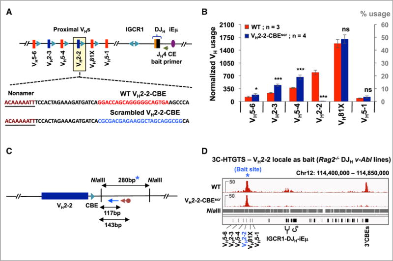
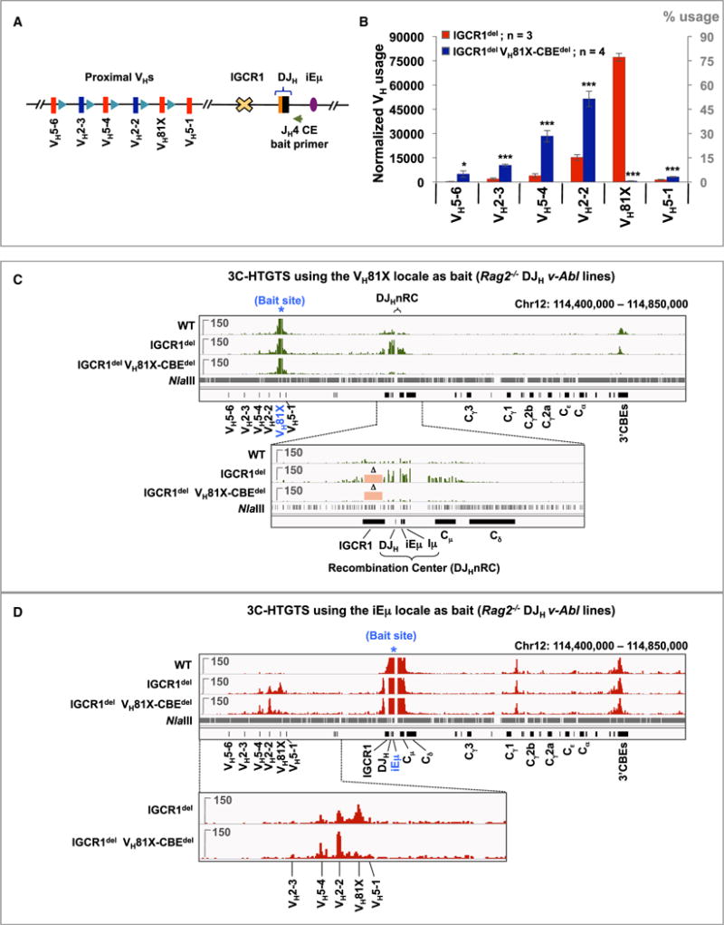
RAG endonuclease initiates antibody heavy chain variable region exon assembly from V, D, and J segments within a chromosomal V(D)J recombination center (RC) by cleaving between paired gene segments and flanking recombination signal sequences (RSSs). The IGCR1 control region promotes DJH intermediate formation by isolating Ds, JHs, and RCs from upstream VHs in a chromatin loop anchored by CTCF-binding elements (CBEs). How VHs access the DJHRC for VH to DJH rearrangement was unknown. We report that CBEs immediately downstream of frequently rearranged VH-RSSs increase recombination potential of their associated VH far beyond that provided by RSSs alone. This CBE activity becomes particularly striking upon IGCR1 inactivation, which allows RAG, likely via loop extrusion, to linearly scan chromatin far upstream. VH-associated CBEs stabilize interactions of D-proximal VHs first encountered by the DJHRC during linear RAG scanning and thereby promote dominant rearrangement of these VHs by an unanticipated chromatin accessibility-enhancing CBE function.

Keywords: 3C-HTGTS; CBE orientation; CTCF-binding elements; HTGTS-Rep-seq; RAG chromatin scanning; V(D)J recombination; V(H) accessibility; V(H)81X; intergenic control region 1; loop extrusion.


Conflict of interest statement

DECLARATION OF INTERESTS

The authors declare no competing interests.

Figures

Figure 1
Figure 1. VH81X-CBE Greatly Enhances VH81X Utilization in Primary Pro-B Cells
(A) Schematic of the murine Igh locus showing proximal VHs, Ds, JHs, CH exons, and regulatory elements (not to scale). Red and blue bars represent members of the IGHV5 (VH7183) and IGHV2 (VHQ52) families, respectively. Teal blue triangles represent position and orientation of CTCF-binding elements (CBEs). Green arrow denotes position of the JH4 coding end bait primer used to generate HTGTS-Rep-seq libraries. (B) Sequence of VH81X-RSS (green) followed by WT (red) or scrambled (blue) VH81X-CBE. (C) Relative VH utilization ± SD in BM pro-B cells from WT (top) or VH81X-CBEscr/scr (bottom) mice. (D) Average utilization frequencies (left axis) and % usage (right axis) of indicated proximal VH segments ± SD. For analysis, each library was normalized to 10,000 VDJH junctions. p values were calculated using unpaired, two-tailed Student’s t test, ns indicates p > 0.05, *p ≤ 0.05, **p ≤ 0.01 and ***p ≤ 0.001. See also Figures S1 and S2 and Tables S1, S3, and S4.
Figure 2
Figure 2. VH81X-CBE Enhances VH81X Utilization in DJH Rearranged v-Abl Pro-B Lines
(A) Schematic representation of the two murine Igh alleles in DJH rearranged v-Abl pro-B cell line (not to scale). One allele (top) harbors a non-productive VDJH rearrangement involving a distal VHJ558 (VH1-2P) that deletes the proximal VH domain and is inert for V(D)J recombination. The other allele (bottom) harbors a DHFL16.1 to JH4 rearrangement (DJH allele) that actively undergoes VH to DJH recombination upon RAG induction via G1 arrest. This DHFL16.1JH4 line served as the parent WT line and was used for all subsequent genetic manipulations. (B) Top line shows the sequence of WT VH81X-CBE (red) while the bottom line shows VH81X-CBE deletion (blue dashed line). (C) Average utilization frequencies (left axis) or % usage (right axis) ± SD of indicated proximal VHs in WT and VH81X-CBEdel v-Abl pro-B lines; libraries were normalized to 3,500 VDJH junctions. As the WT line used for this experiment was the parent of all subsequent VH-CBE mutant lines, we generated WT repeats at several points over the course of these experiments and used the average data, which were highly reproducible, for this and subsequent panels showing comparisons of mutants with WT controls (see STAR Methods for details). (D) Schematic of the 101-kb intergenic deletion extending from 302 bp downstream of VH81X-CBE to 400 bp upstream of the DHFL16.1JH4 RC in the WT DHFL16.1JH4 v-Abl line and its VH81X-CBEdel derivative. (E) Average utilization frequencies (left axis) or % usage (right axis) ± SD of indicated proximal VHs in Intergenicdel and Intergenicdel VH81X-CBEdel v-Abl lines; libraries were normalized to 100,000 VDJH junctions. (F) Sequence of WT (red) and VH81X-CBE inversion mutation (blue). (G) Average utilization frequencies (left axis) or % usage (right axis) ± SD of the indicated proximal VHs in DHFL16.1JH4 WT and VH81X-CBEinv v-Abl lines; libraries were normalized to 3,500 VDJH junctions. Statistical analyses were performed as in Figure 1. See also Figure S3 and Tables S1, S2, S3, and S4.
Figure 3
Figure 3. VH81X-CBE Promotes Interactions of Its Flanking VH with the DJHRC
(A) Schematic representation of the 3C-HTGTS method for studying chromosomal looping interactions of a bait region of interest with the rest of Igh locus (see text and STAR Methods for details). (B) Schematic of the NlaIII restriction fragment (indicated by a blue asterisk) and the relative positions of the biotinylated (cayenne arrow) and nested (blue arrow) PCR primers used for 3C-HTGTS from VH81X bait in (C). (C) Top panel: schematic representation of chromosome interactions of VH81X-CBE containing NlaIII fragment with other Igh locales. Bottom two panels: 3C-HTGTS profiles of Rag2−/− derivatives of control, VH81X-CBEdel, and VH81X-CBEinv DHFL16.1JH4 v-Abl lines using VH81X-CBE locale as bait (blue asterisk). Owing to a DHFL16.1 to JH4 rearrangement in the lines, the region spanning IGCR1, DJH substrate and iEm appears as a broad interaction peak. As v-Abl lines lack locus contraction, we detected few substantial interactions with the upstream Igh locus beyond the most proximal VHs (see legend of Figure S3). Two independent datasets are shown from libraries normalized to 105,638 total junctions. See also Table S4.
Figure 4
Figure 4. V(D)J Recombination of VH2-2 Is Critically Dependent on Its Flanking CBE
(A) Sequence of WT VH2-2-CBE (red) and its scrambled mutation (blue). (B) Average utilization frequencies (left axis) or % usage (right axis) ± SD of indicated proximal VHs in WT and VH2-2-CBEscr v-Abl lines. Each library was normalized to 3,500 VDJH junctions. Statistical analyses were performed as in Figure 1. See also Figure S4A and Table S1. (C) Illustration of NlaIII restriction fragment (blue asterisk) and relative positions of biotinylated (cayenne arrow) and nested (blue arrow) primers used for 3C-HTGTS analyses in (D). Due to repetitive sequences in the restriction fragment that harbors VH2-2-CBE, the downstream flanking restriction fragment was used as bait. (D) Representative 3C-HTGTS interaction profiles of VH2-2 locale (blue asterisk) in Rag2−/− control and VH2-2-CBEscr v-Abl lines, plotted from libraries normalized to 84,578 total junctions. See Figure S4B for an independent repeat. See also Tables S3 and S4.
Figure 5
Figure 5. VH81X-CBE Is Required for Dominant VH81X Usage in the Absence of IGCR1
(A) Schematic of 4.1 kb IGCR1 deletion. (B) Average utilization frequencies (left axis) or % usage (right axis) ± SD of proximal VHs in IGCR1del and IGCR1del VH81X-CBEdel v-Abl lines. Each library was normalized to 100,000 VDJH junctions. Statistical analyses were performed as in Figure 1. See also Figures S5A and S5B and Tables S1 and S2. (C) Representative 3C-HTGTS interaction profiles of VH81X bait (blue asterisk) in Rag2−/− control, IGCR1del, and IGCR1del VH81X-CBEdel DHFL16.1JH4 v-Abl lines performed using the strategy shown in Figure 3B, plotted from libraries normalized to 106,700 total junctions. Bottom panel shows a zoom-in of the region extending from upstream of IGCR1 to downstream of Cd exons. Pink rectangles marked with “D” indicate the IGCR1 region deleted in the IGCR1del and IGCR1del VH81X-CBEdel lines. See Figure S5C for an additional repeat. (D) Representative 3C-HTGTS interaction profiles of iEm bait (blue asterisk) in Rag2−/− v-Abl DJH lines of the indicated genotypes following NlaIII digest using the strategy shown in Figure S5D. Each library was normalized to 273,547 total junctions. Bottom: zoom-in of the proximal VH region. See Figure S5D for an independent repeat and Figure S6 for related repeats. See also Figure S7 and Tables S3 and S4.
Figure 6
Figure 6. Restoration of a CBE Converts VH5-1 into the Most Highly Rearranging VH
(A) Schematic showing the sequence of VH5-1-RSS and its downstream non-functional, “vestigial” CBE. The yellow shaded box highlights the CpG island that is methylated in normal pro-B cells. Bottom sequence shows the four nucleotides mutated (highlighted in blue) to eliminate the CpG island and restore consensus CBE sequence. Two additional nucleotides were mutated just downstream of the CBE to generate a BglII site for screening. (B) Average utilization frequencies (left axis) or % usage (right axis) ± SD of the indicated proximal VHs in WT and VH5-1-CBEins v-Abl lines. Each library was normalized to 3,500 VDJH junctions. Statistical analyses were performed as in Figure 1. See also Figure S4C and Table S1. (C) Illustration of the MseI restriction fragment (blue asterisk) and the relative positions of biotinylated (cayenne arrow) and nested (blue arrow) primers used for 3C-HTGTS analyses in (D). (D) Representative 3C-HTGTS interaction profiles of the VH5-1 locale (blue asterisk) in Rag2−/− control and VH5-1-CBEins v-Abl lines, plotted from libraries normalized to 37,856 total junctions. See Figure S4D for an independent repeat. See also Figure S7 and Tables S3 and S4.
Figure 7
Figure 7. Model for RAG Chromatin Scanning via Loop Extrusion
Figure shows a working model for potential roles of VH-associated CBEs during RAG scanning over chromatin. Numerous variations of the model are conceivable. (A) From its location in the initiating RC, RAG linearly scans cohesin-mediated extrusion loops proceeding through Ds, to allow their utilization; but is largely impeded further upstream by the IGCR1 anchor. After formation of a DJHRC, residual lower level scanning of upstream sequences beyond the IGCR1 impediment allows the most proximal VH-CBEs to mediate direct association with the DJHRC enhancing utilization of their associated VH. VHs further upstream likely access the DJHRC by diffusion with proximal CBEs also enhancing DJHRC interactions and flanking VH utilization. (B) In the absence of IGCR1, loop extrusion progresses upstream allowing RAG to scan the most proximal VHs where associated CBEs promote DJHRC interaction, accessibility, and dominant over-utilization in V(D)J joins. Utilization is most robust for proximal VH81X, which provides the first VH-CBE encountered during linear scanning. VH5-1 is bypassed due to lack of a CBE. Scanning can sometimes bypass VH81X-CBE and continues to the first few upstream VHs, with their CBEs similarly promoting utilization. (C) If both IGCR1 and the VH81X-CBE are mutated, loop-extrusion continues unabated to the VH2-2-CBE and to progressively lesser extents to immediately upstream VH-CBEs. (D–F) CBEs not directly flanking distal VHs theoretically also may augment VH utilization. (D) A distal VH locus CBE associates strongly with chromatin or associated factors (e.g., CTCF/cohesin) at the DJHRC. (E) Cohesin rings load near this DJHRC-associated distal VH locus CBE and initiate loop extrusion. (F) Loop-extrusion allows RAG to scan downstream (or upstream, not illustrated) VHs lacking directly associated CBEs from the DJHRC where the active/transcribed chromatin in which they lie facilitates access for V(D)J recombination (see text for more details and relevant references. See also Figure S1.

References

    1. Aiden EL, Casellas R. Somatic rearrangement in B cells: it’s (mostly) nuclear physics. Cell. 2015;162:708–711. - PMC - PubMed
    1. Alt F, Rosenberg N, Lewis S, Thomas E, Baltimore D. Organization and reorganization of immunoglobulin genes in A-MULV-transformed cells: rearrangement of heavy but not light chain genes. Cell. 1981;27:381–390. - PubMed
    1. Alt FW, Zhang Y, Meng FL, Guo C, Schwer B. Mechanisms of programmed DNA lesions and genomic instability in the immune system. Cell. 2013;152:417–429. - PMC - PubMed
    1. Benner C, Isoda T, Murre C. New roles for DNA cytosine modification, eRNA, anchors, and superanchors in developing B cell progenitors. Proc Natl Acad Sci USA. 2015;112:12776–12781. - PMC - PubMed
    1. Bolland DJ, Koohy H, Wood AL, Matheson LS, Krueger F, Stubbington MJT, Baizan-Edge A, Chovanec P, Stubbs BA, Tabbada K, et al. Two mutually exclusive local chromatin states drive efficient V(D)J recombination. Cell Rep. 2016;15:2475–2487. - PMC - PubMed

Publication types

MeSH terms