. 2018 Jul 12;174(2):312-324.e16.
doi: 10.1016/j.cell.2018.04.029. Epub 2018 May 24.

Structural Basis of Smoothened Activation in Hedgehog Signaling

Affiliations

Structural Basis of Smoothened Activation in Hedgehog Signaling

Pengxiang Huang et al. Cell. .

Erratum in

Abstract

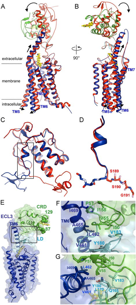
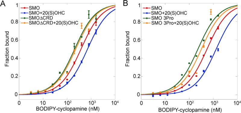
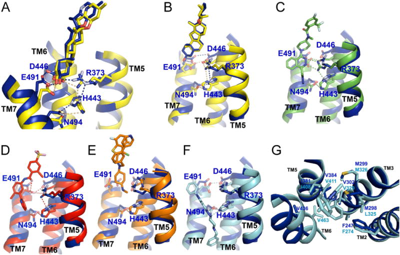
The seven-transmembrane-spanning protein Smoothened is the central transducer in Hedgehog signaling, a pathway fundamental in development and in cancer. Smoothened is activated by cholesterol binding to its extracellular cysteine-rich domain (CRD). How this interaction leads to changes in the transmembrane domain and Smoothened activation is unknown. Here, we report crystal structures of sterol-activated Smoothened. The CRD undergoes a dramatic reorientation, allosterically causing the transmembrane domain to adopt a conformation similar to active G-protein-coupled receptors. We show that Smoothened contains a unique inhibitory π-cation lock, which is broken on activation and is disrupted in constitutively active oncogenic mutants. Smoothened activation opens a hydrophobic tunnel, suggesting a pathway for cholesterol movement from the inner membrane leaflet to the CRD. All Smoothened antagonists bind the transmembrane domain and block tunnel opening, but cyclopamine also binds the CRD, inducing the active transmembrane conformation. Together, these results define the mechanisms of Smoothened activation and inhibition.

Keywords: Hedgehog; Smoothened; cancer; cholesterol; cilia; membrane; oncogene; signaling.


Conflict of interest statement

Declaration of Interests

The authors declare no competing interests.

Figures

Figure 1
Figure 1
Structures of sterol-activated full-length SMO (A) Ribbon model showing overall structure of cyclopamine-bound full-length Xenopus SMO. The CRD is shown in green, LD in cyan, and 7TM domain in blue. The two bound cyclopamine molecules are shown as yellow sticks. Individual transmembrane (TM) helices are numbered. Also shown are the intracellular amphipatic helix H8, and the third extracellular loop (ECL3). The membrane bilayer is indicated by the dotted lines. See also Fig. S1 for an alignment of SMO homologs, showing the various domains and structure elements. (B) As in (A), but showing a view rotated by 90 degrees. (C) Overlay of cyclopamine-bound and cholesterol-bound active xSMO. For the cyclopamine-bound structure, the domains are colored as in (A). The cholesterol-bound structure is shown in light blue. Cyclopamine and cholesterol are shown as yellow sticks. The two structures are nearly identical.
Figure 2
Figure 2
Sterol binding induces dramatic reorientation of the CRD of SMO (A) Ribbon model showing the structure of cyclopamine-bound, active xSMO, superimposed on the structure of vismodegib-bound, inactive hSMO (PDB ID: 5L7I). For the active SMO structure, the CRD is shown in green, LD in cyan, 7TM domain in blue, and cyclopamine is yellow. The inactive SMO structure is in red. CRD rotation and 7TM opening upon SMO activation are indicated with arrows. See also Movie S1 illustrating sterol-induced conformational changes. (B) As in (A), but showing a view rotated by 90 degrees. (C) Overlay of cyclopamine-bound xSMO (blue) and vismodegib-bound hSMO (red), showing their ectodomains, consisting of CRD, connector and LD. The last residues in the connector are responsible for sterol-induced CRD reorientation. See also Fig. S3B for a zoomed out view showing the entire receptors. (D) As in (C), but showing a close up view of the connector, with the SSG switch motif shown in sticks. The residue numbers correspond to hSMO. (E) Overall structure of cyclopamine-bound active xSMO, showing the tri-domain interface between CRD, LD and TM6. Domain colors are as in (A). Shown in sphere are residues 87 and 129, corresponding to residues 114 and 156 in hSMO, where introduction of a glycosylation site leads to constitutive activity. Note that these two positions are completely exposed in active xSMO, compatible with the presence of glycan chains. See also Fig. S3D, showing the same residues buried in inactive hSMO. (F) As in (E), but showing a close up view of the tri-domain interface. The V55 residue of the CRD is at the center of a cavity formed by mostly hydrophobic residues in CRD, LD and the extracellular extension of TM6. (G) As in (F), but showing a view rotated by 90 degrees.
Figure 3
Figure 3
Active conformation of SMO 7TM resembles active classical GPCRs (A) Ribbon model showing the 7TM domain of cyclopamine-bound xSMO (blue), superimposed on the 7TM domain of vismodegib-bound hSMO (red, PDB ID: 5L7I). Note the movement of TM6 and TM5. See also Movie S1 illustrating 7TM conformational changes upon SMO activation. (B) As in (A), but showing a view rotated by 90 degrees, from the cytoplasmic side. (C) As in (A), but showing superposition of active (green, PDB ID: 3PQR) and inactive (pink, PDB ID: 1U19) rhodopsin. For active-inactive comparisons of other Class A and B GPCRs, see also Fig. S4A–D. (D) As in (C), but showing a view rotated by 90 degrees, from the cytoplasmic side.
Figure 4
Figure 4
A conserved π-cation lock essential for stabilizing the inactive state of SMO (A) Ribbon model of the inactive 7TM domain of vismodegib-bound hSMO (red, PDB ID: 5L7I), seen from the cytoplasmic side. Individual transmembrane helices are numbered. The conserved R451 and W535 residues from TM6 and TM7 are involved in a π-cation interaction. (B) As in (A), but showing the active 7TM domain in cyclopamine-bound xSMO. The corresponding R424 and W508 residues have moved apart, breaking the π-cation interaction. (C) Left panel: overall structure comparison between active cyclopamine-bound (blue) and vismodegib-bound (red) SMO, with the R and W residues shown in sticks. Right panel: close up, side view of the π-cation lock, and its breakage upon SMO activation. (D) Mouse SMO (mSMO) constructs, tagged with mCHERRY, were expressed in 293T cells, and binding to BODIPY-cyclopamine was measured by automated fluorescence microscopy and image analysis. Ratio of bound BODIPY-cyclopamine to mCHERRY fluorescence intensities is shown as box plot. The upper and lower bounds of each box correspond to the 75th and 25th percentile of the distribution, and the horizontal line indicates the median. Mutations that disrupt the π-cation interaction between R455 and W539 reduce BODIPY-cyclopamine binding, indicative of mSMO shifting to an active conformation. The oncogenic SMO-M2 mutant corresponds to W539L. Drosophila SMO (dSMO) was used as negative control (Nedelcu et al., 2013). Between 600 and 1000 cells were measured per condition. ** p<5×10⁻⁸. (E) As in (D), but with BODIPY-SANT1. Note the partial rescue of the W539L(M2) mutant by the T542D mutation. ** p<5×10⁻⁸. See also Fig. S5A–C for binding experiments using BODIPY-vismodegib, Fig. S5D for a model of the π-cation lock indicating the T542 residue, and Fig. S5E for a dose-response analysis of BODIPY-SANT1 binding to the partially rescued SMO-M2 mutant.
Figure 5
Figure 5
Allosteric activation of the 7TM domain of SMO by sterol-bound CRD (A) 293T cells expressing HA-tagged mSMO or mSMO CRD were incubated with BODIPY-cyclopamine, in the absence or presence of 20(S)-OHC (10 μM). The cells were then stained with Alexa647-labeled mouse anti-HA antibodies, to reveal mSMO. Bound BODIPY-cyclopamine and mSMO expression were measured by flow cytometry. The graph shows ratios of BODIPY and Alexa647 intensities. Error bars indicate standard deviation. Activation of mSMO by 20(S)-OHC reduces affinity for BODIPY-cyclopamine. MSMO CRD is unaffected by 20(S)-OHC. See also Fig. S5F and S5G, for similar experiments using BODIPY-SANT1 and BODIPY-vismodegib. (B) As in (A), but with an mSMO mutant in which the residues of the SSG “switch” motif were mutated to prolines. The mutant has a greatly reduced response to 20(S)-OHC.
Figure 6
Figure 6
Mechanism of SMO modulation by 7TM ligands (A) Ribbon model showing the active cyclopamine-bound xSMO (blue) superimposed on inactive cyclopamine-bound hSMO (yellow, PDB ID: 4O9R). The 5 residues shown in sticks are connected by an adaptable hydrogen bond network, which stabilizes both inactive and active SMO conformations. The network is remodeled between the two conformations, but the participating residues are the same. Cyclopamine is unique among SMO antagonists, being compatible with both 7TM conformations. Note also the slight upward shift of TM7, and outward opening of TM5 and TM6. Residue numbers are from xSMO. (B) As in (A), but showing a different view. (C) As in (A), but with cyclopamine-bound xSMO (blue) superimposed on inactive LY2940680-bound hSMO (green, PDB ID: 4JKV). (D) As in (A), but with cyclopamine-bound xSMO (blue) superimposed on inactive vismodegib-bound hSMO (red, PDB ID: 5L7I). See also Fig. S6A and S6B, showing inactive hSMO in complex with TC114 and ANTA XV. (E) As in (A), but with cyclopamine-bound xSMO (blue) superimposed on SAG1.5-bound hSMO (orange, PDB ID: 4QIN). SAG1.5. Binding of SAG1.5 breaks the hydrogen bond network, and thus the TM coupling that stabilizes the inactive conformation. (F) As in (A), but with cyclopamine-bound xSMO (blue) superimposed on inactive SANT1-bound hSMO (cyan, PDB ID: 4N4W). Note that SANT1 reaches much deeper into the 7TM site than the other ligands. (G) As in (F), but a different view, showing the insertion of the benzyl moiety of SANT1 in a hydrophobic cavity. This interaction strengthens inactive TM coupling, preventing TM6 from opening. Residues in blue are from xSMO, while residues in cyan are from hSMO. See also Fig. S6C, showing how the inactivating V329F mutation in hSMO mimics the benzyl moiety of SANT1.
Figure 7
Figure 7
A longitudinal tunnel in active SMO connects the membrane to the 7TM orthosteric site (A) Surface of cyclopamine-bound inactive hSMO (yellow, PDB ID: 4O9R). The 7TM cavity is shown in red. See also Figure S7C for the corresponding ribbon model. (B) As in (A), but showing SANT1-bound inactive hSMO (light blue, PDB ID: 4N4W). Note the remodeling of the 7TM site by SANT1. See also Figure S7D for the corresponding ribbon model. (C) As in (A), but showing active cyclopamine-bound xSMO (blue). Note the appearance of a tunnel connecting the membrane to the bottom of the 7TM site. See also Figure S7E for the corresponding ribbon model, and Movie S2 for the 360-degree view of the tunnel. (D) Superimposition of the inactive 7TM cavities for cyclopamine (yellow) and SANT1 (red), and the tunnel (blue). (E) Close up view of the tunnel in active xSMO, superimposed on inactive cyclopamine-bound hSMO (yellow). Residues that obstruct the tunnel in inactive SMO are shown in sticks. Residue numbers are from hSMO. (F) As in (E), but showing active cyclopamine-bound xSMO (blue). Movement of the depicted residues opens the tunnel. Residue numbers are from xSMO. (G) The tunnel superimposed on both inactive (yellow) and active (blue) SMO. Note individual movements of residues involved in tunnel opening upon SMO activation.

References

    1. Alcedo J, Ayzenzon M, Von Ohlen T, Noll M, Hooper JE. The Drosophila smoothened gene encodes a seven-pass membrane protein, a putative receptor for the hedgehog signal. Cell. 1996;86:221–232. - PubMed
    1. Byrne EF, Sircar R, Miller PS, Hedger G, Luchetti G, Nachtergaele S, Tully MD, Mydock-McGrane L, Covey DF, Rambo RP, et al. Structural basis of Smoothened regulation by its extracellular domains. Nature. 2016;535:517–522. - PMC - PubMed
    1. Caffrey M, Cherezov V. Crystallizing membrane proteins using lipidic mesophases. Nature protocols. 2009;4:706–731. - PMC - PubMed
    1. Chen JK, Taipale J, Cooper MK, Beachy PA. Inhibition of Hedgehog signaling by direct binding of cyclopamine to Smoothened. Genes & development. 2002a;16:2743–2748. - PMC - PubMed
    1. Chen JK, Taipale J, Young KE, Maiti T, Beachy PA. Small molecule modulation of Smoothened activity. Proceedings of the National Academy of Sciences of the United States of America. 2002b;99:14071–14076. - PMC - PubMed

Publication types

MeSH terms

LinkOut - more resources