. 2018 Jun 21;25(6):749-760.e9.
doi: 10.1016/j.chembiol.2018.04.017. Epub 2018 May 24.

PDn-3 DPA Pathway Regulates Human Monocyte Differentiation and Macrophage Function

Affiliations

PDn-3 DPA Pathway Regulates Human Monocyte Differentiation and Macrophage Function

Kimberly Pistorius et al. Cell Chem Biol. .

Abstract

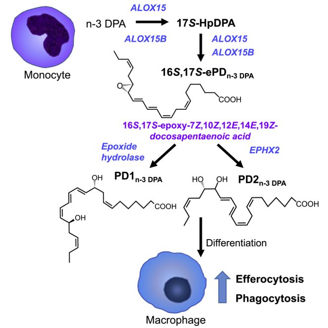
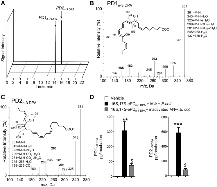
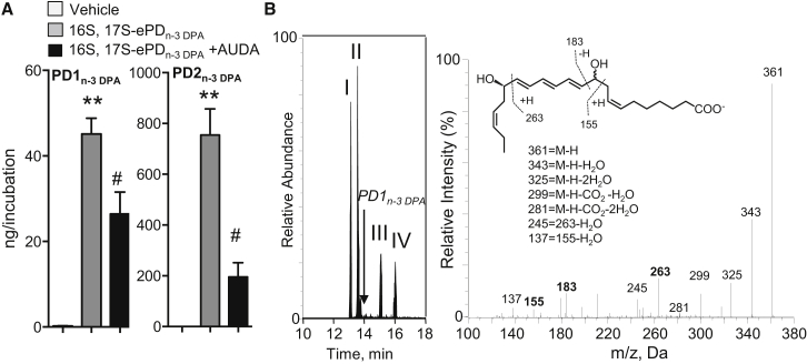
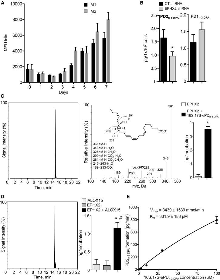
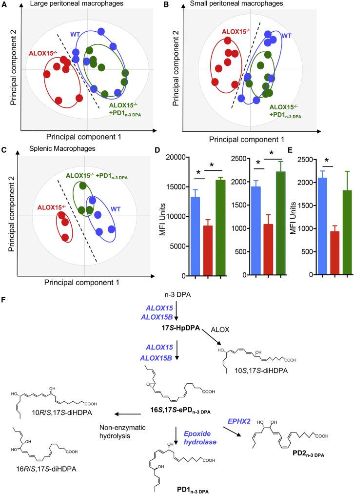
Macrophages are central in orchestrating the clearance of apoptotic cells and cellular debris during inflammation, with the mechanism(s) regulating this process remaining of interest. Herein, we found that the n-3 docosapentaenoic acid-derived protectin (PDn-3 DPA) biosynthetic pathway regulated the differentiation of human monocytes, altering macrophage phenotype, efferocytosis, and bacterial phagocytosis. Using lipid mediator profiling, human primary cells and recombinant enzymes we found that human 15-lipoxygenases initiate the PDn-3 DPA pathway catalyzing the formation of an allylic epoxide. The complete stereochemistry of this epoxide was determined using stereocontrolled total organic synthesis as 16S,17S-epoxy-7Z,10Z,12E,14E,19Z-docosapentaenoic acid (16S,17S-ePDn-3 DPA). This intermediate was enzymatically converted by epoxide hydrolases to PD1n-3 DPA and PD2n-3 DPA, with epoxide hydrolase 2 converting 16S,17S-ePDn-3 DPA to PD2n-3 DPA in human monocytes. Taken together these results establish the PDn-3 DPA biosynthetic pathway in human monocytes and macrophages and its role in regulating macrophage resolution responses.

Keywords: lipid mediators; omega-3; resolution; total organic synthesis.


Figures

None
Graphical abstract
Figure 1
Figure 1
Inhibiting 15-Lipoxygenase Activity Reduces PDn-3 DPA Production Dysregulating Macrophage Phenotype and Function (A) Human monocytes were incubated with M-CSF (20 ng/mL) and either a ALOX15 inhibitor or vehicle (37°C, 5% CO2). On day 7 incubations were quenched, lipid mediators were extracted, identified, and quantified using lipid mediator profiling (see the STAR Methods for details). Results are mean ± SEM. n = 6 donors. *p < 0.05. (B) Human monocytes were isolated and incubated with GM-CSF (20 ng/mL), IFN-γ (20 ng/mL), and LPS (100 ng/mL) to produce M1 or M-CSF (20 ng/mL) and IL-4 (20 ng/mL) to obtain M2 cells, and the expression of ALOX15 and ALOX15B was evaluated during the differentiation time course using flow cytometry. Results are mean ± SEM n = 4–6 donors per interval. (C and D) Human monocytes were incubated with vehicle or ALOX15 inhibitor and then with M-CSF (20 ng/mL) for 7 days, and (C) expression of lineage markers was determined using fluorescently labeled antibodies and flow cytometry on day 7, and interrogated using OPLS-DA. n = 6 donors. (D) Phagocytosis of fluorescently labeled apoptotic cells investigated. Results for are mean ± SEM. n = 6 donors. *p < 0.05. (E) Peritoneal macrophages were harvested from wild-type (WT) and ALOX15−/− mice, and the expression of lineage markers on CD64+ cells was determined using flow cytometry. Results were interrogated using OPLS-DA and are representative of n = 7 mice. (F) Fluorescently labeled apoptotic cells were administered to WT and ALOX15−/− mice via intraperitoneal injection. After 1 hr peritoneal cells were harvested, and phagocytosis of apoptotic cells by CD64+ cells was evaluated using flow cytometry. Results are mean ± SEM. n = 7 mice per group. *p < 0.05. Related to Figures S1 and S2 and Tables S1–S3.
Figure 2
Figure 2
Human ALOX15 and Monocytes Produces a Novel 16,17S-ePDn-3 DPA (A–C) Human monocytes (1 × 108 cells/mL; PBS; 37°C) (A), hr-ALOX15 (0.2 μM, 37°C [pH 8]) (B), and hr-ALOX15B (0.2 μM, 37°C [pH 8]) (C) were incubated with n-3 DPA (10 μM). After 3 min, incubations were quenched using acidified methanol, products extracted and identified using lipid mediator profiling. Left panels: MRM chromatogram for ion pairs m/z 375 > 277. Middle and right panels: MS-MS spectra employed in the identification of (middle panel) 10-methoxy,17S-hydroxy-7Z,11E,13E,15E,19Z-docosapentaenoic acid, (right panel) 16-methoxy,17S-hydroxy-7Z,10Z,12E,14E,19Z-docosapentaenoic acid in monocyte incubations. Results are representative of n = 4 donors and three independent experiments. Related to Figures S3 and S4.
Figure 3
Figure 3
Total Organic Synthesis of 16,17S-ePDn-3 DPA (A) Outline of the synthetic strategy and key precursors employed in the preparation of 16S,17S-ePDn-3 DPA. (B) Z and E stereochemical assignment for C=C using two-dimensional NMR spectroscopy. Contours denote positive and negative contours. Related to Figures S3.
Figure 4
Figure 4
16S,17S-ePDn-3 DPA Is Precursor to PD1n-3 DPA and PD2n-3 DPA 16S,17S-ePDn-3 DPA (10 nM) was incubated with human macrophages (MΦ; 4 × 107 cells/mL) or inactivated human macrophages (i.e., 4 × 107 cells/mL previously been kept at 100°C for 1h). E. coli (2.5 × 108 colony-forming units [CFU]/mL) were added, cells incubated for 15 min, at 37°C, and incubations were quenched using ice-cold methanol. Products were then extracted and profiled using lipid mediator profiling. Vehicle denotes solution containing 0.1% EtOH in PBS. (A) MRM chromatogram for PD1n-3 DPA (m/z 361 > 183) and PD2n-3 DPA (m/z 361 > 233). (B and C) MS-MS spectra employed for identification of (B) PD1n-3 DPA (C) PD2n-3 DPA. (D) PD1n-3 DPA and PD2n-3 DPA concentrations. Results are representative of n = 4 donors from two independent experiments. Results are means ± SEM. **p < 0.001, ***p < 0.0001 versus vehicle incubations. $p < 0.05 versus MΦ + E. coli incubations.
Figure 5
Figure 5
Epoxide Hydrolases Convert 16S,17S-ePDn-3 DPA to PD1n-3 DPA and PD2n-3 DPA (A) Human monocytes (1 × 108 cells/mL) were incubated with vehicle (PBS + 0.1% DMSO) or AUDA (25 μM) for 20 min (at room temperature). Cells were then incubated with either vehicle (PBS + 0.1% EtOH) or 16S,17S-ePDn-3 DPA (10 nM). Incubations were quenched after 15 min and products profiled using LM profiling. Results are mean ± SEM. n = 4 donors and two independent experiments. **p < 0.01 versus vehicle; #p <0.05 versus monocyte incubations. (B) 16S,17S-ePDn-3 DPA (10 nM) was incubated with human recombinant LTA4H (0.2 μM; Tris buffer). Incubations were quenched using ice-cold methanol and products identified using lipid mediator profiling. Left panel: MRM chromatogram m/z 361 > 263 (arrow denotes expected retention time for PD1n-3 DPA); right panel: MS-MS spectrum employed in the identification of 10,17S-hydroxy-7Z,11E,13E,15E,19Z-docosapentanenoic acid. Results are representative of n = 4 independent experiments.
Figure 6
Figure 6
EPHX2 Converts 16S,17S-ePDn-3 DPA to PD2n-3 DPA in Human Monocytes and Macrophages (A) Human monocytes were isolated and differentiated using GM-CSF (20 ng/mL), IFN-γ (20 ng/mL), and LPS (100 ng/mL) to produce M1 or M-CSF (20 ng/mL) and IL-4 (20 ng/mL) to obtain M2 cells and the expression of EPHX2 during the differentiation time course was evaluated using flow cytometry. Results are mean ± SEM. n = 4–6 donors per interval. (B) Human monocytes were transfected with shRNA to EPHX2 or CT shRNA (see the STAR Methods for details), cells were incubated for 10 hr at 37°C, then with E. coli for 45 min, and PDn-3 DPA concentrations evaluated using LM profiling. Results are mean ± SEM. n = 4 donors. *p < 0.05. (C) 16S,17S-ePDn-3 DPA (10 nM) was incubated with hrEPHX2 (0.2 μM; Tris buffer). Incubations were quenched using ice-cold methanol and products identified using lipid mediator profiling. Left panel: MRM chromatogram m/z 361 > 233. Center panel: MS-MS spectrum employed in the identification of PD2n-3 DPA. Right panel: PD2n-3 DPA concentrations. Results are representative of n = 4 independent experiments. *p < 0.01 versus EPHX2 incubations. (D) n-3 DPA (10 μM; Tris buffer) was incubated with hr-ALOX15 (0.2 μM), EPHX2 (0.2 μM), or a combination of the two enzymes. The incubations were quenched after 15 min and products extracted, identified, and quantified using lipid mediator profiling. Left panel: MRM chromatogram m/z 361 > 233; right panel: PD2n-3 DPA concentrations. Results are representative of n = 4 independent experiments. Results for right panels in (C) and (D) are means ± SEM. *p < 0.01 versus EPHX2 incubations; #p < 0.01 versus ALOX15 incubations. (E) EPHX2 (0.2 μM, Tris buffer) was incubated with the indicated concentrations of 16S,17S-ePDn-3 DPA. Incubations were quenched and PD2n-3 DPA concentrations were determined using lipid mediator profiling. Results are mean ± SEM. n = 3 independent experiments.
Figure 7
Figure 7
PD1n-3 DPA Rectifies Murine Resident Macrophage Phenotype and Function in ALOX15-Deficient Mice (A–C) The expression of phenotypic markers was assessed in peritoneal and splenic macrophages from ALOX15−/− mice administered PD1n-3 DPA (10 ng/mouse for 7 days) or vehicle and WT mice using flow cytometry and macrophage phenotype interrogated using PLS-DA in (A) large peritoneal macrophages, (B) small peritoneal macrophages, and (C) splenic macrophages. Results are representative of n = 8 mice per group for (A and B) and n = 3–4 mice per group for (C). (D and E) Mice were treated as in (A–C), and on day 7 administered fluorescently labeled (D) apoptotic cells (6 × 106 cells/mouse) or (E) E. coli (106 CFU/mouse) via an intraperitoneal injection. Peritoneal cells were collected after 1 hr and phagocytosis was assessed in (D) CD64+ large peritoneal macrophages (left panel) and small peritoneal macrophages (right panel), and (E) total CD64+ macrophage population. Results are mean ± SEM. n = 8 mice per group for (D) and n = 4 mice per group for (E). *p < 0.05. (F) Structures are illustrated in most likely configurations based on biosynthetic evidence. The stereochemistries for PD1n-3 DPA and 16S,17S-PDn-3 DPA are established (Aursnes et al., 2015, Aursnes et al., 2014, Dalli et al., 2013a). Related to Figures S5.

References

    1. Aursnes M., Tungen J.E., Colas R.A., Vlasakov I., Dalli J., Serhan C.N., Hansen T.V. Synthesis of the 16S,17S-epoxyprotectin intermediate in the biosynthesis of protectins by human macrophages. J. Nat. Prod. 2015;78:2924–2931. - PMC - PubMed
    1. Aursnes M., Tungen J.E., Vik A., Colas R., Cheng C.Y., Dalli J., Serhan C.N., Hansen T.V. Total synthesis of the lipid mediator PD1n-3 DPA: configurational assignments and anti-inflammatory and pro-resolving actions. J. Nat. Prod. 2014;77:910–916. - PMC - PubMed
    1. Borgeat P., Fruteau de Laclos B., Picard S., Drapeau J., Vallerand P., Corey E.J. Studies on the mechanism of formation of the 5S, 12S-dihydroxy-6,8,10,14(E, Z,E,Z)-icosatetraenoic acid in leukocytes. Prostaglandins. 1982;23:713–724. - PubMed
    1. Bystrom J., Evans I., Newson J., Stables M., Toor I., van Rooijen N., Crawford M., Colville-Nash P., Farrow S., Gilroy D.W. Resolution-phase macrophages possess a unique inflammatory phenotype that is controlled by cAMP. Blood. 2008;112:4117–4127. - PMC - PubMed
    1. Chiang N., Serhan C.N. Structural elucidation and physiologic functions of specialized pro-resolving mediators and their receptors. Mol. Aspects Med. 2017;58:114–129. - PMC - PubMed

Publication types

MeSH terms

LinkOut - more resources