. 2018 Jun 7;102(6):1143-1157.
doi: 10.1016/j.ajhg.2018.04.009. Epub 2018 May 24.

Mutations in the Epithelial Cadherin-p120-Catenin Complex Cause Mendelian Non-Syndromic Cleft Lip with or without Cleft Palate

Affiliations

Mutations in the Epithelial Cadherin-p120-Catenin Complex Cause Mendelian Non-Syndromic Cleft Lip with or without Cleft Palate

Liza L Cox et al. Am J Hum Genet. .

Abstract

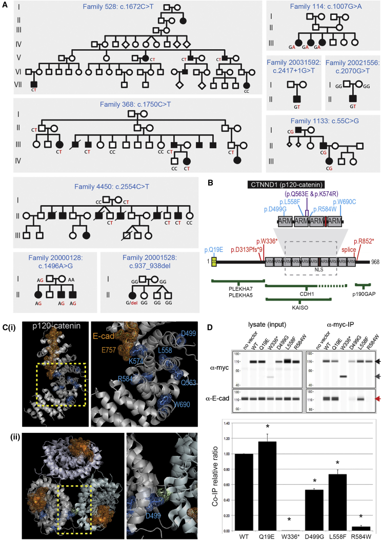
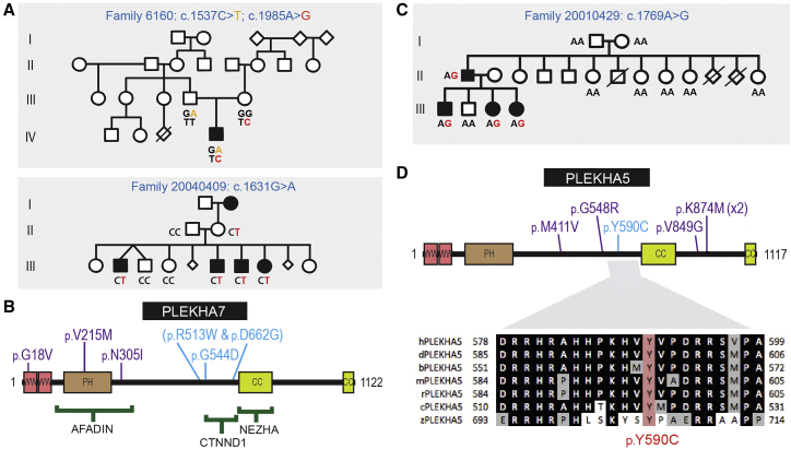
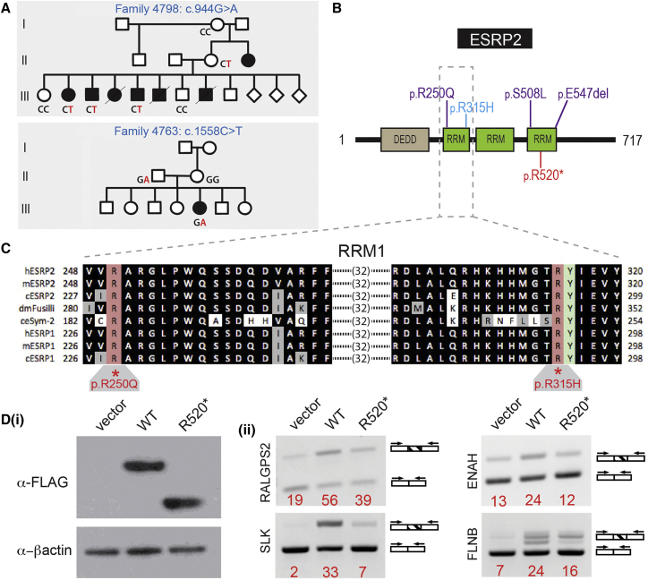
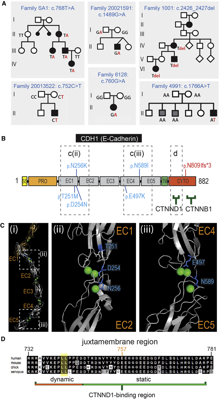
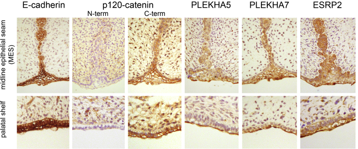
Non-syndromic cleft lip with or without cleft palate (NS-CL/P) is one of the most common human birth defects and is generally considered a complex trait. Despite numerous loci identified by genome-wide association studies, the effect sizes of common variants are relatively small, with much of the presumed genetic contribution remaining elusive. We report exome-sequencing results in 209 people from 72 multi-affected families with pedigree structures consistent with autosomal-dominant inheritance and variable penetrance. Herein, pathogenic variants are described in four genes encoding components of the p120-catenin complex (CTNND1, PLEKHA7, PLEKHA5) and an epithelial splicing regulator (ESRP2), in addition to the known CL/P-associated gene, CDH1, which encodes E-cadherin. The findings were also validated in a second cohort of 497 people with NS-CL/P, comprising small families and singletons with pathogenic variants in these genes identified in 14% of multi-affected families and 2% of the replication cohort of smaller families. Enriched expression of each gene/protein in human and mouse embryonic oro-palatal epithelia, demonstration of functional impact of CTNND1 and ESRP2 variants, and recapitulation of the CL/P spectrum in Ctnnd1 knockout mice support a causative role in CL/P pathogenesis. These data show that primary defects in regulators of epithelial cell adhesion are the most significant contributors to NS-CL/P identified to date and that inherited and de novo single gene variants explain a substantial proportion of NS-CL/P.

Keywords: adherens junction; cadherin; catenin; cell adhesion; cleft lip; cleft lip/palate; cleft palate; epithelia; exome sequencing; knockout.


Figures

Figure 1
Figure 1
CTNND1 Variants (A) Pedigrees showing segregation of CTNND1 variants. Segregation of indicated variants was performed by Sanger sequencing in all individuals for whom DNA was available. (B) CTNND1 (p120Ctn) protein domain structure showing variants (blue, pathogenic missense variants; purple, missense variants of unknown significance; red, nonsense/frameshift variants). Regions required for binding partner interaction are shown below as green bars. (C) X-ray structure of the p120Ctn ARM domains (silver) complexed with the E-cadherin tail juxtamembrane core (gold dots and sticks) (PDB: 3l6y). The dashed box in (B) and the left image in (i) highlight the enlarged image to the right in (i). Mutated residues are identified in blue (dots/sticks). The E-cadherin E757 juxtamembrane core residue, which interacts with p120Ctn Lys574, is indicated in gold. (ii) Top-down view of the packing of the p120Ctn monomer structure. Asp499 is positioned at the monomer interaction surface. The unlabeled residue (chartreuse dots and sticks) on the p120Ctn monomer contacts Asp499. (D) Co-immunoprecipitation of endogenous E-cadherin with myc-tagged ectopically expressed wild-type and mutant p120Ctn proteins (top). Western blots showing each ectopically expressed p120Ctn protein (detected using an anti-myc antibody) and endogenous E-cadherin in each lysate and following p120Ctn immunoprecipitation. Full-length p120Ctn (black arrow), p.Trp336 protein (dark gray arrow), and E-cadherin (red arrow, lower panel). The relative amounts of E-cadherin pulled down are shown relative to the amount of the respective immunoprecipitated p120Ctn proteins, following normalization to the wild-type p120Ctn:E-cadherin ratio. Data are reported as mean E-cadherin:c-Myc tagged p120Ctn ratios + SD; p < 0.05 (n = 3).
Figure 2
Figure 2
Rare Variants Identified in PLEKHA7 and PLEKHA5 (A) Pedigrees showing segregation of PLEKHA7 variants. Segregation of indicated variants was performed by Sanger sequencing in all individuals for whom DNA was available. (B) PLEKHA7 protein structure showing variants (blue, pathogenic missense variants; purple, missense variants of unknown significance). (C) Family 20010429 showing de novo PLEKHA5 c.1769A>G variant (p.Tyr590Cys) in the second generation and subsequent segregation (PDB: 3q2v). (D) PLEKHA5 protein structure showing variants (blue, pathogenic missense variants; purple, missense variants of unknown significance). Lower panel: Evolutionary conservation of Tyr590, a phosphorylation site. Sequences shown from human (h), dog (d), cow (b), mouse (m), rat (r), chick (c), and zebrafish (z).
Figure 3
Figure 3
ESRP2 Variants (A) Pedigrees showing segregation of ESRP2 variants. Segregation of indicated variants was performed by Sanger sequencing in all individuals for whom DNA was available. (B) ESRP2 protein structure variants (blue, pathogenic missense variants; purple, missense variants of unknown significance; red, nonsense variant). (C) Evolutionary conservation of the first RNA recognition motif (RRM1) in ESRP1 and ESRP2 with the orthologs of ESRP2 in D. melanogaster and C. elegans. The locations and conservation of two residues mutated in NS-CL/P, Arg250 and Arg315, are shown. Arg315 is adjacent the invariant Tyr316 that is critical for RNA recognition. (D) Expression of the ESRP2 nonsense mutant, p.Arg520, in HEK293T cells alters the amount of splice isoforms of ESRP2 target genes (ii) compared to cells expressing comparable ectopic wild-type ESRP2 protein (i).
Figure 4
Figure 4
CDH1 Variants (A) Pedigrees showing segregation of CDH1 variants. Segregation of indicated variants was performed by Sanger sequencing in all individuals for whom DNA was available. Family 4991 shows a de novo pathogenic variant in the individual presenting with CL/P. Two siblings (gray squares) have secondary palate clefting and do not carry the CDH1 variant; sibling phenotype due to a second unidentified cause. (B) CDH1 (E-cadherin) protein structure showing variants (blue, missense variants; red, frameshift variant). Binding partner protein regions shown as green bars. The dashed boxes in (B) and (Ci) are represented in the X-ray structure shown in (Cii) and (Ciii), and the alignment in (D). (C) (i) The X-ray structure of the extracellular region of E-cadherin. Dashed boxes mark the regions enlarged to the right: (ii) the Ca2+-binding EC1-EC2 hinge region, and (iii) the Ca2+-binding EC4-EC5 hinge region. In all cases the CL/P missense mutations involve residues (blue) surrounding the Ca2+ ions (green spheres) with some residues participating in chelation. (D) Sequence conservation of the E-cadherin juxtamembrane core region essential for binding to CTNND1/p120Ctn. The dileucine endocytic motif (yellow) and the conserved Glu757, which interacts with p120Ctn Lys574 in the X-ray structure (Figure 1), are indicated.
Figure 5
Figure 5
Immunohistochemical Detection of CL/P Candidate Proteins in 67–72 day Human Embryonic Oral and Secondary Palatal Epithelia Strong E-cadherin staining seen in all oral epithelia (basal and periderm layers). p120Ctn (pan-isoform antibody; C-term), PLEKHA5, PLEKHA7, and ESRP2 are also seen in all oral epithelia but show stronger staining in the peridermal layer of the palatal shelves as well as throughout the midline epithelial seam (MES). Staining of p120Ctn isoform 1 (N-term) is seen throughout the oral mesenchyme and weakly in the basal epithelial layer but is absent from the peridermal layer of the palate and MES.
Figure 6
Figure 6
Conditional Ablation of Ctnnd1 in the Embryonic Mouse Oral Epithelium Results in CL/P (A) Mouse embryos homozygous (hom) for a Ctnnd1 oral epithelial-specific deletion displayed variable clefts—unilateral CL only (top left), unilateral CL/P (top center), bilateral CL/P (top right), or cleft secondary palate only (far right)—similar to the human spectrum (lower panel). (B) 3D OPT-imaged littermates of different genotypes. Conditional heterozygotes and non-cleft presenting conditional homozygotes () are distinguished by a delay in medial growth of the maxillary prominences shown by the increased gap marked by the red line. Non-cleft presenting homozygotes showed dysmorphic nasal tips and nares.

References

    1. Dixon M.J., Marazita M.L., Beaty T.H., Murray J.C. Cleft lip and palate: understanding genetic and environmental influences. Nat. Rev. Genet. 2011;12:167–178. - PMC - PubMed
    1. Ghoumid J., Stichelbout M., Jourdain A.S., Frenois F., Lejeune-Dumoulin S., Alex-Cordier M.P., Lebrun M., Guerreschi P., Duquennoy-Martinot V., Vinchon M. Blepharocheilodontic syndrome is a CDH1 pathway-related disorder due to mutations in CDH1 and CTNND1. Genet. Med. 2017;19:1013–1021. - PubMed
    1. Rahimov F., Jugessur A., Murray J.C. Genetics of nonsyndromic orofacial clefts. Cleft Palate Craniofac. J. 2012;49:73–91. - PMC - PubMed
    1. Mitchell L.E., Risch N. Mode of inheritance of nonsyndromic cleft lip with or without cleft palate: a reanalysis. Am. J. Hum. Genet. 1992;51:323–332. - PMC - PubMed
    1. Beaty T.H., Marazita M.L., Leslie E.J. Genetic factors influencing risk to orofacial clefts: today’s challenges and tomorrow’s opportunities. F1000Res. 2016;5:2800. - PMC - PubMed

Publication types

MeSH terms

LinkOut - more resources