. 2018 Jun 5;10(6):1705-1720.
doi: 10.1016/j.stemcr.2018.04.024. Epub 2018 May 24.

Retinoic Acid Is Required for Neural Stem and Progenitor Cell Proliferation in the Adult Hippocampus

Affiliations

Retinoic Acid Is Required for Neural Stem and Progenitor Cell Proliferation in the Adult Hippocampus

Swati Mishra et al. Stem Cell Reports. .

Abstract

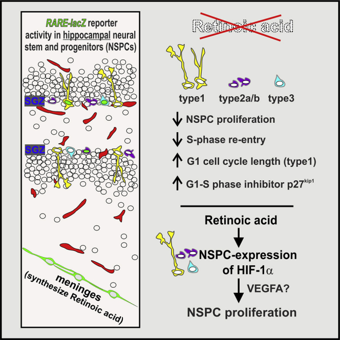
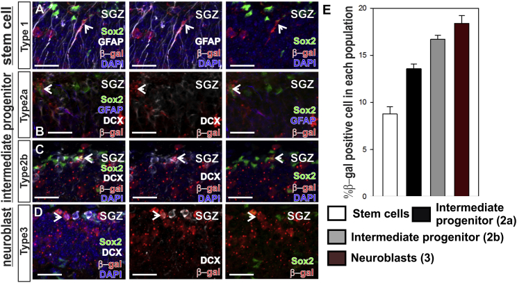
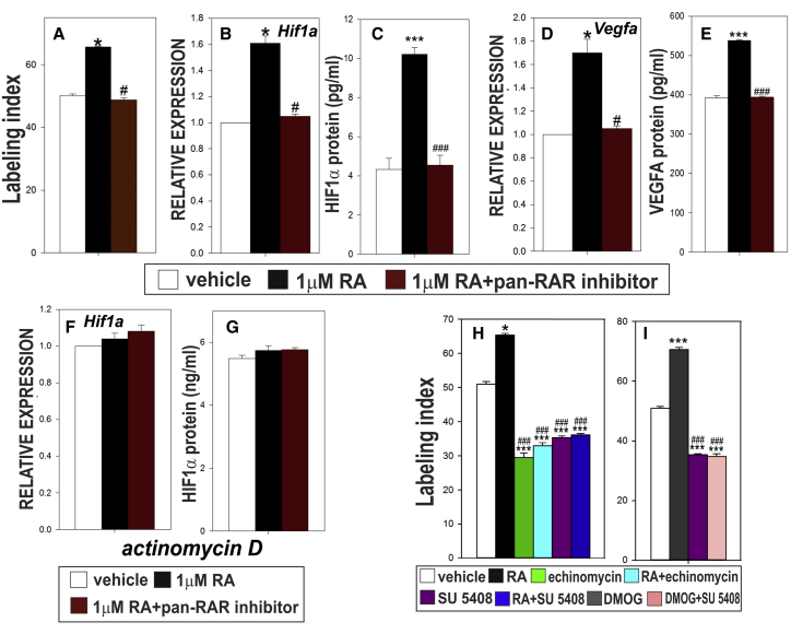
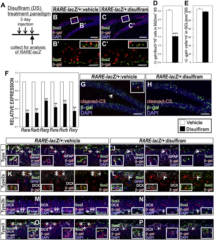
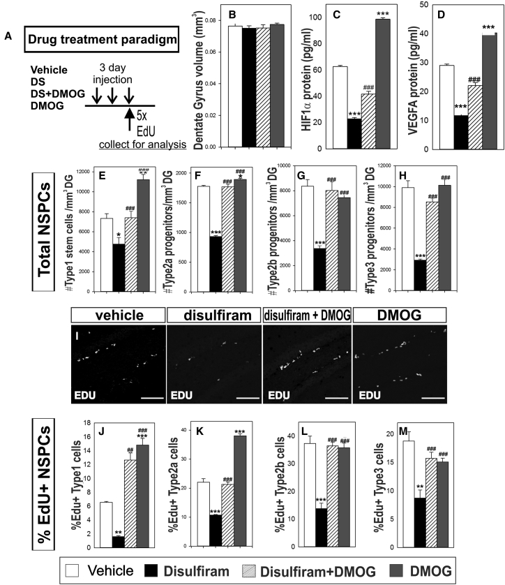
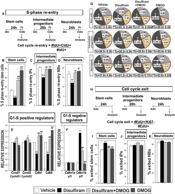
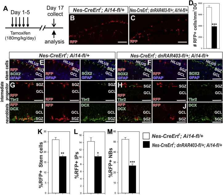
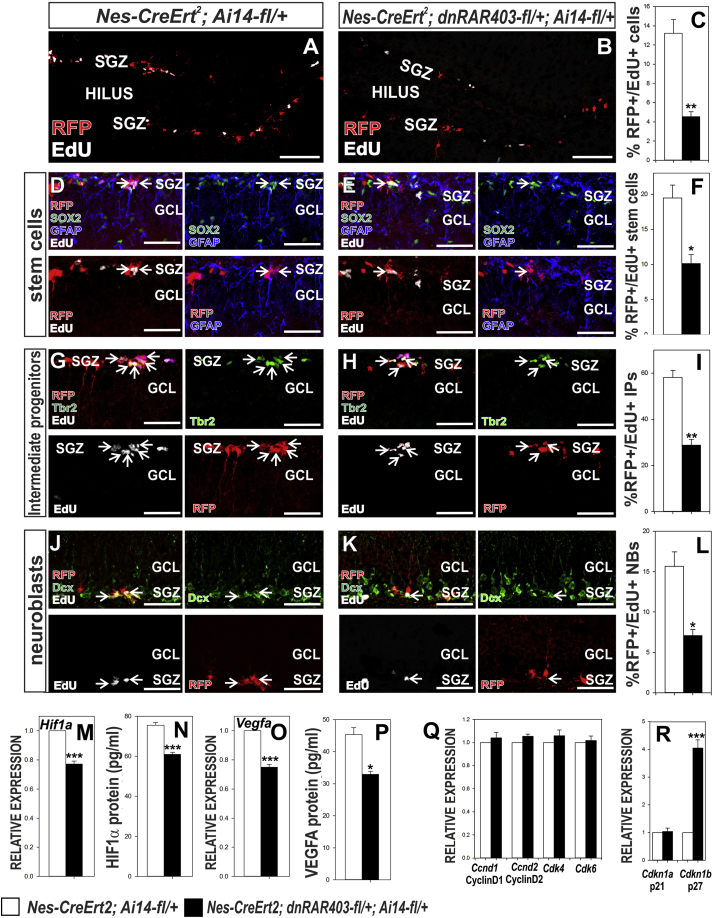
Neural stem and precursor cell (NSPC) proliferation in the rodent adult hippocampus is essential to maintain stem cell populations and produce new neurons. Retinoic acid (RA) signaling is implicated in regulation of adult hippocampal neurogenesis, but its exact role in control of NSPC behavior has not been examined. We show RA signaling in all hippocampal NSPC subtypes and that inhibition of RA synthesis or signaling significantly decreases NSPC proliferation via abrogation of cell-cycle kinetics and cell-cycle regulators. RA signaling controls NSPC proliferation through hypoxia inducible factor-1α (HIF1α), where stabilization of HIF1α concurrent with disruption of RA signaling can prevent NSPC defects. These studies demonstrate a cell-autonomous role for RA signaling in hippocampal NSPCs that substantially broadens RA's function beyond its well-described role in neuronal differentiation.

Keywords: HIF1α; VEGFA; adult neurogenesis; cell cycle; hippocampus; neural stem cells; retinoic acid.


Figures

None
Graphical abstract
Figure 1
Figure 1
Retinoic Acid Signaling in Adult Hippocampal NSPCs (A–D) Arrows indicate β-gal (red) in subgranular zone (SGZ) of RARE-lacZ mice in (A) type 1 stem cells SOX2+ (green) and GFAP+ (white), (B) type 2a progenitors SOX2+GFAP−DCX− (GFAP, blue; DCX, white), (C) type 2b progenitors SOX2+DCX+, and (D) type 3 progenitors SOX2−DCX+. Scale bar, 20 μm. (E) Quantification of %β-gal-positive NSPCs. Data represented as means ± SEM, n = 3.
Figure 2
Figure 2
RA Is Sufficient to Stimulate Adult Hippocampal NSPC Proliferation and HIF1α Mediates This Effect In Vitro (A–G) Quantification (A) of labeling index (LI) in cultures of NSPCs. RT-PCR for (B) Hif1a and (D) Vegfa. ELISA quantification of (C) HIF1α and (E) VEGFA protein levels. Transcriptional inhibitor actinomycin D ablates RA-induced increase in (F) Hif1a gene expression and (G) HIF1α protein level. (H) Quantification of LI for NSPC cultures treated with vehicle, RA, echinomycin, SU 5,408, RA + echinomycin or RA + SU 5,408. (I) Quantification of LI for NSPC cultures treated with vehicle, DMOG, SU 5408, or DMOG + SU 5408. Data represented as means ± SEM, ,#p ≤ 0.05, ∗∗∗,###p ≤ 0.001, n = 3.
Figure 3
Figure 3
Systemic Manipulation of RA Synthesis to Test Effects of RA on Adult Hippocampal NSPCs (A) Schematic of experimental paradigm. (B–C′). Confocal images of hippocampi immunolabeled with SOX2 (green), β-gal (red), and DAPI. Scale bars: (B and C) 100 μm, (B′ and C′) 50 μm. (D and E) Quantification of β-gal+/SOX2+ cells in the SGZ representing NSPCs with RA signaling (D) and (E) β-gal+ cells in the GCL representing granule neurons. (F–H) RT-PCR analysis (F) of Rar and Rxr genes in vehicle and DS-treated mice (G and H) Confocal images of immunolabeling for cleaved caspase-3 (C3; red) and β-gal (green) in RARE-lacZ SGZ from vehicle or DS-treated animals. Arrow in (G) indicates C3+/β-gal+ cells. Scale bar, 100 μm. (I–P) Confocal images from vehicle and DS-treated RARE-lacZ mice depicting (I and J) β-gal+ type 1 stem cells (arrows), (K and L) β-gal+ type 2a intermediate progenitor (arrows), (M and N) β-gal+ type 2b intermediate progenitors (arrows), and (O and P) β-gal+ type 3 neuroblasts (arrows). Scale bar, 50 μm. DS, disulfiram; SGZ, subgranular zone; GCL, granule cell layer. Data represented as mean ± SEM, p ≤ 0.05, ∗∗p ≤ 0.01, ∗∗∗p ≤ 0.001, n = 3.
Figure 4
Figure 4
RA-HIF1A Regulates Adult Hippocampal NSPC Proliferation In Vivo (A) Schematic of experimental paradigm. (B) Quantification of dentate gyrus (DG) volume in vehicle-, DS-, DS + DMOG-, or DMOG-treated mice. (C and D) ELISA for HIF1α and VEGFA protein in hippocampus from vehicle-, DS-, DS + DMOG-, or DMOG-treated mice. (E–H) Quantification of total number of NSPC subtypes type 1 (E), type 2a (F), type 2b (G) and type 3 (H) across all treatments. (I) Confocal images showing EdU (white) labeling in the SGZ across all treatments. (J–M) Quantification of percentage proliferating NSPC subtypes type 1 (J), type 2a (K), type 2b (L) and type 3 (M) in vehicle-, DS-, DS + DMOG-, or DMOG-treated mice. Scale bar, 100 μm. Data represented as mean ± SEM, p ≤ 0.05, ∗∗,##p ≤ 0.01, ∗∗∗,###p ≤ 0.001, n = 3.
Figure 5
Figure 5
RA-HIF1A Regulates Adult Hippocampal NSPC Cell-Cycle Kinetics (A) Experimental paradigm for calculating %S-phase re-entry. (B–D) Quantification for %S-phase re-entry in (B) stem cells, (C) Tbr2+ intermediate progenitors, and (D) DCX+ neuroblasts. (E and F) RT-PCR gene expression analysis (E) of G1-S positive regulators and (F) G1-S negative regulators. (G) Cell-cycle length analysis using double thymidine analog method in NSPC subtypes. (H) Schema of % cell-cycle exit. (I–K) Quantification of cell-cycle exit in (I) stem cells, (J) intermediate progenitors, and (K) neuroblasts. Data represented as mean ± SEM, p ≤ 0.05, ∗∗,##p ≤ 0.01, ∗∗∗,###p ≤ 0.001, n = 3.
Figure 6
Figure 6
Disruption of RA Signaling in Adult NSPCs Decreases Their Population (A) Paradigm of tamoxifen injection and collection of experimental animals. (B–D) Representative images depicting RFP+ cells in the (B) control and (C) mutant SGZ. Quantification of RFP+ cells in the DG of each genotype is in (D). Scale bar, 100 μm. (E–M) Fewer RFP+ (E and F) stem cells, intermediate progenitors (G and H ), and (I and J) neuroblasts are obsevred in the mutant SGZ as in compared to control animals. Quantification of RFP+ cells (K) stem cells, (L) intermediate progenitors (IP) and (M) neuroblasts (NB) in the two genotypes. Scale bar, 50 μm. Data represented as mean ± SEM, p ≤ 0.05, ∗∗p ≤ 0.01, ∗∗∗p ≤ 0.001, n = 3.
Figure 7
Figure 7
Cell-Autonomous Disruption of NSPC RA Signaling Affects Proliferation and HIF1α, VEGFA, and p27 Expression (A–C) Representaitive images of RFP+ (red)/EdU+ (white) cells in SGZ of (A) control and (B) mutant hippocampus. Quantification of %RFP+/EdU+ in each genotype is depicted in (C) Scale bar, 100 μm. (D–L) Representaitive images of RFP+/EdU+ (D and E) stem cells, (G and H) intermediate progenitors and (J and K) neuroblasts in control and mutant animals. Quatification of %RFP+/EdU+ (F) stem cells, (I) intermediate progenitors (IP), and (L) neuoblasts (NB) in the DG of control and mutant animals. Arrows in (D), (E), (G), (H), (J), and (K) indicate EdU+ cells expression subtype markers. Scale bar, 50 μm. (M–R) Graphs depict quantification of HIF1α (M) gene (RT-PCR) and (N) protein (ELISA) expression in whole hippocampus of indicated genotypes. VEGFA (O) gene and (P) protein expression in whole hippocampus. Gene expression of (Q) G1-S positive and (R) negative regulators. Data represented as mean ± SEM, p ≤ 0.05, ∗∗p ≤ 0.01, ∗∗∗p ≤ 0.001, n = 3.

References

    1. Amati F., Diano L., Campagnolo L., Vecchione L., Cipollone D., Bueno S., Prosperini G., Desideri A., Siracusa G., Chillemi G. Hif1α down-regulation is associated with transposition of great arteries in mice treated with a retinoic acid antagonist. BMC Genomics. 2010;11:497. - PMC - PubMed
    1. Andreu Z., Khan M.A., González-Gómez P., Negueruela S., Hortigüela R., San Emeterio J., Ferrón S.R., Martínez G., Vidal A., Fariñas I. The cyclin-dependent kinase inhibitor p27kip1 regulates radial stem cell quiescence and neurogenesis in the adult hippocampus. Stem Cells. 2015;33:219–229. - PubMed
    1. Antequera D., Portero A., Bolos M., Orive G., Hernández R.M., Pedraz J.L., Carro E. Encapsulated VEGF-secreting cells enhance proliferation of neuronal progenitors in the hippocampus of AβPP/Ps1 mice. J. Alzheimers Dis. 2012;29:187–200. - PubMed
    1. Aoto J., Nam C.I., Poon M.M., Ting P., Chen L. Synaptic signaling by all-trans retinoic acid in homeostatic synaptic plasticity. Neuron. 2008;60:308–320. - PMC - PubMed
    1. Artegiani B., Lindemann D., Calegari F. Overexpression of Cdk4 and CyclinD1 triggers greater expansion of neural stem cells in the adult mouse brain. J. Exp. Med. 2011;208:937–948. - PMC - PubMed

Publication types

Substances

LinkOut - more resources