. 2018 Jun;15(6):8695-8702.
doi: 10.3892/ol.2018.8410. Epub 2018 Apr 2.

Upregulation of let-7f-5p promotes chemotherapeutic resistance in colorectal cancer by directly repressing several pro-apoptotic proteins

Affiliations

Upregulation of let-7f-5p promotes chemotherapeutic resistance in colorectal cancer by directly repressing several pro-apoptotic proteins

Yateng Tie et al. Oncol Lett. 2018 Jun.

Abstract

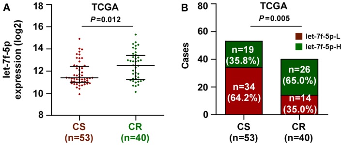
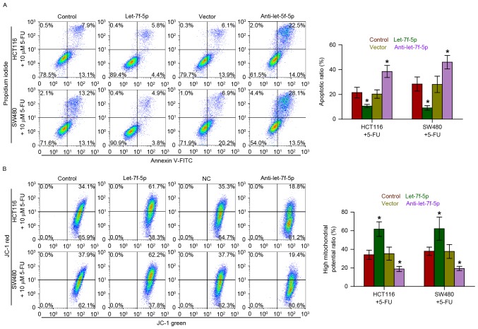
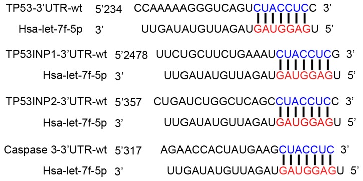
Colorectal cancer (CRC) is one of the most frequently occurring primary malignant tumors worldwide. Chemotherapeutic resistance is a major clinical problem in the treatment of CRC. Therefore, it is of great importance to investigate novel biomarkers that may predict chemoresistance and facilitate the development of individualized treatment for patients with CRC. The present study reported that let-7f-5p expression was elevated in chemotherapy-resistant CRC tissues compared with chemotherapy-sensitive tissues. Furthermore, upregulating let-7f-5p increased the expression levels of the anti-apoptotic proteins, B-cell lymphoma 2 (Bcl-2) and B-cell lymphoma-extra large (Bcl-xL), and decreased the activity of caspase-3 and caspase-9 in CRC cells. By contrast, downregulating let-7f-5p yielded the opposite effect. Notably, the results indicated that let-7f-5p promoted chemotherapeutic resistance by directly repressing the expression of several pro-apoptotic proteins, including tumor protein p53, tumor protein p53-inducible nuclear protein 1, tumor protein p53-inducible nuclear protein 2 and caspase-3. Therefore, a novel mechanism by which let-7f-5p enhances the resistance of CRC cells to chemotherapeutics has been revealed, indicating that silencing let-7f-5p may become an effective therapeutic strategy against CRC.

Keywords: B-cell lymphoma-2; B-cell lymphoma-extra large; caspase-3/9; chemotherapeutic resistance; colorectal cancer; let-7f-5p; tumor protein p53; tumor protein p53-inducible nuclear protein 1; tumor protein p53-inducible nuclear protein 2.


Figures

Figure 1.
Figure 1.
Upregulation of let-7f-5p is associated with poor chemotherapeutic response in CRC. (A) In the TCGA dataset, let-7f-5p expression levels were markedly higher in CRC patients with a poor chemotherapy response (CR) than those with a favorable chemotherapy response (CS). A Mann-Whitney U test was used to determine statistical differences between two groups. The data are presented as the median ± interquartile range. (B) Percentages and numbers of samples exhibiting high or low let-7f-5p expression with different chemotherapeutic responses in the TCGA dataset. The χ2 test was used to determine statistical differences between two groups. CRC, colorectal cancer; TCGA, The Cancer Genome Atlas; CR, chemoresistant; CS, chemosensitive.
Figure 2.
Figure 2.
let-7f-5p increases the expression of the anti-apoptotic proteins, Bcl-2 and Bcl-xL, and decreases the activity of caspase-3 and caspase-9 in CRC cells. (A) Reverse transcription-quantitative polymerase chain reaction analysis of let-7f-5p expression in HCT116 and SW480 cells transfected with a let-7f-5p mimic or inhibitor, compared with controls. Transcript levels were normalized to U6 expression. Error bars represent the mean ± standard deviation of three independent experiments. *P<0.05. One-way ANOVA was used to determine statistical differences among multiple groups. (B) Western blot analysis of Bcl-2 and Bcl-xL in the indicated cells. The activities of (C) caspase-3 and (D) caspase-9 were detected by the cleaved forms of these two proteins. Error bars represent the mean ± standard deviation of three independent experiments. *P<0.05. One-way ANOVA was used to determine statistical differences among multiple groups. CRC, colorectal cancer; Bcl-2, B-cell lymphoma 2; Bcl-xL, B-cell lymphoma-extra large; ANOVA, analysis of variance.
Figure 3.
Figure 3.
let-7f-5p decreases the apoptotic rate and increases the mitochondrial potential of CRC cells. (A) Annexin V-FITC/PI staining of the indicated cells treated with 5-FU for 36 h. Error bars represent the mean ± standard deviation of three independent experiments. *P<0.05. One-way ANOVA was used to determine statistical differences among multiple groups. (B) The JC-1 staining revealed that upregulating let-7f-5p enhanced the mitochondrial potential of the CRC cells, while silencing let-7f-5p decreased the mitochondrial potential of these cells. Error bars represent the mean ± standard deviation of three independent experiments. *P<0.05. One-way ANOVA was used to determine statistical differences among multiple groups. CRC, colorectal cancer; FITC, fluorescein isothiocyanate; PI, propidium iodide; 5-FU, fluorouracil; ANOVA, analysis of variance.
Figure 4.
Figure 4.
let-7f-5p targets multiple pro-apoptotic proteins. Predicted let-7f-5p targeting sequence in 3′-UTRs of TP53, TP53INP1, TP53INP2 and caspase-3. TP53, tumor protein p53; TP53INP1, TP53-inducible nuclear protein 1; TP53INP2, TP53-inducible nuclear protein p53 2; UTR, untranslated region.
Figure 5.
Figure 5.
let-7f-5p decreases the mRNA expression levels of TP53, TP53INP1, TP53INP2 and caspase-3. Reverse transcription-quantitative polymerase chain reaction analysis of TP53, TP53INP1, TP53INP2 and caspase-3 following transfection with a let-7f-5p mimic or inhibitor in (A) HCT116 and (B) SW480 cells. Transcript levels were normalized to U6 expression. Error bars represent the mean ± standard deviation of three independent experiments. *P<0.05. One-way analysis of variance was used to determine statistical differences among multiple groups. TP53, tumor protein p53; TP53INP1, TP53-inducible nuclear protein 1; TP53INP2, TP53-inducible nuclear protein p53 2; UTR, untranslated region.
Figure 6.
Figure 6.
let-7f-5p decreases the luciferase activity of the 3′-UTRs of TP53, TP53INP1, TP53INP2 and caspase-3. Luciferase assay of cells transfected with pmiRGLO-3′UTR reporter of TP53, TP53INP1, TP53INP2 and caspase-3 in let-7f-5p-overexpressing and silenced (A) HCT116 and (B) SW480 cells. One-way analysis of variance was used to determine statistical differences among multiple groups. TP53, tumor protein p53; TP53INP1, TP53-inducible nuclear protein 1; TP53INP2, TP53-inducible nuclear protein p53 2; UTR, untranslated region. Error bars represent the mean ± standard deviation of three independent experiments. *P<0.05 vs. control cells.

References

    1. Jemal A, Siegel R, Ward E, Hao Y, Xu J, Thun MJ. Cancer statistics, 2009. CA Cancer J Clin. 2009;59:225–249. doi: 10.3322/caac.20006. - DOI - PubMed
    1. Ricci-Vitiani L, Fabrizi E, Palio E, De Maria R. Colon cancer stem cells. J Mol Med (Berl) 2009;87:1097–1104. doi: 10.1007/s00109-009-0518-4. - DOI - PubMed
    1. Johnston PG, Kaye S. Capecitabine: A novel agent for the treatment of solid tumors. Anticancer Drugs. 2001;12:639–646. doi: 10.1097/00001813-200109000-00001. - DOI - PubMed
    1. Giacchetti S, Perpoint B, Zidani R, Le Bail N, Faggiuolo R, Focan C, Chollet P, Llory JF, Letourneau Y, et al. Phase III multicenter randomized trial of oxaliplatin added to chronomodulated fluorouracil-leucovorin as first-line treatment of metastatic colorectal cancer. J Clin Oncol. 2000;18:136–147. doi: 10.1200/JCO.2000.18.1.136. - DOI - PubMed
    1. Douillard JY, Cunningham D, Roth AD, Navarro M, James RD, Karasek P, Jandik P, Iveson T, Carmichael J, Alakl M, et al. Irinotecan combined with fluorouracil compared with fluorouracil alone as first-line treatment for metastatic colorectal cancer: A multicenter randomised trial. Lancet. 2000;355:1041–1047. doi: 10.1016/S0140-6736(00)02034-1. - DOI - PubMed