

Magnetic resonance features of meningeal solitary fibrous tumors
- PMID: 29805622
- PMCID: PMC5950533
- DOI: 10.3892/ol.2018.8426
Magnetic resonance features of meningeal solitary fibrous tumors
Abstract
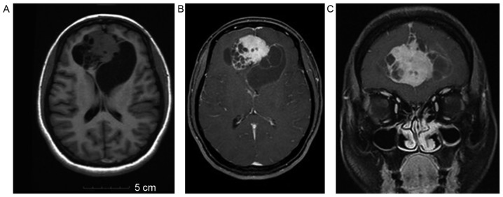
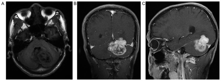
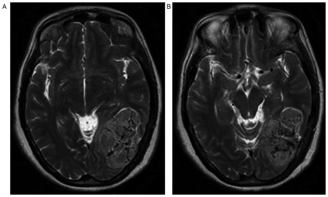
The aim of the present study was to investigate magnetic resonance (MR) features of meningeal solitary fibrous tumors (SFTs) in order to improve their recognition. The study retrospectively analyzed MR manifestations in 12 cases of meningeal SFTs confirmed by surgery. The lesions were analyzed in terms of the site of their growth, growth mode, morphology, size, changes in the MR signal and the edge of the focus. The tumors were malignant in 3 cases, benign in 7 cases and borderline in 2 cases. A total of 3 cases spanned the tentorium cerebelli, 3 spanned the falx and another case was close to the falx. Overall, 4 superficial tumors were accompanied by adjacent bony destruction. One case was located at the triangular area created by the right lateral ventricles. The tumors were all lobular, with a large diameter and more cystic areas in the center of the focus. The signals were often heterogeneous. The parenchyma in the tumors was significantly enhanced. For certain tumors, the signal was usually inhomogeneous and not uniform. The MR features of a SFT were often similar to other intracranial tumors and easily misdiagnosed. There were certain innate characteristics, for example, the tumor was often a solitary large soft-tissue mass with an irregular edge and a clear boundary, with a lobulated contour, and was widely involved with other tissues. The tumors often grew across either the falx or tentorium cerebelli. A significantly inhomogeneous signal was found and either a low or low-high mixed signal on T2-weighted images in particular. These features are useful for the differential diagnosis of SFTs and other tumors.
Keywords: magnetic resonance imaging; meningeal; solitary fibrous tumor.
Figures
References
-
- Wagner E. Das tuberkelahnliche lymphadenom (der cytogene oder reticulirte tuberkel) Arch Heilk (Leipzig) 1870;11:497. (In German)
-
- Guinee DG, Allen TC. Primary pleural neoplasia: Entities other than diffuse malignant mesothelioma. Arch Pathol Lab Med. 2008;132:1149–1170. - PubMed
LinkOut - more resources
Full Text Sources
Other Literature Sources