. 2018 May 4;9(34):23519-23531.
doi: 10.18632/oncotarget.25240.

Breast cancer stem cell-like cells generated during TGFβ-induced EMT are radioresistant

Affiliations

Breast cancer stem cell-like cells generated during TGFβ-induced EMT are radioresistant

Julie Konge et al. Oncotarget. .

Abstract

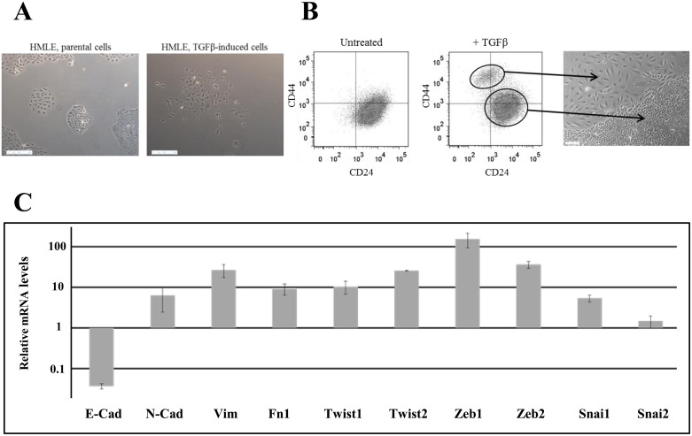
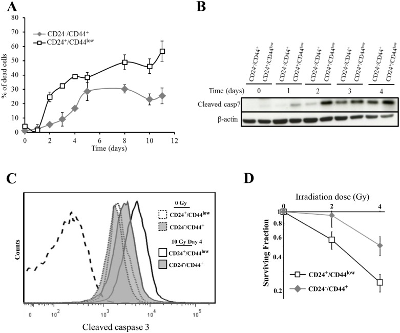
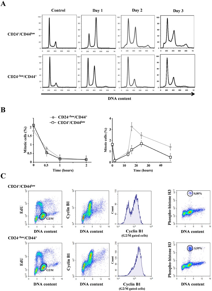
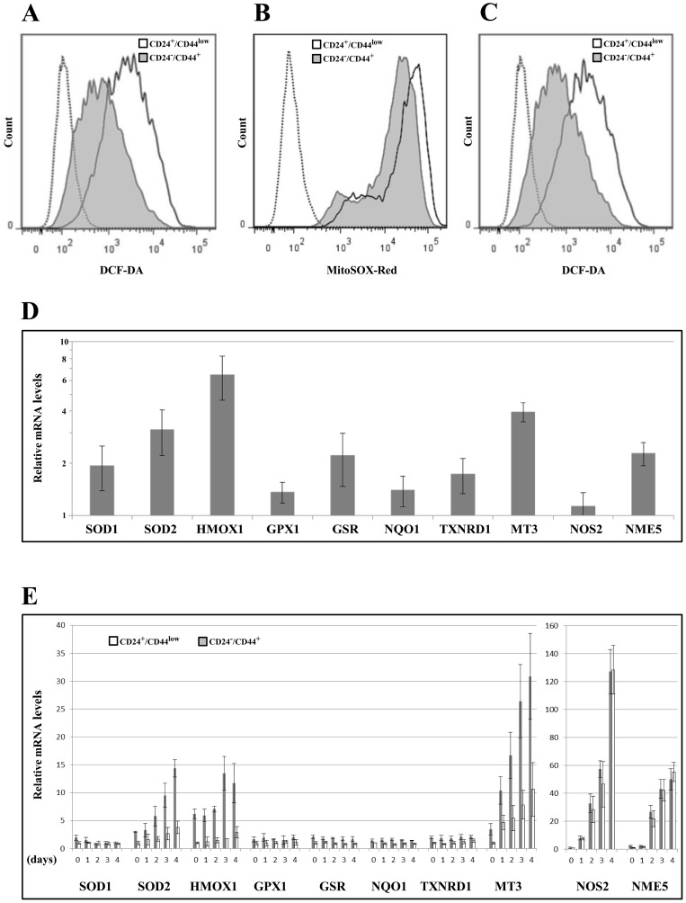
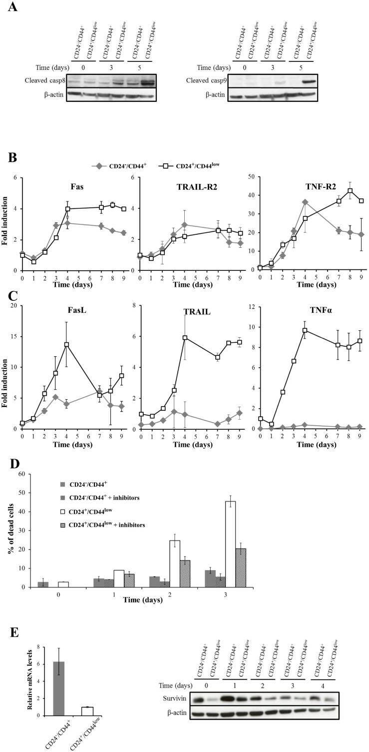
Failure of conventional antitumor therapy is commonly associated with cancer stem cells (CSCs), which are often defined as inherently resistant to radiation and chemotherapeutic agents. However, controversy about the mechanisms involved in the radiation response remains and the inherent intrinsic radioresistance of CSCs has also been questioned. These discrepancies observed in the literature are strongly associated with the cell models used. In order to clarify these contradictory observations, we studied the radiosensitivity of breast CSCs using purified CD24-/low/CD44+ CSCs and their corresponding CD24+/CD44low non-stem cells. These cells were generated after induction of the epithelial-mesenchymal transition (EMT) by transforming growth factor β (TGFβ) in immortalized human mammary epithelial cells (HMLE). Consequently, these 2 cellular subpopulations have an identical genetic background, their differences being related exclusively to TGFβ-induced cell reprogramming. We showed that mesenchymal CD24-/low/CD44+ CSCs are more resistant to radiation compared with CD24+/CD44low parental cells. Cell cycle distribution and free radical scavengers, but not DNA repair efficiency, appeared to be intrinsic determinants of cellular radiosensitivity. Finally, for the first time, we showed that reduced radiation-induced activation of the death receptor pathways (FasL, TRAIL and TNF-α) at the transcriptional level was a key causal event in the radioresistance of CD24-/low/CD44+ cells acquired during EMT.

Keywords: breast cancer; cancer stem cells; epithelial-mesenchymal transition; radioresistance.


Conflict of interest statement

CONFLICTS OF INTEREST None declared.

Figures

Figure 1
Figure 1. Characterization of TGFβ-induced CD24−/low/CD44+ cells
(A) Phase-contrast image of HMLE parental cells (left) and HMLE TGFβ-induced cells (right). (B) Flow cytometry characterization of HMLE parental and TGFβ-induced cells. Parental cells display essentially CD24+/CD44low labeling, and TGFβ treatment led to the appearance of a CD24−/low/CD44+ cell population. CD24+/CD44low cells were morphologically cuboidal-like epithelial cells, and CD24−/low/CD44+ cells were fibroblast-like, mesenchymal cells. (C) Analysis by qRT-PCR of the relative expression of the mRNAs encoding E-cadherin, N-cadherin, vimentin, fibronectin 1, Twist1, Twist2, Zeb1, Zeb2, Snail1 and Snail2. Normalization was performed as indicated in Materials and Methods. For each gene, expression in CD24+/CD44low cells was normalized to 1 and the ratio of relative mRNA level of CD24−/low/CD44+ cells to CD24+/CD44low cells was presented. Each value corresponds to the mean value of at least 2 independent PCRs performed from 3 independent experiments. Error bars correspond to SEM.
Figure 2
Figure 2. Analysis of cell death of CD24+/CD44low cells and CD24−/low/CD44+ cells after 10 Gy irradiation
(A) Time course of cell death of 10 Gy-irradiated CD24+/CD44low cells and CD24−/low/CD44+ cells. Results correspond to the mean ± SD of 2 to 4 independent experiments. (B) Kinetics of cleavage of caspase 7 at different times after 10 Gy irradiation. The Western blot shown is representative of 3 independent experiments. (C) Flow cytometry characterization of cleaved caspase 3 before and 4 days after irradiation. Dotted line corresponds to isotypic control. Similar results were obtained in 3 different experiments. (D) Clonogenic cell survival curves for CD24+/CD44low cells and CD24−/low/CD44+ cells exposed to 2 and 4 Gy irradiation. Error bars represent standard error from the mean for 3 separate experiments.
Figure 3
Figure 3. Analysis of cell cycle distribution in CD24+/CD44low cells and CD24−/low/CD44+ cells after 10 Gy irradiation
(A) Cell cycle analysis of 10 Gy-irradiated cells over 3 days. Similar results were obtained in at least 3 different experiments. (B) Mitotic index using phospho-histone H3 labelling at different times after 10 Gy irradiation: left 0–2 hours and right 0–48 hours after irradiation. Mean and SD of 4 independent experiments are presented. (C) Cyclin B1 and EdU labellings 16 hours after irradiation. Histograms of cyclin B1 were obtained from G2M cells (gated in the EdU/DNA content panel). Similar results were obtained in 2 different experiments. On the right, from the same sample, mitosis characterization using phospho-histone H3 labelling.
Figure 4
Figure 4. Analysis of global DSB repair in CD24+/CD44low cells and CD24−/low/CD44+ cells after 4 Gy irradiation
Number of γ-H2AX foci as a function of time upon 4 Gy irradiation.
Figure 5
Figure 5. Analyses of ROS levels and expression of oxidative stress-related genes in CD24+/CD44low cells and CD24−/low/CD44+ cells
(A) Basal intracellular ROS concentrations were measured by DCF-DA staining, (---- negative control without DCF-DA probe). The flow cytometry analysis shown is representative of 3 independent experiments. (B) As in (A) but using MitoSOX red instead of DCF-DA (---- negative control without MitoSOX red probe). (C) As in (A) but 2 days after 10 Gy irradiation. (D) Analysis by qRT-PCR of the relative expression of the mRNAs encoding stress-related genes. Normalization was performed as indicated in Materials and Methods. For each gene, expression in CD24+/CD44low cells was normalized to 1 and the ratio of relative mRNA level of CD24−/low/CD44+ cells to CD24+/CD44low cells was presented. Each value corresponds to the mean value of at least 2 independent PCRs performed from 3 independent experiments. Error bars correspond to SEM. (E) Analysis by qRT-PCR of the relative expression of the mRNAs encoding stress-related genes during 4 days after 10 Gy irradiation. Normalization was performed as indicated in Materials and Methods. For each gene, expression at day 0 in CD24+/CD44low cells was normalized to 1.
Figure 6
Figure 6. Role of death receptor pathways in radiation-induced cell death of CD24+/CD44low cells and CD24−/low/CD44+ cells
(A) Kinetics of cleavage of caspases 8 and 9 at different times after 10 Gy irradiation. The Western blots shown are representative of 3 independent experiments. (B) and (C) mRNA levels of death receptors and corresponding ligands after a 10 Gy irradiation of CD24+/CD44low cells and CD24−/low/CD44+ cells. These mRNA levels were determined by quantitative RT-PCR. Normalization was performed as indicated in Materials and Methods and the basal expression of CD24+/CD44low cells on day 0 was normalized to 1. Each value corresponds to the mean value of at least 2 independent PCRs performed from 3 independent experiments. Error bars correspond to standard deviation. (D) Neutralization of the death receptor pathways reduces radiation-induced apoptosis. CD24+/CD44low cells and CD24−/low/CD44+ cells were 10 Gy-irradiated and treated or not with a combination of Fas/Fc, TRAIL-R1/Fc and TNF-R1/Fc chimera (250 ng/mL, 100 ng/mL and 100 ng/mL, respectively). These chimeras, which are cytokines designed to neutralize apoptosis induced by the corresponding ligand, were added to the culture medium at days 0 and 2. Viable and dead cells were counted after trypan blue staining. Results correspond to the mean ± standard deviation of 3 independent experiments. (E) Survivin expression by quantitative RT-PCR (left) and Western blot (right) after a 10 Gy irradiation of CD24+/CD44low cells and CD24−/low/CD44+ cells. For RT-PCR, normalization was performed as indicated above, and the Western blot shown is representative of 3 independent experiments.

References

    1. Mani SA, Guo W, Liao MJ, Eaton EN, Ayyanan A, Zhou AY, Brooks M, Reinhard F, Zhang CC, Shipitsin M, Campbell LL, Polyak K, Brisken C, et al. The epithelial-mesenchymal transition generates cells with properties of stem cells. Cell. 2008;133:704–715. - PMC - PubMed
    1. Morel AP, Lievre M, Thomas C, Hinkal G, Ansieau S, Puisieux A. Generation of breast cancer stem cells through epithelial-mesenchymal transition. PLoS One. 2008;3:e2888. - PMC - PubMed
    1. Gupta PB, Chaffer CL, Weinberg RA. Cancer stem cells: mirage or reality? Nat Med. 2009;15:1010–1012. - PubMed
    1. Bao S, Wu Q, McLendon RE, Hao Y, Shi Q, Hjelmeland AB, Dewhirst MW, Bigner DD, Rich JN. Glioma stem cells promote radioresistance by preferential activation of the DNA damage response. Nature. 2006;444:756–760. - PubMed
    1. Li X, Lewis MT, Huang J, Gutierrez C, Osborne CK, Wu MF, Hilsenbeck SG, Pavlick A, Zhang X, Chamness GC, Wong H, Rosen J, Chang JC. Intrinsic resistance of tumorigenic breast cancer cells to chemotherapy. J Natl Cancer Inst. 2008;100:672–679. - PubMed

LinkOut - more resources