. 2018 May 4;9(34):23636-23660.
doi: 10.18632/oncotarget.25318.

Changes of signal transductivity and robustness of gene regulatory network in the carcinogenesis of leukemic subtypes via microarray sample data

Affiliations

Changes of signal transductivity and robustness of gene regulatory network in the carcinogenesis of leukemic subtypes via microarray sample data

Cheng-Wei Li et al. Oncotarget. .

Abstract

Mutation accumulation and epigenetic alterations in genes are important for carcinogenesis. Because leukemogenesis-related signal pathways have been investigated and microarray sample data have been produced in acute myeloid leukemia (AML), myelodysplastic syndromes (MDS) and normal cells, systems analysis in coupling pathways becomes possible. Based on system modeling and identification, we could construct the coupling pathways and their associated gene regulatory networks using microarray sample data. By applying system theory to the estimated system model in coupling pathways, we can then obtain transductivity sensitivity, basal sensitivity and error sensitivity of each protein to identify the potential impact of genetic mutations, epigenetic alterations and the coupling of other pathways from the perspective of energy, respectively. By comparing the results in AML, MDS and normal cells, we investigated the potential critical genetic mutations and epigenetic alterations that activate or repress specific cellular functions to promote MDS or AML leukemogenesis. We suggested that epigenetic modification of β-catenin and signal integration of CSLs, AP-2α, STATs, c-Jun and β-catenin could contribute to cell proliferation at AML and MDS. Epigenetic regulation of ERK and genetic mutation of p53 could lead to the repressed apoptosis, cell cycle arrest and DNA repair in leukemic cells. Genetic mutation of JAK, epigenetic regulation of ERK, and signal integration of C/EBPα could result in the promotion of MDS cell differentiation. According to the results, we proposed three drugs, decitabine, genistein, and monorden for preventing AML leukemogenesis, while three drugs, decitabine, thalidomide, and geldanamycin, for preventing MDS leukemogenesis.

Keywords: basal sensitivity; error sensitivity; network robustness; transductivity; transductivity sensitivity.


Conflict of interest statement

CONFLICTS OF INTEREST The authors declare that they have no competing interests.

Figures

Figure 1
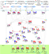
Figure 1. Flowchart of the proposed method for estimating network robustness of the GRNs and for predicting the impact of genetic mutations, epigenetic alterations and the coupling of other pathways of the proteins in the coupling STPs (Supplementary Figure 1)
Figure 2
Figure 2. The network robustness of Supplementary Figure 2 across seven leukemic subtypes and one normal type
The normal type is with the smallest robustness (0.18) by comparison to other leukemic subtypes. It means that normal type is less tolerable to intrinsic perturbation such as genetic mutation or epigenetic alteration and more responsible to external molecular signals than leukemic subtypes. Among these leukemic subtypes, the GRNs of AML+nok/abn and MDS with higher robustness than other leukemic subtypes can give harbor to a diversity of intrinsic genetic mutation and epigenetic alteration and further develop a variety of evolutionary process to leukemogenesis than other leukemic subtypes.
Figure 3
Figure 3
Comparison of correlation sensitivities (A) and transductivity sensitivities (B) for 28 TFs between AML+nok/abn and MDS in the same dataset (GSE6891). Comparison of transductivity sensitivities for 28 TFs at AML+nok/abn (C) or at MDS (D) between two datasets, i.e., between GSE13159 and GSE6891 at AML+nok/abn or between GSE13159 and GSE15061 at MDS. According to top 3 TFs with the largest differences between two datasets at AML+nok/abn (C) and at MDS (D) including FOXO3a, NF-κBs, HIF-1α, E2Fs, and PU.1, the TFs also have large differences between AML+nok/abn and MDS in (B). Therefore, we inferred that the 5 TFs with the largest differences in transductivity sensitivity result from the effect of individual difference on the induction of redundant cellular functions to promote AML/MDS leukemogenesis. We suggested that the proposed results (B) to compare the transductivity sensitivities between AML+nok/abn and MDS in the same dataset (GSE13159) are reliable.
Figure 4
Figure 4
Gain or lose of cellular functions of five proliferation-related TFs (CSLs, AP-2α, STATs, c-Jun and β-catenin) (A), two apoptosis-related TFs (p53, FOXO3a) (B) and three differentiation-related TFs (C/EBPα, AML1, and ETO) (C) in AML/MDS leukemogenesis. If the transductivity sensitivity of a TF, such as CSLs in (A), in a leukemic subtype was positive, Δρ > 0 (Red bar), its activity was increased in the leukemic subtype when comparing to normal type. In contrast, the activity of p53 in (B) was decreased at leukemic subtypes. The large error sensitivity of a TF in a leukemic subtype, such as AP-2α in (A), and C/EBPα in (C) in both AML+nok/abn, and MDS, and c-Jun in (A) in AML+nok/abn, represents the probable impact of pathways other than those in Supplementary Figure 1. Additionally, the large basal sensitivity of a TF in a leukemic subtype, such as AML1, C/EBPα, β-catenin, and FOXO3a in (A–C) in both AML+nok/abn and MDS, implicates the probable impact of epigenetic regulation on the TF-coding gene.
Figure 5
Figure 5. The illustration for identifying the impact of genetic mutation, epigenetic regulation and the coupling of other pathways on the downstream protein
The sign change of the transductivity sensitivities between two proteins implicated the impact of genetic mutation, epigenetic regulation or the coupling of other pathways on the downstream protein B. The black bar denotes the absolute value of the error sensitivity ΔE or basal sensitivity ΔB. The absolute values of the basal and error sensitivities in protein B were higher than 0.5 indicated epigenetic regulation ((B) at AML and (E) at MDS) and the impact of other pathways ((C) at AML and (F) at MDS) on B, respectively. Otherwise, genetic mutation occurred in protein B ((A) at AML and (D) at MDS).
Figure 6
Figure 6. Transductivity sensitivity, error sensitivity, and basal sensitivity of the coupled pathways contributing to the loss of transductivity of p53 at AML+nok/abn and MDS cells when comparing to the normal type
The identified genetic mutations on the genes TRAF2, and MKK4 at AML+nok/abn cell, and MDM2 at MDS cell were by the sign change of the transductivity sensitivity at leukemic subtype when comparing to the upstream protein. The identified effect of epigenetic regulation on the gene ERK at both leukemic subtypes was by not only the sign change of the transductivity sensitivity at leukemic subtype, but also the large basal sensitivity of ERK at both leukemic subtypes. The proteins shown in red symbols with underline in the STPs are analyzed as the main causes of dysfunction of TF.
Figure 7
Figure 7. Transductivity sensitivity, error sensitivity, and basal sensitivity of the coupled pathways contributing to gain of transductivity of STATs and C/EBPα at AML+nok/abn and MDS cells when comparing to the normal type
The identified genetic mutations on the genes JAK, MKK4, and ETO at AML+nok/abn cell, and PTEN, and IKKs at MDS cell were due to the sign change of the transductivity sensitivity at leukemic subtype when comparing to the upstream protein. The identified effect of epigenetic regulation on the gene ERK, and AML1 at both leukemic subtypes was due to not only the sign change of the transductivity sensitivity at leukemic subtype, but also the large basal sensitivities of ERK, and AML1 at both leukemic subtypes. The identified impact of other pathways on the gene c-Jun at AML+nok/abn cell was due to not only the sign change of the transductivity sensitivity at leukemic subtype, but also the large error sensitivity of c-Jun at AML+nok/abn cell.
Figure 8
Figure 8. Transductivity sensitivity, error sensitivity, and basal sensitivity of the coupled pathways contributing to the gain of transductivity of CSLs at AML+nok/abn and MDS cells when comparing to the normal type
The identified genetic mutations on the genes CSLs at AML+nok/abn cell, Numb, Deltex, and PSE2 at MDS cell, and DVL, CTBPs, and SMRT at both cells were by the sign change of the transductivity sensitivity at leukemic subtype when comparing to the upstream protein.
Figure 9
Figure 9. Transductivity sensitivity, error sensitivity, and basal sensitivity of the coupled pathways associated with β-catenin and FOXO3a at AML+nok/abn and MDS cells when comparing to the normal type
The gain of function in β-catenin attributed to the genetic mutations in DVL and the interaction site between AKT and GSK3β in AML and the genetic mutations in DVA and the impact of other pathways on Axin in MDS. The loss of function in FOXO3a in AML was due to the genetic mutations in TRADD and TRAF2.
Figure 10
Figure 10. Transductivity sensitivities of 28 TFs in AML, MDS, and the proposed drugs
We proposed three drugs, decitabine, genistein, and monorden, with effectiveness (Ej = 2.058, 3.085, and 3.132, respectively) for treating patients with AML, while three drugs, decitabine, thalidomide, and geldanamycin, with effectiveness (Ej = 3.846, 5.057, and 3.392, respectively) for treating patients with MDS. The transductivity sensitivities of 28 TFs in three drugs are inversely correlated with the transductivity sensitivities of 28 TFs in the corresponding leukemic subtype. The full table of effectiveness of drugs for treating patients with AML/MDS is shown in Supplementary Table 4.

Similar articles

References

    1. Sands WA, Copland M, Wheadon H. Cell Communication and Signaling. 2013. p. 11. - PMC - PubMed
    1. Steelman LS, Abrams SL, Whelan J, Bertrand FE, Ludwig DE, Bäsecke J, Libra M, Stivala F, Milella M, Tafuri A, Lunghi P, Bonati A, Martelli AM, McCubrey JA. Contributions of the Raf/MEK/ERK, PI3K/PTEN/Akt/mTOR and Jak/STAT pathways to leukemia. Leukemia. 2008;22:686–707. https://doi.org/10.1038/leu.2008.26. - DOI - PubMed
    1. Takahashi S. Downstream molecular pathways of FLT3 in the pathogenesis of acute myeloid leukemia: biology and therapeutic implications. J Hematol Oncol. 2011;4:13. https://doi.org/10.1186/1756-8722-4-13. - DOI - PMC - PubMed
    1. Koschmieder SKU. Myeloid Leukemia - Basic Mechanisms of Leukemogenesis. InTech. 2011.
    1. Zhang R, Shah MV, Yang J, Nyland SB, Liu X, Yun JK, Albert R, Loughran TP., Jr Network model of survival signaling in large granular lymphocyte leukemia. Proc Natl Acad Sci USA. 2008;105:16308–13. https://doi.org/10.1073/pnas.0806447105. - DOI - PMC - PubMed