. 2017:1:889-901.
doi: 10.1038/s41551-017-0137-2. Epub 2017 Oct 2.

Nanoparticle delivery of Cas9 ribonucleoprotein and donor DNA in vivo induces homology-directed DNA repair

Affiliations

Nanoparticle delivery of Cas9 ribonucleoprotein and donor DNA in vivo induces homology-directed DNA repair

Kunwoo Lee et al. Nat Biomed Eng. 2017.

Abstract

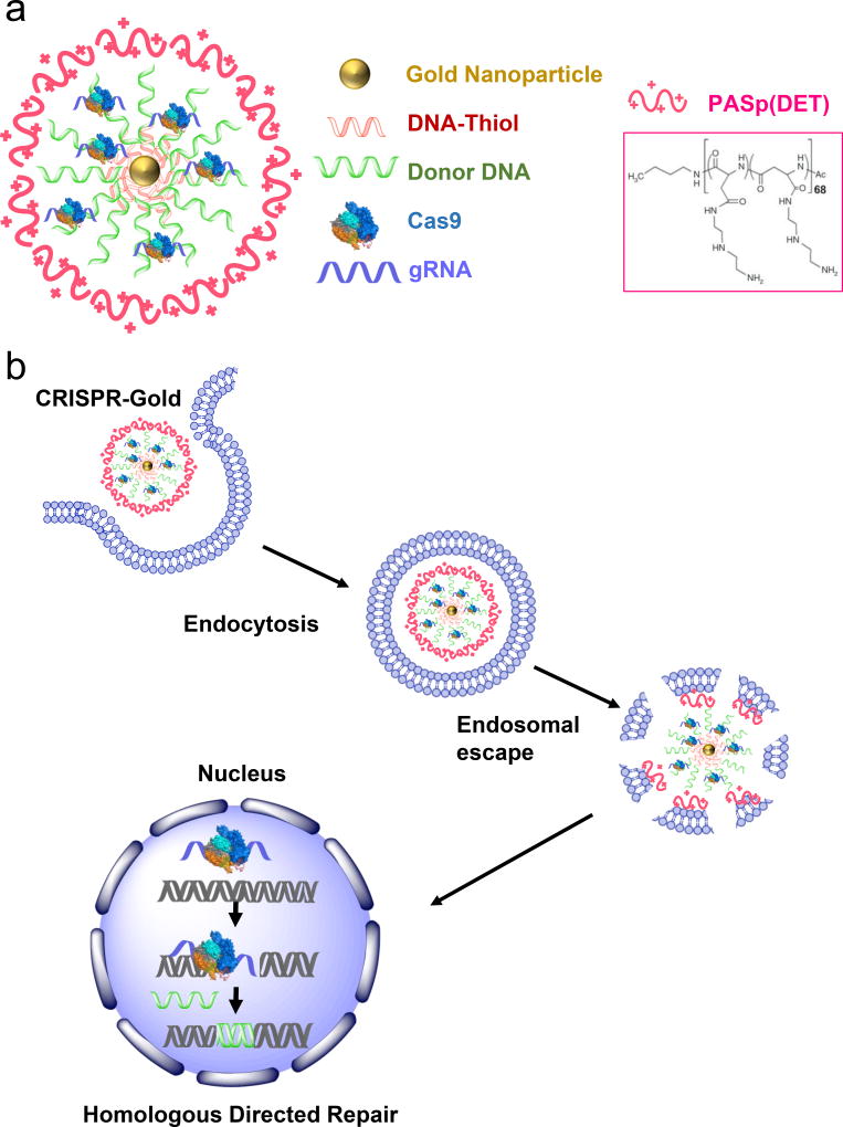
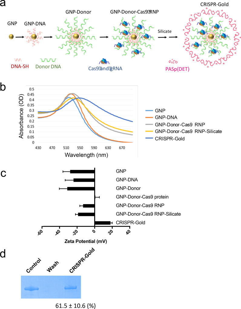
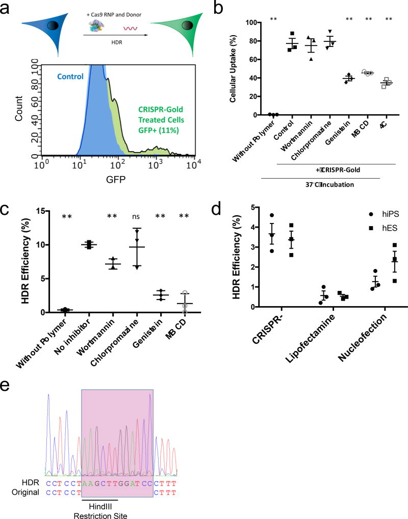
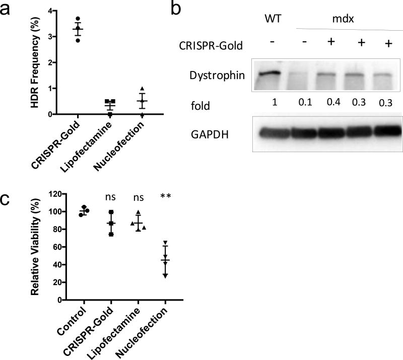
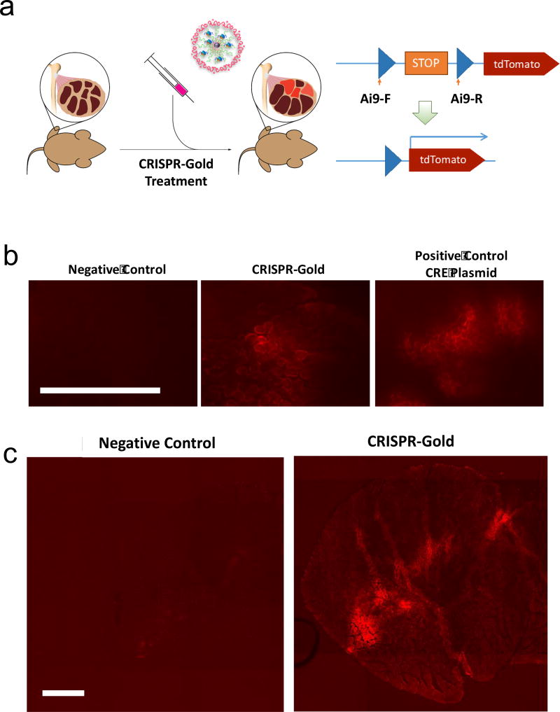
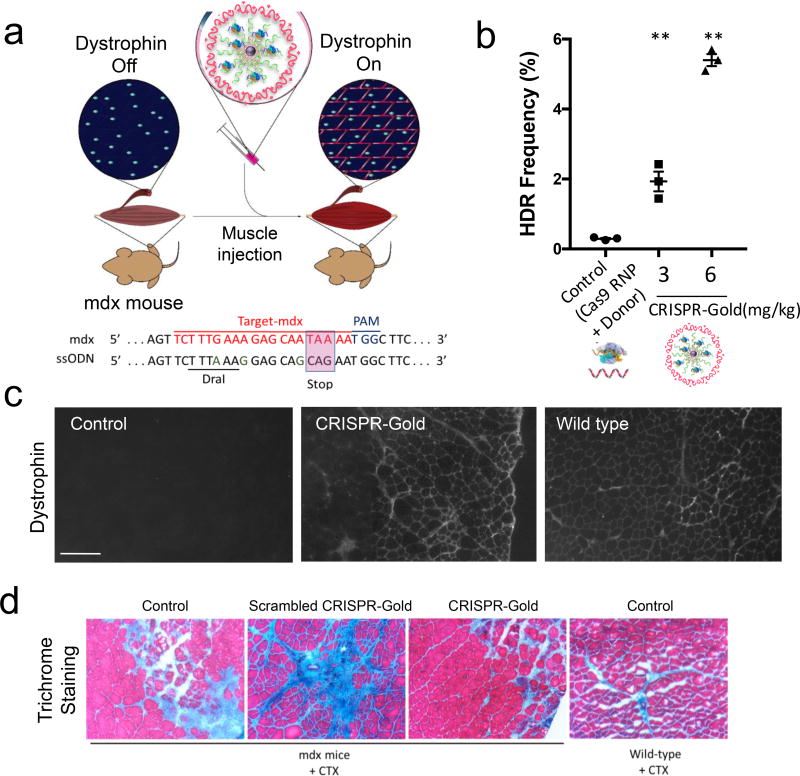
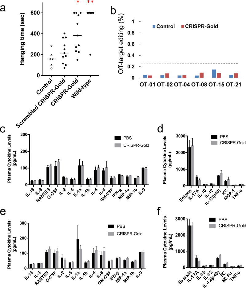
CRISPR/Cas9-based therapeutics, especially those that can correct gene mutations via homology directed repair (HDR), have the potential to revolutionize the treatment of genetic diseases. However, HDR-based therapeutics are challenging to develop because they require simultaneous in vivo delivery of Cas9 protein, guide RNA and donor DNA. Here, we demonstrate that a delivery vehicle composed of gold nanoparticles conjugated to DNA and complexed with cationic endosomal disruptive polymers can deliver Cas9 ribonucleoprotein and donor DNA into a wide variety of cell types, and efficiently correct the DNA mutation that causes Duchenne muscular dystrophy in mice via local injection, with minimal off-target DNA damage.


Conflict of interest statement

Competing interests: K.L., H. P, and N.M. are co-founders of GenEdit Inc. J.A.D. is a co-founder of Caribou Biosciences, Editas Medicine, and Intellia Therapeutics.

Figures

Figure 1
Figure 1. CRISPR-Gold can deliver Cas9 ribonucleoprotein and donor DNA in vivo and induce homology directed DNA (HDR) repair
a) CRISPR-Gold is composed of 15 nm gold nanoparticles conjugated to thiol modified oligonucleotides, which are hybridized with donor ssODN and subsequently complexed with Cas9 RNP, and the endosomal disruptive polymer PAsp(DET). b) CRISPR-Gold is internalized by cells in vitro and in vivo via endocytosis, triggers endosomal disruption, and releases Cas9 RNP and donor DNA into the cytoplasm. Nuclear delivery results in HDR.
Figure 2
Figure 2. Synthesis and characterization of CRISPR-Gold
a) Synthesis of CRISPR-Gold. Gold nanoparticles 15 nm in diameter were conjugated with a 5′ thiol modified single stranded DNA (DNA-SH), and hybridized with single stranded donor DNA. Cas9 and gRNA are loaded and then a silicate and PAsp(DET) polymer coating are added. b) Absorbance spectra analysis. The absorption maxima of CRISPR-Gold intermediates are red shifted as CRISPR-Gold is sequentially constructed. Unmodified GNPs had an absorbance peak at 518 nm and the peak shifts to 522 nm for GNP-DNA, 528 nm for GNP-DNA-Cas9 RNP, and 546 nm for CRISPR-Gold. c) Zeta potential analysis. Zeta potential measurements were performed on CRISPR-Gold and all of the synthetic intermediates generated during the construction of CRISPR-Gold. Zeta potential changes demonstrate the sequential synthesis of CRISPR-Gold. d) Cas9 loading analysis. CRISPR-Gold was formulated and the unbound Cas9 RNP was removed via spin filtration. Gel electrophoresis was performed on CRISPR-Gold and the SDS gel was stained with Coomassie blue to quantify the amount of Cas9 RNP bound to CRISPR-Gold. The control represents the original amount of Cas9 RNP added to the CRISPR-Gold formulation. The wash represents the unbound Cas9 RNP, removed by filtration.
Figure 3
Figure 3. CRISPR-Gold induces HDR in vitro
a) CRISPR-Gold can induce HDR in BFP-HEK cells by delivering Cas9 RNP and a donor DNA. 11.3% of BFP-HEK cells were converted to GFP expressing cells, after treatment with CRISPR-Gold. GFP expression due to BFP sequence editing was determined by flow cytometry. b) The uptake of CRISPR-Gold in HEK293 cells is dependent on non-clathrin mediated endocytosis. CRISPR-Gold containing Alexa647 labeled sgRNA was added to HEK293 cells and the uptake efficiency was measured using flow cytometry. Various inhibitors and low temperature were used to study the cellular uptake mechanism. Also, the uptake of CRISPR-Gold without the PAsp(DET) polymer coating was investigated. Genistein, MBCD, and 4°C all significantly inhibited the uptake of CRISPR-Gold, whereas clathrin based endocytosis inhibitors had no effect. Mean ± S.E, n=3. **, p < 0.01. c) Non-clathrin mediated endocytosis plays a critical role in CRISPR-Gold mediated HDR. The inhibitor of clathrin mediated endocytosis, chlorpromazine, has minimal effects on CRISPR-Gold mediated HDR, whereas the caveolae/raft mediated endocytosis inhibitor MBCD and genistein reduced CRISPR-Gold mediated HDR by 80%. Mean ± S.E, n=3. **, p < 0.01, ns= not significant. d) CRISPR-Gold induces HDR in hES and hiPS cells with higher efficiency than lipofectamine or nucleofection. Cells were treated with CRISPR-Gold that contained a donor sequence with a HindIII cleavage site, and a guide RNA that targeted the CXCR4 gene. The CXCR4 gene was amplified by PCR and the HDR efficiency was determined by quantifying HindIII cleavage of the CXCR4 PCR amplicon. Mean ± S.E, n=3. A one-way ANOVA test had a p= 0.0066 for hES samples and p=0.0023 for hiPS samples. e) Sanger sequencing demonstrates that CRISPR-Gold induces HDR in hES cells. PCR of the CXCR4 gene was performed on CRISPR-Gold treated hES cells, and sequencing confirms the presence of the 12 bp insertion (pink box).
Figure 4
Figure 4. CRISPR-Gold induces HDR and promotes the expression of dystrophin protein in primary myoblasts
a) The dystrophin gene was edited with CRISPR-Gold in primary myoblasts from the mdx mouse. CRISPR-Gold corrected the nonsense mutation in the dystrophin gene of mdx myoblasts with an HDR efficiency of 3.3%, which is significantly higher than either nucleofection or lipofectamine. No correction was observed in the negative control, composed of CRISPR-Gold without gRNA (data not shown). Mean ± S.E, n=3. A one-way ANOVA test had a p= 0.0002. b) Dystrophin protein is expressed in myotubes that were differentiated from CRISPR-Gold edited primary mdx myoblasts. Western blot analysis was conducted to quantify the levels of dystrophin protein in muscle cells. The fold expression was determined by dividing the band pixel density of each group with the dystrophin band intensity from WT (C57.B6) myotubes (n=1). GAPDH was used as a loading control. c) CRISPR-Gold causes minimal toxicity to primary myoblasts, whereas nucleofection caused significant toxicity. Primary mdx myoblasts were transfected with the indicated methods and cell viability was measured 2 days after the transfections with the cell counting kit-8. Relative viability to control, mean ± S.E, n=6. *, p < 0.05, ns = statistically not significant to control.
Figure 5
Figure 5. CRISPR-Gold induces gene editing in the muscle tissue of Ai9 mice
a) CRISPR-Gold was injected into Ai9 mice and gene deletion of the stop sequence in the Ai9 gene was determined by tdTomato expression. b) Representative images of tdTomato fluorescence in the gastrocnemius muscle after a single CRISPR-Gold injection. A plasmid expressing Cre recombinase was injected with lipofectamine as a positive control. Scale bar is 500 µm. c) tdTomato expression is observed in a broad area of the muscle after injection with CRISPR-Gold. The entire TA muscle cross-section image shows tdTomato expression after CRISPR-Gold injection. Scale bar is 500 µm.
Figure 6
Figure 6. CRISPR-Gold promotes HDR in the dystrophin gene and dystrophin protein expression, and reduces muscle fibrosis in mdx mice, with CTX stimulation
a) CRISPR-Gold was injected into the hind leg muscle of 8-week-old mdx mice (n=3), simultaneously with CTX. Two weeks later, the mice were sacrificed and analyzed for dystrophin expression by immunofluorescence, for HDR by deep sequencing and for their degree of fibrosis. Bottom panel: dystrophin mutation sequence and donor DNA design. Donor DNA sequence designed to repair the non-sense mutation are marked in the pink box, nucleotides marked in green (A, G, G) are silent mutations that prevent Cas9 activity on the edited sequence. b) CRISPR-Gold induced genome editing in the dystrophin gene was confirmed by deep sequencing. Deep sequencing of genomic DNA from muscle tissues injected with CRISPR-Gold revealed a 5.4% HDR efficiency. The negative control composed of Cas9 RNP and donor DNA had an HDR efficiency of only 0.3%. Mean ± S.E, n=3. **, p < 0.01. c) CRISPR-Gold causes dystrophin expression in muscle tissue of mdx mice. The CRISPR-Gold injected muscle shows dystrophin expression (immuno-fluorescence), whereas control mdx mice did not express dystrophin protein. The scale bar is 200 µm. d) CRISPR-Gold reduces muscle fibrosis in mdx mice. Trichrome staining was performed on the TA muscle cryosectioned to 10 µm, 2 weeks after the injection of CRISPR-Gold. CTX was co-injected in all three groups of mdx mice: No treatment control, scrambled CRISPR-Gold, and CRISPR-Gold. Images were acquired at the areas of muscle injury and regeneration. Fibrotic tissue appears blue, while muscle fibers appear red. Wild-type mice treated with CTX were analyzed 5 days after injection. The width of each image is 0.7 mm.
Figure 7
Figure 7. CRISPR-Gold induced dystrophin editing enhances muscle function with minimal off-target effects and without elevation of systemic inflammatory cytokines
a) CRISPR-Gold improves strength and agility in mdx mice without CTX. A four-limb hanging test was conducted in mdx mice treated with CRISPR-Gold, two weeks after the initial CRISPR-Gold injection. CRISPR-Gold significantly enhanced muscle strength in comparison to mice treated with scrambled CRISPR-Gold. Mean ± S.E, n=11 for injected mdx mice, n=6 for control mdx mice and wild-type mice, *, p < 0.05, **, p < 0.01, statistically significant to scrambled CRISPR-Gold group. b) In vivo off-target effects are insignificant in CRISPR-Gold injected mouse muscle tissue. The major predicted and reported off-target sites for the mdx gRNA were analyzed with deep sequencing. Control: scrambled CRISPR-Gold injected mouse and CRISPR-Gold: CRISPR-Gold injected mouse without CTX. Each read-out was more than 30,000 reads and all off-target mutations were within the range of deep sequencing error. c–f) CRISPR-Gold does not elevate plasma cytokine levels as compared to PBS injections. c–d) Plasma cytokine levels after a single injection of CRISPR-Gold. e–f) Plasma cytokine levels after two injections of CRISPR-Gold. c, e) group of high plasma level cytokines. d, f) group of low plasma level cytokines.

References

    1. Jinek M, et al. A Programmable Dual-RNA – Guided DNA Endonuclease in Adaptive Bacterial Immunity. Science. 2012;337:816–822. - PMC - PubMed
    1. Cong L, et al. Multiplex Genome Engineering Using CRISPR/Cas System. Science. 2013;339:819–823. - PMC - PubMed
    1. Mali P, et al. RNA-guided human genome engineering via Cas9. Science. 2013;339:823–826. - PMC - PubMed
    1. Cho SW, Kim S, Kim JM, Kim J-S. Targeted genome engineering in human cells with the Cas9 RNA-guided endonuclease. Nat. Biotechnol. 2013;31:230–2. - PubMed
    1. Ran FA, et al. In vivo genome editing using Staphylococcus aureus Cas9. Nature. 2015;520:186–190. - PMC - PubMed