

[Dopamine modified and cartilage derived morphogenetic protein 1 laden polycaprolactone-hydroxyapatite composite scaffolds fabricated by three-dimensional printing improve chondrogenic differentiation of human bone marrow mesenchymal stem cells]
- PMID: 29806415
- PMCID: PMC8414101
- DOI: 10.7507/1002-1892.201708017
[Dopamine modified and cartilage derived morphogenetic protein 1 laden polycaprolactone-hydroxyapatite composite scaffolds fabricated by three-dimensional printing improve chondrogenic differentiation of human bone marrow mesenchymal stem cells]
Abstract
Objective: To prepare dopamine modified and cartilage derived morphogenetic protein 1 (CDMP1) laden polycaprolactone-hydroxyapatite (PCL-HA) composite scaffolds by three-dimensional (3D) printing and evaluate the effect of 3D scaffolds on in vitro chondrogenic differentiation of human bone marrow mesenchymal stem cells (hBMSCs).
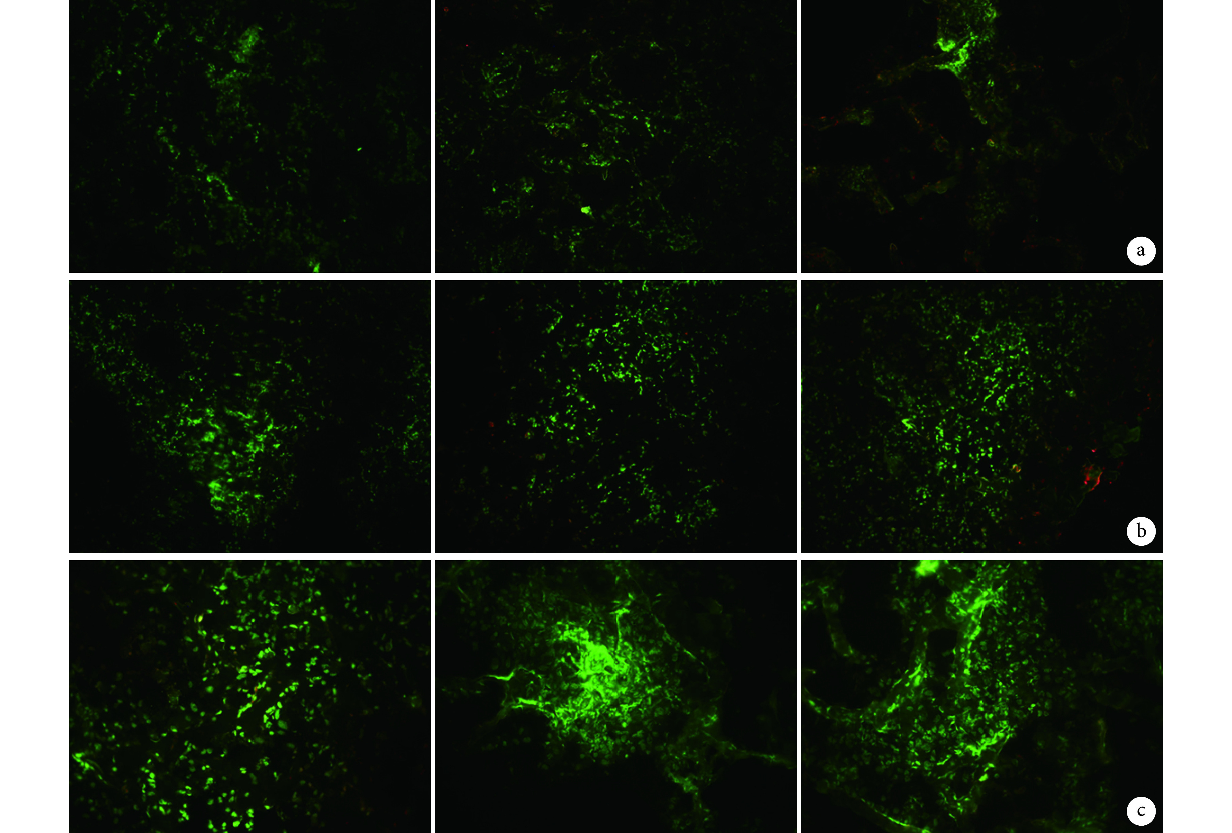
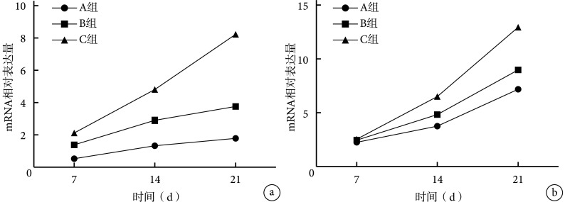
Methods: A dimensional porous PCL-HA scaffold was fabricated by 3D printing. Dopamine was used to modify the surface of PCL-HA and then CDMP-1 was loaded into scaffolds. The surface microstructure was observed by scanning electron microscope (SEM) and porosity and water static contact angle were also detected. The cytological experiment in vitro were randomly divided into 3 groups: group A (PCL-HA scaffolds), group B (dopamine modified PCL-HA scaffolds), and group C (dopamine modified and CDMP-1 laden PCL-HA scaffolds). The hBMSCs were seeded into three scaffolds, in chondrogenic culture conditions, the cell adhesive rate, the cell proliferation (MTT assay), and cell activity (Live-Dead staining) were analyzed; and the gene expressions of collagen type Ⅱ and Aggrecan were detected by real-time fluorescent quantitative PCR.
Results: The scaffolds in 3 groups were all showed a cross-linked and pore interconnected with pore size of 400-500 μm, porosity of 56%, and fiber orientation of 0°/90°. For dopamine modification, the scaffolds in groups B and C were dark brown while in group A was white. Similarly, water static contact angle was from 76° of group A to 0° of groups B and C. After cultured for 24 hours, the cell adhesion rate of groups A, B, and C was 34.3%±3.5%, 48.3%±1.5%, and 57.4%±2.5% respectively, showing significant differences between groups ( P<0.05). Live/Dead staining showed good cell activity of cells in 3 groups. MTT test showed that hBMSCs proliferated well in 3 groups and the absorbance ( A) value was increased with time. The A value in group C was significantly higher than that in groups B and A, and in group B than in group A after cultured for 4, 7, 14, and 21 days, all showing significant differences ( P<0.05). The mRNA relative expression of collagen type Ⅱ and Aggrecan increased gradually with time in 3 groups. The mRNA relative expression of collagen type Ⅱafter cultured for 7, 14, and 21 days, and the mRNA relative expression of Aggrecan after cultured for 14 and 21 days in group C were significantly higher than those in groups A and B, and in group B than in group A, all showing significant differences ( P<0.05).
Conclusion: Co-culture of dopamine modified and CDMP1 laden PCL-HA scaffolds and hBMSCs in vitro can promote hBMSCs' adhesion, proliferation, and chondrogenic differentiation.
目的: 探讨利用 3D 打印技术制备、经多巴胺表面修饰及负载软骨源性形态发生蛋白 1(cartilage derived morphogenetic protein 1,CDMP1)的聚己内酯(polycaprolactone,PCL)-羟基磷灰石(hydroxyapatite,HA)三维多孔支架,体外诱导人 BMSCs(human BMSCs,hBMSCs)成软骨分化的可行性。.
方法: 采用 3D 打印技术制备 PCL-HA 支架,经多巴胺表面修饰后,将 CDMP-1 负载于支架上,扫描电镜观察支架表面微结构,并检测孔隙率和水静态接触角。体外成软骨分化实验:分为 A 、B、C 3 组,A 组为 PCL-HA 支架,B 组为多巴胺表面修饰的 PCL-HA 支架,C 组为多巴胺表面修饰及负载 CDMP-1 的 PCL-HA 支架;将 hBMSCs 植入 3 组支架,成软骨诱导培养后比较细胞黏附率、细胞增殖(MTT 法)和细胞活性(Live/Dead 染色法),并采用实时荧光定量 PCR 检测Ⅱ型胶原和聚集蛋白聚糖(Aggrecan)的基因表达。.
结果: 3 组支架均呈三维多孔圆柱体状,孔洞相互联通,孔径为 400~500 μm,孔隙率为 56%,材料纤维走向为 0°/90°。经多巴胺表面修饰后,支架颜色由初始的白色变为棕色;水静态接触角也由 76° 降为 0°。体外培养 24 h,A、B、C 组细胞黏附率分别为 34.3%±3.5%、48.3%±1.5%、57.4%±2.5%,比较差异均有统计学意义( P<0.05)。Live/Dead 染色显示 3 组细胞均有较好的细胞活性。MTT 检测显示各组 hBMSCs 均生长良好,吸光度( A)值随培养时间延长而增大;培养 4、7、14、21 d 时,C 组 A 值显著高于 A、B 组,B 组高于 A 组,差异均有统计学意义( P<0.05)。随培养时间延长,3 组Ⅱ型胶原 mRNA 和 Aggrecan mRNA 表达均持续增加;培养 7、14、21 d Ⅱ型胶原 mRNA 相对表达量及培养 14、21 d Aggrecan mRNA 相对表达量,C 组均显著高于 A、B 组,B 组高于 A 组,比较差异有统计学意义( P<0.05)。.
结论: 采用3D 打印技术制备、经多巴胺表面修饰及负载 CDMP-1 的 PCL-HA 三维多孔支架,体外与 hBMSCs 共培养,可促进细胞黏附、增殖及成软骨分化。.
Keywords: Three-dimensional printing technology; bone marrow mesenchymal stem cells; cartilage derived morphogenetic protein 1; cartilage tissue engineering; composite scaffold; dopamine; hydroxyapatite; polycaprolactone.
Figures


References
-
- 裴福兴 中国髋、膝关节置换的现状及展望. 中国骨与关节杂志. 2012;1(1):4–8.
-
- Poh CK, Shi Z, Lim TY, et al The effect of VEGF functionalization of titanium on endothelial cells in vitro. Biomaterials. 2010;31(7):1578–1585. - PubMed
MeSH terms
Substances
LinkOut - more resources
Full Text Sources
Research Materials