. 2018 May 29:7:e34349.
doi: 10.7554/eLife.34349.

Neurocranial anatomy of an enigmatic Early Devonian fish sheds light on early osteichthyan evolution

Affiliations

Neurocranial anatomy of an enigmatic Early Devonian fish sheds light on early osteichthyan evolution

Alice M Clement et al. Elife. .

Abstract

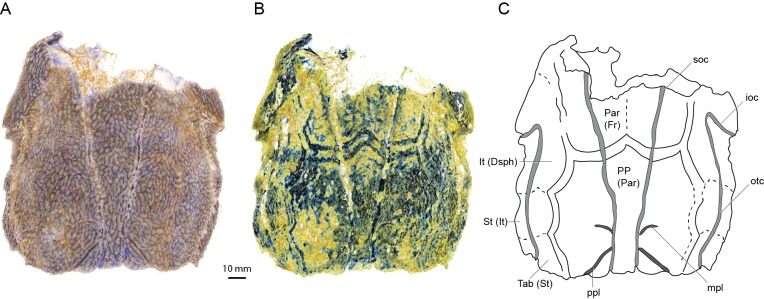
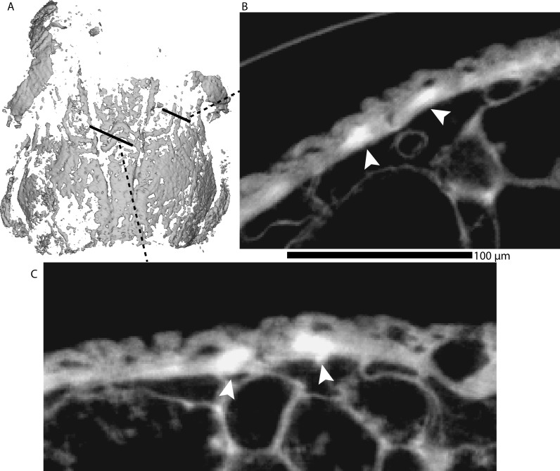
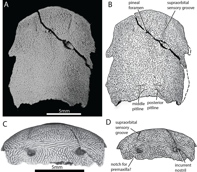
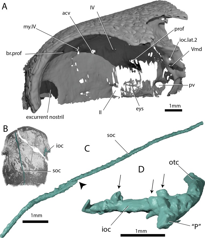
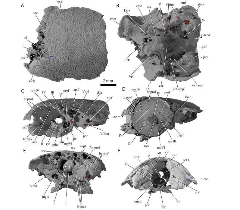
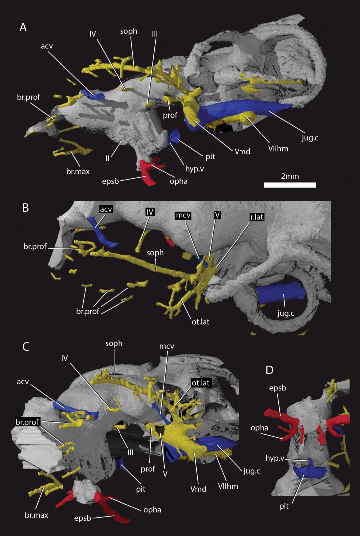
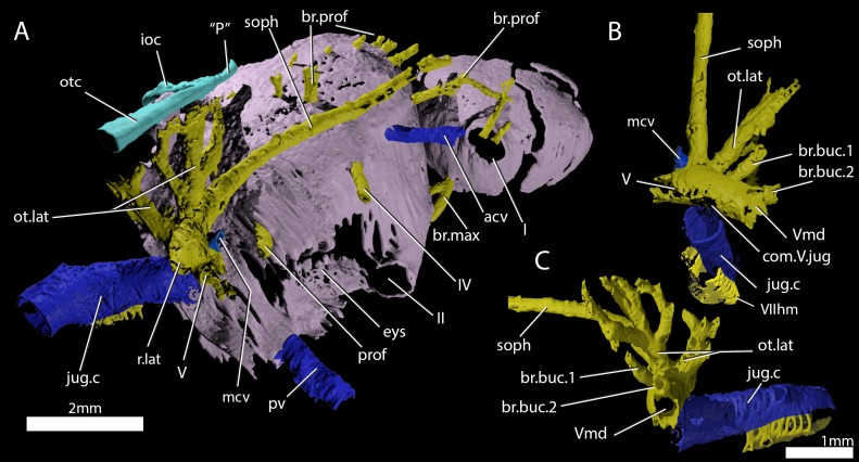
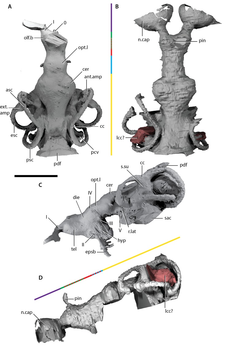
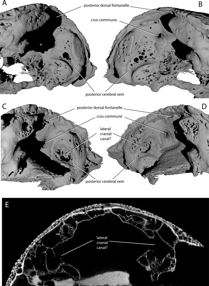
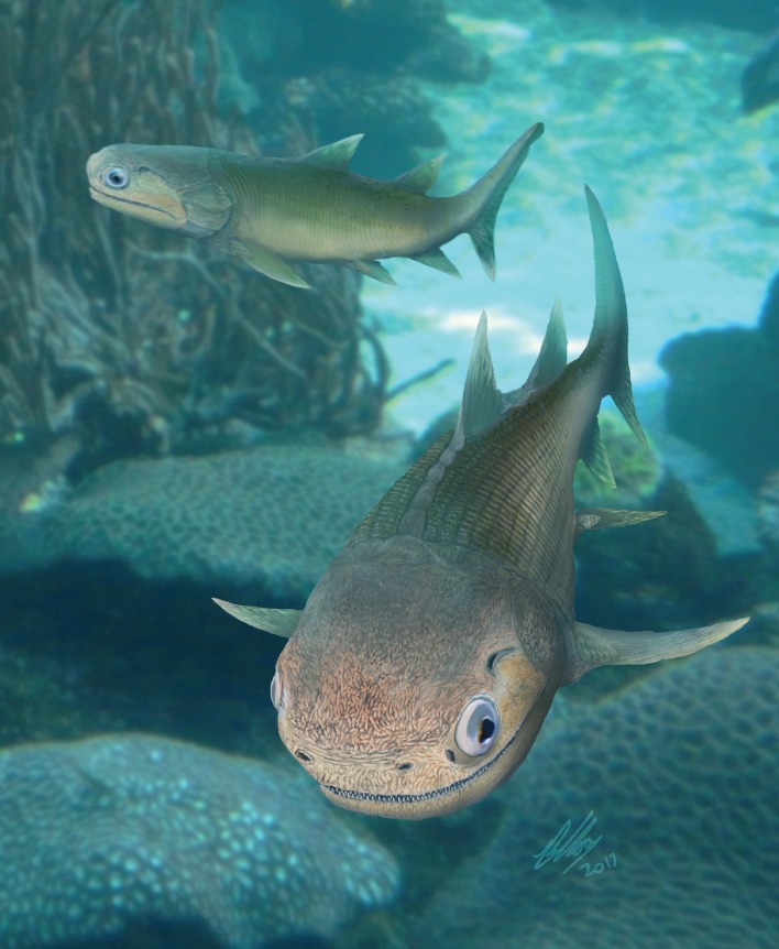
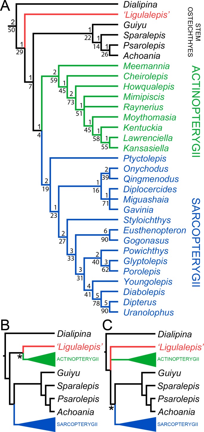
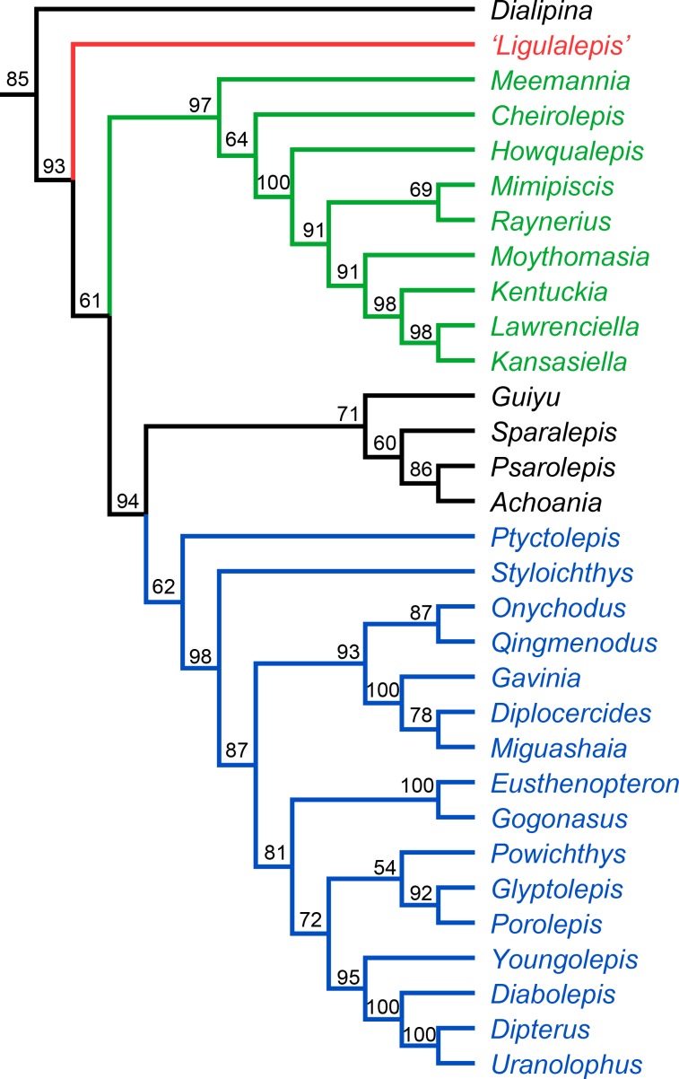
The skull of 'Ligulalepis' from the Early Devonian of Australia (AM-F101607) has significantly expanded our knowledge of early osteichthyan anatomy, but its phylogenetic position has remained uncertain. We herein describe a second skull of 'Ligulalepis' and present micro-CT data on both specimens to reveal novel anatomical features, including cranial endocasts. Several features previously considered to link 'Ligulalepis' with actinopterygians are now considered generalized osteichthyan characters or of uncertain polarity. The presence of a lateral cranial canal is shown to be variable in its development between specimens. Other notable new features include the presence of a pineal foramen, the some detail of skull roof sutures, the shape of the nasal capsules, a placoderm-like hypophysial vein, and a chondrichthyan-like labyrinth system. New phylogenetic analyses place 'Ligulalepis' as a stem osteichthyan, specifically as the sister taxon to 'psarolepids' plus crown osteichthyans. The precise position of 'psarolepids' differs between parsimony and Bayesian analyses.

Keywords: Braincase; Computed Tomography; Devonian; Ligulalepis; Osteichthyes; Phylogeny; genetics; genomics.


Conflict of interest statement

AC No competing interests declared

Figures

Figure 1.
Figure 1.. Skull roof of ‘Ligulalepis’ AM-F101607 in dorsal view.
Artificial colouration added in Drishti to highlight (A) sensory canals; and (B) bone sutures. (C) Interpretive diagram showing skull roof pattern; patterns of sensory canals inferred from both specimens. Bone names use sarcopterygian conventions, with actinopterygian conventions in brackets.
Figure 1—figure supplement 1
Figure 1—figure supplement 1. Bands of high-density bone in the basal layer of the skull roof dermal bone are assumed to follow sutures in AM-F101607.
(A) Mimics rendering of a mask with a high minimum threshold, thereby showing only the highest density bone, including the double bands that we presume occurred either side of sutures. (B-C) Cross sections, with these high-density bands marked with arrows. Note that they appear to be confined to the basal part of the bone.
Figure 2.
Figure 2.. Skull of ‘Ligulalepis’ ANU V3628.
(A) Dorsal view, photograph of specimen whitened with ammonium chloride. (B) Line drawing of A. (C) Anterior view, imaged using Drishti to reveal parts embedded in resin. (D) Line drawing of C.
Figure 3.
Figure 3.. Skull and sensory canals of ‘Ligulalepis’ ANU V3628.
(A) Segmented model of dermal and perichondral bone of the left orbit, showing the posterior nostril within the orbit and endochondral bone in the eyestalk. (B) Position of supraorbital canal (soc) and infraorbital canal (ioc) on the skull. (C) Left supraorbital canal in left lateral view. Arrow indicates point where anterior and posterior canal sections overlap. (D) Right infraorbital and postotic canal in anterior view. Arrows indicate tubules that connect the canal to the surface.
Figure 4.
Figure 4.. Cranium of ‘Ligulalepis’ AM-F101607.
(A) dorsal; (B) ventral; (C) left lateral; (D) left anterolateral showing details of orbit; (E) anterior; and (F) posterior view.
Figure 5.
Figure 5.. Cranial nerves and vessels of ‘Ligulalepis’ AM-F101607.
(A) left lateral; (B) dorsal; (C) left anterolateral and (D) ventral view of anterior section only. Cranial endocast in grey, nerves in yellow, veins in blue and arteries in red.
Figure 6.
Figure 6.. Cranial nerves and vessels of ‘Ligulalepis’ ANU V3628.
(A) ANU V3628, segmentation of the interior of the left orbital region, viewed from a postero-dorsal-medial viewpoint. The cranial endocast is not shown. Perichondral bone lining the orbit and nasal capsules is in lilac. Nerves are yellow, veins blue and sensory canals are in turquoise. The trigeminal, lateralis and facial nerves and their branches and the jugular vein, viewed from an anterior-ventral (B) and left lateral (C) viewpoints.
Figure 7.
Figure 7.. Endocast of ‘Ligulalepis’.
(A) AM-F101607, dorsal view. (B) ANU V3628, dorsal view. (C) AM-F101607 lateral view. (D) ANU V3628 lateral view with possible lateral cranial canal in red. Major brain regions indicated by coloured bars: nasal capsules (purple), telencephalon (green), diencephalon (red), mesencephalon (blue), metencephalon and myelencephalon (yellow).
Figure 8.
Figure 8.. Variability in the development of a lateral cranial canal in ‘Ligulalepis’.
(A–B) Ventrolateral view of AM-F101607, showing internal view of the otic region on the right hand side (A) and the left hand side (B). (C–D) Ventrolateral view of ANU V3628, showing internal view of the otic region on the right hand side (C) and the left hand side (D). (E) CT scan cross-section of ANU V3628 showing diverticula that may represent lateral cranial canals.
Figure 9.
Figure 9.. Life reconstruction of ‘Ligulalepis’.
Based on the skull roof morphology of AM-F101607 and ANU V3628, other features remain hypothetical.
Figure 10.
Figure 10.. Results of parsimony phylogenetic analysis.
(A) Strict consensus tree. Numbers above nodes refer to bremer support, numbers below nodes represent bootstrap support. (B) Strict consensus tree after enforcing ‘Ligulalepis’ as a stem actinopterygian. (C) Strict consensus tree after constraining ‘psarolepids’ (Guiyu, Sparalepis, Psarolepis, Achoania) as stem sarcopterygians. Asterisks indicate constrained nodes.
Figure 11.
Figure 11.. Results of Bayesian phylogenetic analysis.
Maximum clade credibility tree. Numbers represent posterior probabilities, displayed as percentages for presentation purposes.

References

    1. Basden AM, Young GC, Coates MI, Ritchie A. The most primitive osteichthyan braincase? Nature. 2000;403:185-8. doi: 10.1038/35003183. - DOI - PubMed
    1. Basden AM, Young GC. A primitive actinopterygian neurocranium from the Early Devonian of southeastern Australia. Journal of Vertebrate Paleontology. 2001;21:754–766. doi: 10.1671/0272-4634(2001)021[0754:APANFT]2.0.CO;2. - DOI
    1. Bjerring HC. Morphological observations on the exoskeletal skull roof of an osteolepiform from the carboniferous of Scotland. Acta Zoologica. 1972;53:73–92. doi: 10.1111/j.1463-6395.1972.tb00575.x. - DOI
    1. Brazeau MD, de Winter V. The hyoid arch and braincase anatomy of Acanthodes support chondrichthyan affinity of 'Acanthodians'. Proceedings. Biological Sciences. 2015;282:20152210. doi: 10.1098/rspb.2015.2210. - DOI - PMC - PubMed
    1. Brazeau MD. The braincase and jaws of a devonian 'acanthodian' and modern gnathostome origins. Nature. 2009;457:305–308. doi: 10.1038/nature07436. - DOI - PubMed

Publication types

MeSH terms