Clinical Trial
. 2018 Jul 5;26(7):1855-1866.
doi: 10.1016/j.ymthe.2018.05.003. Epub 2018 May 8.

Dominant-Negative TGF-β Receptor Enhances PSMA-Targeted Human CAR T Cell Proliferation And Augments Prostate Cancer Eradication

Affiliations
Clinical Trial

Dominant-Negative TGF-β Receptor Enhances PSMA-Targeted Human CAR T Cell Proliferation And Augments Prostate Cancer Eradication

Christopher C Kloss et al. Mol Ther. .

Abstract

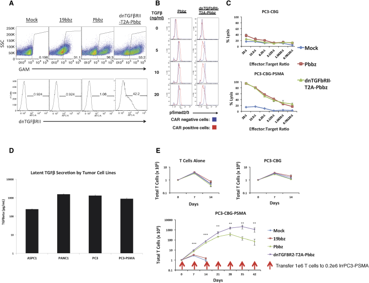
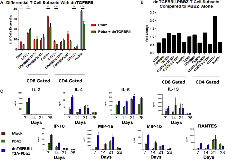
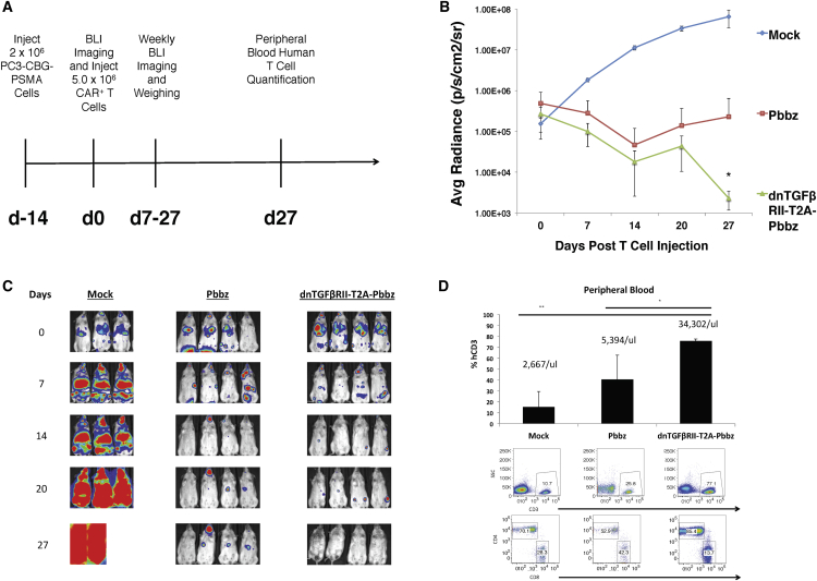
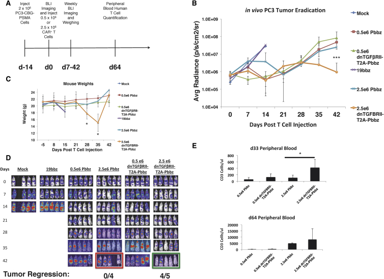
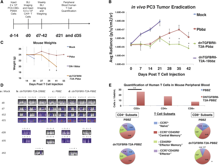
Cancer has an impressive ability to evolve multiple processes to evade therapies. While immunotherapies and vaccines have shown great promise, particularly in certain solid tumors such as prostate cancer, they have been met with resistance from tumors that use a multitude of mechanisms of immunosuppression to limit effectiveness. Prostate cancer, in particular, secretes transforming growth factor β (TGF-β) as a means to inhibit immunity while allowing for cancer progression. Blocking TGF-β signaling in T cells increases their ability to infiltrate, proliferate, and mediate antitumor responses in prostate cancer models. We tested whether the potency of chimeric antigen receptor (CAR) T cells directed to prostate-specific membrane antigen (PSMA) could be enhanced by the co-expression of a dominant-negative TGF-βRII (dnTGF-βRII). Upon expression of the dominant-negative TGF-βRII in CAR T cells, we observed increased proliferation of these lymphocytes, enhanced cytokine secretion, resistance to exhaustion, long-term in vivo persistence, and the induction of tumor eradication in aggressive human prostate cancer mouse models. Based on our observations, we initiated a phase I clinical trial to assess these CAR T cells as a novel approach for patients with relapsed and refractory metastatic prostate cancer (ClinicalTrials.gov: NCT03089203).

Keywords: TGF-β; chimeric antigen receptor; prostate cancer.


Figures

Figure 1
Figure 1
In Vitro Functionality of Pbbz CAR and dnTGF-βRII-T2A-Pbbz CAR (A) Lentiviral transduction allows for efficient expression of 19bbz, Pbbz alone, or dnTGF-βRII-T2A-Pbbz in primary human T cells. (B) Expression of dnTGF-βRII prevents TGF-β signal induction through Smad2/3. (C) T cells expressing Pbbz specifically lyse PSMA+ PC3 cells in 24-hr luciferase-based lysis assays. (D) ELISA determination of the secretion of the latent form of TGF-β by tumor lines. (E) Expression of dnTGF-βRII-T2A-Pbbz enhances antigen-specific proliferation of CAR T cells upon co-culture with PSMA+ PC3 cells (y axis log10 scale). The error bars represent ± SD. **p < 0.01, and ***p < 0.001.
Figure 2
Figure 2
Flow Cytometric and Multiplex Cytokine Profiling of CAR T Cell Subsets from PC3-PSMA Co-culture (A and B) T cells were analyzed with flow cytometry at day 21 post co-culture of Pbbz or dnTGF-βRII-T2A-Pbbz CAR T cells. (A) shows the differential percentage of various T cell subsets, which is further represented as fold change of T cell subsets found in the dnTGF-βRII-T2A-Pbbz versus Pbbz CAR T cells alone (B). (C) Luminex 30-Plex cytokine analysis was performed using T cell supernatants isolated at days 7, 14, 21, and 28 from Pbbz or dnTGF-βRII-T2A-Pbbz CAR T cells, as shown in Figure 1E. Pbbz-alone T cells, green bars; dnTGF-βRII-T2A-Pbbz T cells, blue bars. The error bars represent ± SD. *p < 0.05, **p < 0.01, and ***p < 0.001.
Figure 3
Figure 3
Protein-Protein Interactions in PSMA dnTGF-βRII-T2A-Pbbz CAR T Cells A protein-protein interaction network was constructed using STRING, version 10.5, based on differentially expressed genes on days 14 (A) and 21 (B) of culture. Network nodes represent proteins and the edges represent protein-protein interactions (physical or functional). In (A), the network has 43 nodes and 91 edges, with a p value of 1.3 × 10−14 compared to random interactions. In (B), the network has 69 nodes and 182 edges, with a p value of <1.0 × 10−16 compared to random interactions. The error bars represent ± SD.
Figure 4
Figure 4
dnTGF-βRII-T2A-Pbbz CAR T Cells Have Augmented Proliferation and Eradicate Metastatic Prostate Cancer In Vivo Systemic PC3-PSMA tumors were established by tail vein infusions of 2 × 106 tumor cells with the timeline described in (A) treated by infusing 5.0 × 106 CAR T cells or PBS (mock) per mouse 2 weeks later. Mice were assessed with bioluminescent imaging (BLI) weekly to assess tumor burden (B). Images of this BLI assessment are shown to demonstrate location and burden of tumors (C). Mice were bled at the end of the experiment at day 27 to assess the phenotype and the amounts of human CD3 T cells within the blood of these mice (D) (n = 4 mice/group). The error bars represent ± SD. *p < 0.05 and **p < 0.01, determined using a Student’s two-tailed t test.
Figure 5
Figure 5
Dose-Dependent Augmentation of CAR T Cell Proliferation and Antitumor Efficacy of dnTGF-βRII CAR T Cells Compared to PSMA CAR T Cells In Vivo Systemic PC3-PSMA tumors were established from intravenous (i.v.) infusions with the timeline described in (A), with varied doses of 0.5 × 106 and 2.5 × 106 CAR T cells per mouse infused 2 weeks later. Mice were assessed with BLI weekly to assess tumor burden (B). Body weight of the groups of mice is shown in (C). Images of this BLI assessment are shown to demonstrate location and burden of tumors (D). Mice were bled at days 33 and 64 to assess the amounts of human CD3 T cells within the blood of these mice (E) (n = 5 mice/group). The error bars represent ± SD. *p < 0.05 and ***p < 0.001.
Figure 6
Figure 6
Enhanced Survival and Central Memory PSMA dnTGF-βRII-T2A-Pbbz CAR T Cells In Vivo in Mice with Metastatic Prostate Cancer Systemic PC3-PSMA tumors were established by tail vein infusion with the timeline described in (A) with a dose of 5.0 × 106 CAR T cells per mouse infused 2 weeks later. Mice were assessed with BLI weekly to assess tumor burden (B). Body weight is shown in (C) and images of this BLI assessment are shown to demonstrate location and burden of tumors (D). (E) Mice were bled at the end of the experiment at day 35 to assess the amounts of human CD3+ T cells within the blood of these mice. In depth analysis to assess T cell subsets was performed based on memory markers (CCR7, CD45R0, and CD127) (n = 10 mice/group). The error bars represent ± SD. *p < 0.05 and **p < 0.01.

References

    1. Litwin M.S., Tan H.J. The Diagnosis and Treatment of Prostate Cancer: A Review. JAMA. 2017;317:2532–2542. - PubMed
    1. Kwon E.D., Drake C.G., Scher H.I., Fizazi K., Bossi A., van den Eertwegh A.J., Krainer M., Houede N., Santos R., Mahammedi H., CA184-043 Investigators Ipilimumab versus placebo after radiotherapy in patients with metastatic castration-resistant prostate cancer that had progressed after docetaxel chemotherapy (CA184-043): a multicentre, randomised, double-blind, phase 3 trial. Lancet Oncol. 2014;15:700–712. - PMC - PubMed
    1. Chen D.S., Mellman I. Elements of cancer immunity and the cancer-immune set point. Nature. 2017;541:321–330. - PubMed
    1. Huang A.C., Postow M.A., Orlowski R.J., Mick R., Bengsch B., Manne S., Xu W., Harmon S., Giles J.R., Wenz B. T-cell invigoration to tumour burden ratio associated with anti-PD-1 response. Nature. 2017;545:60–65. - PMC - PubMed
    1. Verdegaal E.M., de Miranda N.F., Visser M., Harryvan T., van Buuren M.M., Andersen R.S., Hadrup S.R., van der Minne C.E., Schotte R., Spits H. Neoantigen landscape dynamics during human melanoma-T cell interactions. Nature. 2016;536:91–95. - PubMed

Publication types

MeSH terms

Substances

Associated data