. 2018 Jul;2(7):1155-1160.
doi: 10.1038/s41559-018-0569-4. Epub 2018 May 28.

An epigenetic mechanism for cavefish eye degeneration

Affiliations

An epigenetic mechanism for cavefish eye degeneration

Aniket V Gore et al. Nat Ecol Evol. 2018 Jul.

Abstract

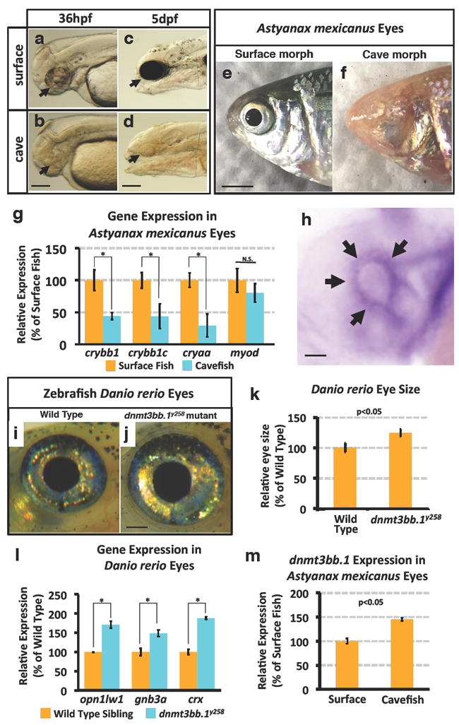
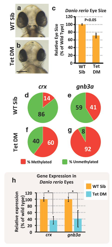
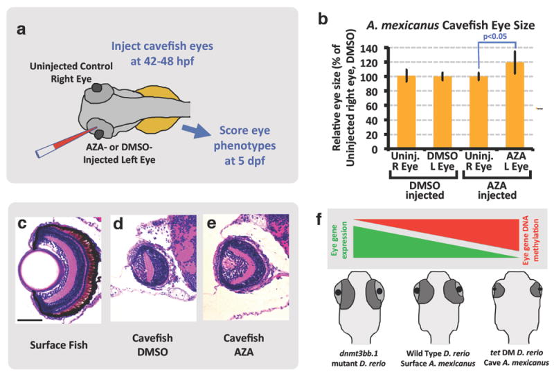
Coding and non-coding mutations in DNA contribute significantly to phenotypic variability during evolution. However, less is known about the role of epigenetics in this process. Although previous studies have identified eye development genes associated with the loss-of-eyes phenotype in the Pachón blind cave morph of the Mexican tetra Astyanax mexicanus, no inactivating mutations have been found in any of these genes. Here, we show that excess DNA methylation-based epigenetic silencing promotes eye degeneration in blind cave A. mexicanus. By performing parallel analyses in A. mexicanus cave and surface morphs, and in the zebrafish Danio rerio, we have discovered that DNA methylation mediates eye-specific gene repression and globally regulates early eye development. The most significantly hypermethylated and downregulated genes in the cave morph are also linked to human eye disorders, suggesting that the function of these genes is conserved across vertebrates. Our results show that changes in DNA methylation-based gene repression can serve as an important molecular mechanism generating phenotypic diversity during development and evolution.


Conflict of interest statement

The authors declare no competing financial interests.

Figures

Figure 1
Figure 1. Eye phenotypes in Astyanax mexicanus surface and cave fish, and in zebrafish Danio rerio wild type and dnmt3bb.1y258 mutant animals
a-d, Transmitted light photomicrographs of the heads of 36 hpf (a,b) and 5 dpf (c,d) surface (a,c) and cavefish (b,d) morphs of A. mexicanus. Black arrow mark the developing eyes in CF and SF. e,f, Photographic images of the heads of adult surface (e, with eyes) and cave (f, eyeless) morphs of A. mexicanus. g, Quantitative RT-PCR analysis of the percent relative expression of crybb1, crybb1c, cryaa, and myod in isolated heads from 54 hpf surface (orange columns) and cave (blue columns) morphs of A. mexicanus, normalized to surface fish levels. Histograms show mean, error bars represent s.e.m of three biological replicates; two-tailed t-test, *p<0.005. h, Whole mount in situ hybridization of a 36 hpf zebrafish eye probed for dnmt3bb.1, showing expression in the ciliary marginal zone (CMZ, arrows). i,j, Photographic images of the eyes of adult wild type sibling (i) and dnmt3bb.1y258 mutant (j) zebrafish. k, Quantitation of eye size in three week old wild type sibling and dnmt3bb.1y258 mutant animals. Histograms show mean, error bars represent s.d. in the relative eye size (n =18) two-tailed t-test, *p<0.05. l, Quantitative RT-PCR analysis of the percent relative expression of opn1lw1, gnb3a, and crx in adult wild type sibling (orange columns) and dnmt3bb.1y258 mutant (blue columns) zebrafish eyes, normalized to wild type sibling levels. Histograms show mean, error bars represent s.e.m of three biological replicates; two-tailed t-test, *p<0.005. m, Quantitative RT-PCR analysis of the percent relative expression of dnmt3bb.1 in surface and cave morphs of A. mexicanus, normalized to surface fish levels. All images are lateral views, rostral to the left. Histograms show mean, error bars represent s.e.m of three biological replicates, p<0.05 using t-test. Scale bars 100μM in b for a,b, 250μM in d for c,d, 3mm in e for e,h, 50μM for h and 1mm in j for i,j.
Figure 2
Figure 2. Gene expression changes in cave versus surface fish morphs of Astyanax mexicanus
a, Diagram showing the workflow for obtaining larval eyes from A. mexicanus and for co-isolating eye DNA and RNA for whole genome assessment of DNA methylation and gene expression, respectively. b, Percent relative expression of dnmt3bb.1 in the eyes of A. mexicanus cave vs. surface fish morphs by comparison of their respective RNAseq data sets (2-replicates), normalized to surface fish levels, FPKM p<0.05. c, Log2 fold differential expression (p<0.05) of genes in A. mexicanus cave vs. surface fish morph RNAseq data sets, with down-regulated (red) or up-regulated (blue) expression of selected genes in CF noted. d, Listing of the Gene Ontology (GO) terms showing the greatest down-regulation in cave fish compared to surface fish. e-p, Assessment of opn1lw1(e-h), gnb3a (i-l), and crx (m-p) promoter DNA methylation (e,g,i,k,m,o) and gene expression (f,h,j,l,n,p) in 54 hpf surface (e,f,i,j,m,n) or cave (g,h,k,l,o,p) morphs of A. mexicanus. Panels show pie chart graphical representation of the percentage methylation of the promoter CpG (e,g,i,k,m,o), and representative whole mount in situ hybridization of the larval heads (f,h,j,l,n,p) using the probes noted (ventral views, rostral up, minimum 20-25 embryos were analyzed by in situ hybridization). Scale bar 100μM in p for f,h,j,l,n, and p.
Figure 3
Figure 3. Eye phenotype and associated gene expression changes in wild type and DNA methylation-deficient Danio rerio
a-b, Transmitted light photomicrographs of the heads of 48 hpf wild type sibling (a) and tet2-/-, tet3-/- double mutant (b) embryos. c, Quantitation of eye size in 48 hpf wild type sibling and tet2-/-, tet3-/- double mutant embryos. Histograms show mean, error bars represent s.d. in the relative eye size (n =10), two-tailed t-test, *p<0.05. d-g, Assessment of crx (d,f) and gnb3a (e,g), promoter DNA methylation in isolated eyes from 48 hpf wild type sibling (d,e) and tet2-/-, tet3-/- double mutant (f,g) animals. h, Quantitative RT-PCR analysis of the percent relative expression of crx and gnb3a in isolated eyes from 48 hpf wild type sibling (orange columns) and tet2-/-, tet3-/- double mutant (blue columns) animals. Histograms show mean, error bars represent s.e.m of three biological replicates; two-tailed t-test, *p<0.005. Scale bar 100μM in b for a and b.
Figure 4
Figure 4. Partial rescue of cavefish eyes by AZA-mediated inhibition of eye DNA methylation
a, Schematic diagram showing the experimental procedure for injection of DMSO or AZA into 42-48 hpf cavefish embryo eyes. b, Quantitation of eye size in 5 dpf cavefish embryos injected in the left eye with DMSO or AZA. Histograms show mean, error bars represent s.d. in the relative eye size (18 DMSO and 12 Aza injected embryos) two-tailed t-test, *p<0.05. c-e, Histological analysis of H&E stained 5 dpf surface fish eye (c), DMSO injected cavefish eye (d) and AZA injected cavefish eye (e) (four to eight embryos per condition were sectioned and analyzed by H&E staining). f, Model depicting the role of DNA methylation in teleost eye development and degeneration. Hypermethylation and down-regulation of eye gene expression in cavefish and zebrafish tet2/3 double mutants leads to eye degeneration. Scale bar 100μM in a for a-c.

Comment in

References

    1. Gross JB, Meyer B, Perkins M. The rise of Astyanax cavefish. Dev Dyn. 2015 doi: 10.1002/dvdy.24253. - DOI - PMC - PubMed
    1. Moran D, Softley R, Warrant EJ. The energetic cost of vision and the evolution of eyeless Mexican cavefish. Sci Adv. 2015;1:e1500363. doi: 10.1126/sciadv.1500363. - DOI - PMC - PubMed
    1. Hinaux H, et al. Lens defects in Astyanax mexicanus Cavefish: evolution of crystallins and a role for alphaA-crystallin. Dev Neurobiol. 2015;75:505–521. doi: 10.1002/dneu.22239. - DOI - PubMed
    1. Casane D, Retaux S. Evolutionary Genetics of the Cavefish Astyanax mexicanus. Adv Genet. 2016;95:117–159. doi: 10.1016/bs.adgen.2016.03.001. - DOI - PubMed
    1. Ma L, Parkhurst A, Jeffery WR. The role of a lens survival pathway including sox2 and alphaA-crystallin in the evolution of cavefish eye degeneration. Evodevo. 2014;5:28. doi: 10.1186/2041-9139-5-28. - DOI - PMC - PubMed

Publication types