. 2019 Feb;56(2):954-962.
doi: 10.1007/s12035-018-1137-2. Epub 2018 May 28.

Multimodal Molecular Imaging Demonstrates Myeloperoxidase Regulation of Matrix Metalloproteinase Activity in Neuroinflammation

Affiliations

Multimodal Molecular Imaging Demonstrates Myeloperoxidase Regulation of Matrix Metalloproteinase Activity in Neuroinflammation

Yinian Zhang et al. Mol Neurobiol. 2019 Feb.

Abstract

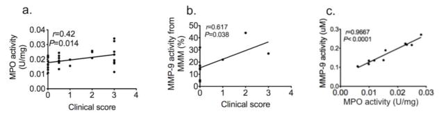
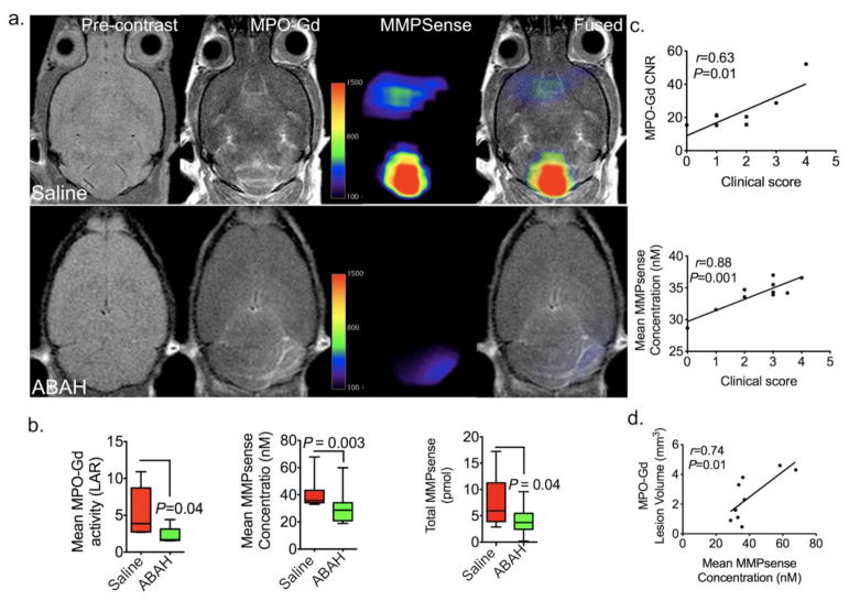
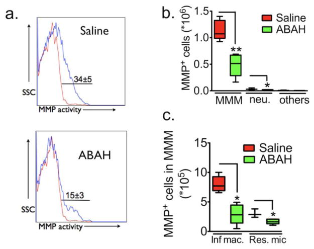
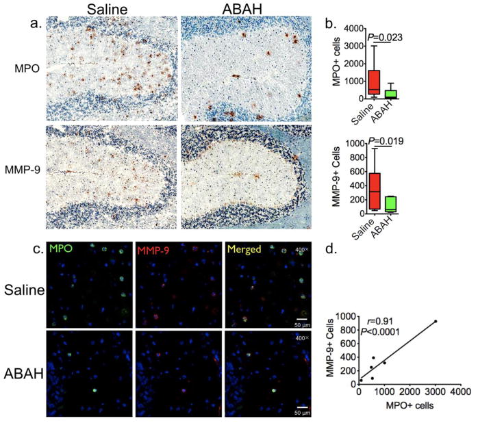
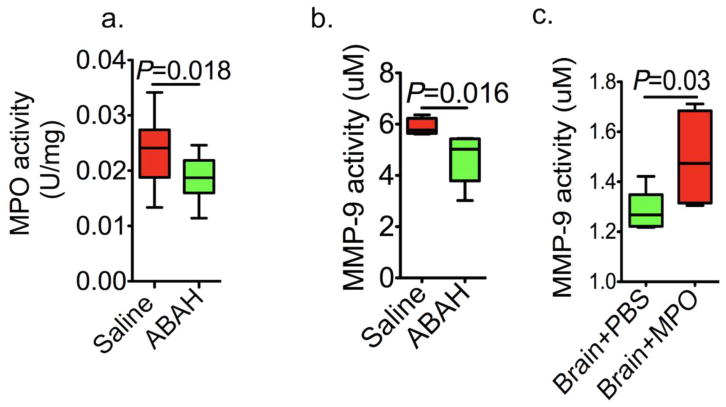
Myeloperoxidase (MPO) has paradoxically been found to be able to both activate matrix metalloproteinases (MMPs) as well as inhibit MMPs. However, these regulatory effects have not yet been observed in vivo, and it is unclear which pathway is relevant in vivo. We aim to track MPO regulation of MMP activity in living animals in neuroinflammation. Mice induced with experimental autoimmune encephalomyelitis (EAE), a mouse model of neuroinflammation and multiple sclerosis, were treated with either the MPO-specific inhibitor 4-aminobenzoic acid hydrazide or saline as control. Mice underwent concurrent magnetic resonance imaging (MRI) with the MPO-specific molecular imaging agent MPO-Gd and fluorescence molecular tomography (FMT) with the MMP-targeting agent MMPsense on day 12 after induction. Biochemical and histopathological correlations were performed. Utilizing concurrent MRI and FMT imaging, we found reduced MMP activity in the brain with MPO inhibition, demonstrating MPO activity positively regulates MMP activity in vivo. In vivo MMPSense activation and MMP-9 activity correlated with MPO-Gd+ lesion volume and disease severity. This was corroborated by in vitro assays and histopathological analyses that showed MMP activity and MMP-9+ cells correlated with MPO activity and MPO+ cells. In conclusion, multimodal molecular imaging demonstrates for the first time MPO regulation of MMP activity in living animals. This approach could serve as a model to study the interactions of other biologically interesting molecules in living organisms.

Keywords: Experimental autoimmune encephalomyelitis; Fluorescence molecular tomography; Magnetic resonance imaging; Matrix metalloproteinases; Myeloperoxidase; Neuroinflammation.


Figures

Fig. 1
Fig. 1. Myeloid cells are the main source of matrix metalloproteinase (MMP) activity in EAE
a Cell surface markers used to subdivide cells into various subsets of leukocytes in the brain of saline-treated control EAE mice 12 days after immunization. Numbers denote percent MMP-positive cells in the saline (34%± 5%) group (Red line: percent MMP-positive cells in sham mice; Blue line: percent MMP-positive cells in EAE mice). b Flow cytometric analysis revealed MMP activity mainly derived from microglia/monocytes/macrophages (MMM) in brains of EAE mice (53%). c MMP mean fluorescence intensity (MFI) per cell for different cell types in brain, as identified by flow cytometry. MMM cells contribute to most of the MMP activity (96%). d The percentage of MMP positive cells in inflammatory macrophages and resident microglia, 73% and 27% respectively. SSC = side scatter; neu. = neutrophils; MMM =microglia/monocytes/macrophages; MFI = mean fluorescence intensity; Inf. mac = inflammatory macrophages; Res. Mic = resident microglia
Fig. 2
Fig. 2. MPO and MMP-9 activity correlate with disease severity and that the two are strongly correlated
a Peroxidase activity correlated with clinical score (n = 30). b MMP activity from microglia/monocytes/macrophages in vitro correlated with clinical score (n = 8). c MMP-9 activity positively correlated to peroxidase activity (n=10, data were obtained by using Pearson correlation analysis, r indicated Pearson correlation coefficient). MPO=myeloperoxidase, MMP=matrix metalloproteinase.
Fig. 3
Fig. 3
In vivo mapping and regulation of MPO on MMP activity using MRI/FMT multimodal imaging. a Representative MRI/FMT fusion images show that in vivo MPO and MMP activity both increased in EAE mice and both MPO and MMP activity were partially blocked with the MPO inhibitor 4-aminobenzoic acid hydrazide (ABAH). b MPO-Gd activation from MRI decreased with MPO inhibition, n=10 per group, Mann-Whitney test, left panel). Mean fluorescence of MMPsense signal between saline-treated and ABAH-treated EAE mice (n=10 per group, Mann-Whitney test, middle panel). Total activated MMPsense probe significantly decreased with MPO inhibition in EAE mice (Mann-Whitney test, right panel). c Contrast-to-noise ratio (CNR) from MRI images correlated with clinical score (n=7) (upper panel). MMP activity from FMT images (mean MMPsense concentration) positively correlated with clinical score. (n=10, Pearson correlation analysis) (low panel). d MPO-Gd+ lesion volume positively correlated with MMPsense concentration (n=9, data were obtained by using Pearson correlation analysis, r indicated Pearson correlation coefficient). All data are means ± standard errors of measurement. CNR = contrast noise ratio, LAR = lesion activation ratio, MPO=myeloperoxidase, MMP=matrix metalloproteinase.
Fig. 4
Fig. 4. The effects of 4-aminobenzoic acid hydrazide (ABAH) on MMP+ cells
a Cell surface markers used to subdivide cells into various subsets of leukocytes in the brain of saline-treated control and ABAH-treated EAE mice 12 days after immunization. Numbers denote percent MMP-positive cells in saline (34%± 5%) versus ABAH group (15%±3%) (P=0.009, n=5 per group, two-tailed student t-test) (Red line: percent MMP-positive cells in sham mice; Blue line: percent MMP-positive cells in EAE mice). b Total MMP positive cell counts in brain. There are significant differences between saline and ABAH group in cell number of microglia/monocytes/macrophages and neutrophils (Mann-Whitney test, **P = 0.007 and **P = 0.04). c Contribution of cell types to MMP activity in EAE based on results in a and b. MMP=matrix metalloproteinase.
Fig. 5
Fig. 5. MPO and MMP-9 positive cells are reduced with MPO inhibition
a Photomicrographs (immunohistochemistry, scale bar = 200 μm [original magnification, × 100]) from the cerebellum showed that MPO (upper panel) and MMP-9 (low panel) positive cells are decreased with MPO inhibition. b Quantification of (a) (n=5 per group, Mann-Whitney test). The average total number of positive cells from the entire brain on three different sections for each mouse was used for quantification. c Photomicrographs (immunofluorescence staining, scale bar = 500 μm [original magnification, × 400], cerebellum) showed most of MPO positive cells co-localized with MMP positive cells. d MPO-positive cells correlated with MMP positive cells (n=6, Pearson correlation analysis, r indicated Pearson correlation coefficient). MPO=myeloperoxidase, MMP=matrix metalloproteinase.
Fig. 6
Fig. 6. In vitro validation of MPO regulation of matrix metalloproteinases
a MPO activity decreased with MPO inhibition (P = 0.018, n=5 per group, Mann-Whitney test). b MMP-9 activity decreased with MPO inhibition in EAE (P = 0.016, n = 5 per group, Mann-Whitney test). c MMP-9 activity was significantly elevated with MPO (10ug/ml) stimulation in vivo (P = 0.03, n = 5 per group, Mann-Whitney test). All data are means ± standard errors of measurement. MPO=myeloperoxidase, MMP=matrix metalloproteinase, ABAH=4-aminobenzoic acid hydrazide.

References

    1. Hu J, Van den Steen PE, Sang QX, Opdenakker G. Matrix metalloproteinase inhibitors as therapy for inflammatory and vascular diseases. Nat Rev Drug Discov. 2007;6(6):480–498. doi: 10.1038/nrd2308. - DOI - PubMed
    1. Maier CM, Hsieh L, Yu F, Bracci P, Chan PH. Matrix metalloproteinase-9 and myeloperoxidase expression: quantitative analysis by antigen immunohistochemistry in a model of transient focal cerebral ischemia. Stroke. 2004;35(5):1169–1174. doi: 10.1161/01.STR.0000125861.55804.f2. - DOI - PubMed
    1. Parks WC, Wilson CL, Lopez-Boado YS. Matrix metalloproteinases as modulators of inflammation and innate immunity. Nature reviews. 2004;4(8):617–629. - PubMed
    1. Gerwien H, Hermann S, Zhang X, Korpos E, Song J, Kopka K, Faust A, Wenning C, Gross CC, Honold L, Melzer N, Opdenakker G, Wiendl H, Schafers M, Sorokin L. Imaging matrix metalloproteinase activity in multiple sclerosis as a specific marker of leukocyte penetration of the blood-brain barrier. Sci Transl Med. 2016;8(364):364ra152. doi: 10.1126/scitranslmed.aaf8020. - DOI - PubMed
    1. Sorokin L. The impact of the extracellular matrix on inflammation. Nature reviews. 2010;10(10):712–723. - PubMed

MeSH terms

Substances

LinkOut - more resources