. 2018 Jun 7;10(22):10596-10608.
doi: 10.1039/c7nr09606b.

111In-labelled polymeric nanoparticles incorporating a ruthenium-based radiosensitizer for EGFR-targeted combination therapy in oesophageal cancer cells

Affiliations

111In-labelled polymeric nanoparticles incorporating a ruthenium-based radiosensitizer for EGFR-targeted combination therapy in oesophageal cancer cells

Martin R Gill et al. Nanoscale. .

Abstract

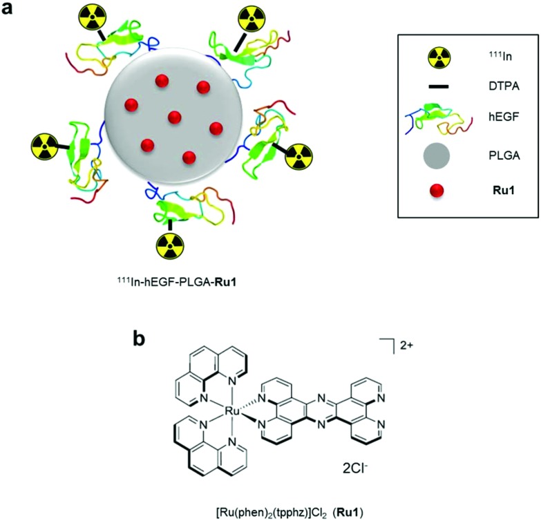
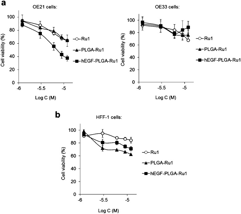
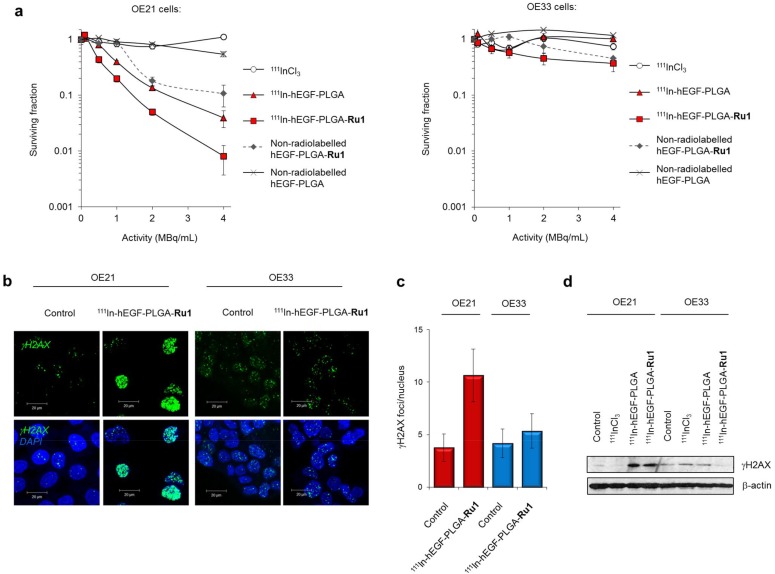
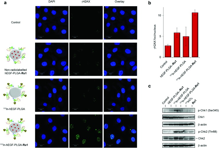
Radiolabelled, drug-loaded nanoparticles may combine the theranostic properties of radionuclides, the controlled release of chemotherapy and cancer cell targeting. Here, we report the preparation of poly(lactic-co-glycolic acid) (PLGA) nanoparticles surface conjugated to DTPA-hEGF (DTPA = diethylenetriaminepentaacetic acid, hEGF = human epidermal growth factor) and encapsulating the ruthenium-based DNA replication inhibitor and radiosensitizer Ru(phen)2(tpphz)2+ (phen = 1,10-phenanthroline, tpphz = tetrapyridophenazine) Ru1. The functionalized PLGA surface incorporates the metal ion chelator DTPA for radiolabelling and the targeting ligand for EGF receptor (EGFR). Nanoparticles radiolabelled with 111In are taken up preferentially by EGFR-overexpressing oesophageal cancer cells, where they exhibit radiotoxicity through the generation of cellular DNA damage. Moreover, nanoparticle co-delivery of Ru1 alongside 111In results in decreased cell survival compared to single-agent formulations; an effect that occurs through DNA damage enhancement and an additive relationship between 111In and Ru1. Substantially decreased uptake and radiotoxicity of nanoparticles towards normal human fibroblasts and oesophageal cancer cells with normal EGFR levels is observed. This work demonstrates nanoparticle co-delivery of a therapeutic radionuclide plus a ruthenium-based radiosensitizer can achieve combinational and targeted therapeutic effects in cancer cells that overexpress EGFR.


Figures

Scheme 1
Scheme 1. a) Radiolabelled nanoparticles employed in this study. The ruthenium(ii) metallo-intercalator and radiosensitizer, Ru1, is encapsulated within a PLGA core and nanoparticles are surface labelled with 111In-DTPA-hEGF. PLGA = poly(lactic-co-glycolic acid, Ru1 = Ru(phen)2(tpphz)2+ (phen = 1,10-phenanthroline, tpphz = tetrapyrido[3,2-a:2′,3′-c:3′′,2′′-h:2′′′,3′′′-j]phenazine), hEGF = human epidermal growth factor, DTPA = diethylenetriaminepentaacetic acid. (b) Chemical structure of Ru1.
Fig. 1
Fig. 1. (a) TEM images of unloaded (left) and Ru1-containing (right) PLGA nanoparticles. For unloaded PLGA nanoparticles (left) uranyl acetate contrast stain was employed. For Ru1-loaded particles (right), no TEM contrast stain was used, thereby allowing direct visualisation of Ru1 contrast within the PLGA core. (b) Coomassie blue stained SDS-PAGE gel of free hEGF (1 μg, left lane) or PLGA nanoparticles (1 mg) added to hEGF in the presence (middle lane) or absence (right lane) of NHS/EDC crosslinking agents. Nanoparticles were separated from unreacted hEGF by centrifugation before loading. *hEGF-PLGA, #hEGF. (c) Release kinetics of hEGF-PLGA-Ru1 showing biphasic release profile of Ru1. (d) Representative instant thin layer chromatograms (iTLC) of purified 111In-hEGF-PLGA and 111In-hEGF-PLGA-Ru1 nanoparticles in citrate buffer using EDTA (0.5 M, pH = 7.6) as the mobile phase.
Fig. 2
Fig. 2. (a) EGFR levels in HFF-1 normal human fibroblasts and OE21, OE33 and FLO-1 oesophageal cancer cells, as determined by immunoblotting of whole-cell lysates with anti-EGFR antibodies (top) and quantified by densitometry (bottom). Data expressed as a ratio of EGFR to β-actin levels and are normalised to results for HFF-1 cells. Mean of two technical repeats ± S.D. (b) Uptake of 111In-labelled hEGF-PLGA nanoparticles by HFF-1, OE21, OE33 or FLO-1 cells, as assessed by internalised cellular radioactivity (2 h incubation). Data expressed as counts per minute (CPM) per μg cell protein. Mean of triplicates ± S.D. Results for each cell line treated with equivalent amounts of added radioactivity of 111InCl3 included for comparison (grey). (c) Western blot analysis of total EGFR and pEGFR (EGFR phosphorylated at Tyr1068) levels in OE21 cell lysates. Cells were serum-starved (24 h) before treatment with hEGF (50 ng mL–1), hEGF-PLGA (0.5 mg mL–1) or PLGA (0.5 mg mL–1) for 5 or 15 minutes. (d) Effect of EGFR blocking on 111In-hEGF-PLGA uptake in OE21 cells. Cells were pre-incubated with a concentration range of non-radiolabelled hEGF (1 h) before addition of 111In-hEGF-PLGA (0.5 MBq mL–1) plus non-radiolabelled hEGF (2 h co-incubation). The internalised radioactivity was measured and normalised to control (i.e. unblocked) conditions. Data are the mean of two independent experiments ± S.D., where each experiment was performed in triplicate.
Fig. 3
Fig. 3. (a) Sub-cellular radioactivity content of OE21 or OE33 cells treated with 111In-hEGF-PLGA (0.125–0.5 MBq mL–1, 2 h). Isolated cytosol (Cyt) and nuclear (Nuc) fractions were obtained. The amount of accumulated radioactivity was measured by gamma-counting and normalised to protein content of each fraction (experiment performed in triplicate ± S.D.). See ESI for verification of efficient sub-cellular fractionation and data expressed as % of total radioactivity added. (b) Sub-cellular ruthenium content of OE21 or OE33 cells treated with hEGF-PLGA-Ru1 (1 mg mL–1, 24 h), as determined by ICP-MS. Data for cells treated with equivalent concentration of free Ru1 (12 μM, 24 h) included for comparison. Data are normalised to protein concentration and are the mean of two independent experiments ± S.D. (c) Confocal microscopy (CLSM) of OE21 or OE33 cells treated with hEGF-PLGA-Ru1 (1 mg mL–1, 24 h) showing intracellular MLCT (metal to ligand charge-transfer) emission of Ru1. Live cell imaging (top row) or the same cells visualised immediately after 4% formaldehyde fixation (bottom row). Identical imaging parameters were used for all images shown. Arrows indicate nuclear MLCT emission.
Fig. 4
Fig. 4. (a) Impact of Ru1-containing hEGF-PLGA or PLGA nanoparticles on cell viability of OE21 (overexpressed EGFR) or OE33 (normal EGFR levels) oesophageal cancer cells. Cell viabilities determined by MTT assay (24 h incubation) and expressed as a function of Ru1 concentration. The equivalent concentration of free Ru1 is included for comparison. (b) Impact of Ru1-containing nanoparticles on cell viability of HFF-1 normal human fibroblasts. Data for (a,b) are the mean of two or three independent experiments ± S.D, where each experiment was performed in triplicate.
Fig. 5
Fig. 5. (a) Clonogenic survival assays of OE21 or OE33 cells exposed to 111In radiolabelled hEGF-PLGA nanoparticles with or without Ru1-loading (24 h incubation time). Non-radiolabelled nanoparticles (in equivalent concentrations; 0–1000 μg mL–1) and free 111InCl3 (in equivalent specific activity) are included as controls. Data points are the mean of triplicates ± S.D. (b) Representative CLSM images of OE21 or OE33 cells treated with 111In-hEGF-PLGA-Ru1 (2 MBq mL–1, 24 h) followed by immunofluorescence staining for γH2AX (green). DNA was counterstained with DAPI (blue). See ESI for full micrographs. (c) Quantification of γH2AX foci/nucleus (in plane of view) for cells treated as in (b). Data are the average of two independent experiments ± SD. A minimum of 100 nuclei were counted for each treatment group. (d) Immunoblotting of OE21 or OE33 whole-cell lysates for γH2AX expression after treatment with 111In-hEGF-PLGA or 111In-hEGF-PLGA-Ru1 (2 MBq mL–1, 24 h). β-Actin was used as a loading control. Cells treated with free 111InCl3 (specific activity equivalent) were included for comparison.
Fig. 6
Fig. 6. (a) CLSM images of OE21 cells treated with 111In-hEGF-PLGA or 111In-hEGF-PLGA-Ru1 (1 MBq mL–1, 24 h) followed by immunofluorescence staining for γH2AX (green). DNA was stained with DAPI (blue). Equivalent non-radiolabelled hEGF-PLGA-Ru1 treatment was included for comparison. (b) Quantification of γH2AX foci/nucleus (in plane of view) for cells treated as in (a). Data average of two independent repeats ± S.D. A minimum of 100 nuclei per condition were counted. (c) Immunoblotting of OE21 whole-cell extracts after 24 h treatment with non-radiolabelled hEGF-PLGA-Ru1, 111In-hEGF-PLGA or 111In-hEGF-PLGA-Ru1 (1 MBq mL–1) using anti-pChk2 (Thr68) or pChk1 (Ser345) antibodies, as indicated. Total Chk2 and Chk1 protein content is provided. β-Actin was used as a loading control. Cells treated with free 111InCl3 (specific activity equivalent) or free Ru1 (concentration equivalent) were included for comparison.

References

    1. Smyth E. C., Lagergren J., Fitzgerald R. C., Lordick F., Shah M. A., Lagergren P., Cunningham D. Nat. Rev. Dis. Primers. 2017;3:17048. - PMC - PubMed
    1. Wei Q., Chen L., Sheng L., Nordgren H., Wester K., Carlsson J. Int. J. Oncol. 2007;31:493. - PubMed
    1. Gibson M. K., Abraham S. C., Wu T. T., Burtness B., Heitmiller R. F., Heath E., Forastiere A. Clin. Cancer Res. 2003;9:6461. - PubMed
    1. Liang K., Ang K. K., Milas L., Hunter N., Fan Z. Int. J. Radiat. Oncol., Biol., Phys. 2003;57:246. - PubMed
    1. Wang V. E., Grandis J. R., Ko A. H. Clin. Cancer Res. 2016;22:4283. - PubMed