Chondrocyte and mesenchymal stem cell derived engineered cartilage exhibits differential sensitivity to pro-inflammatory cytokines
- PMID: 29809295
- PMCID: PMC7735382
- DOI: 10.1002/jor.24061
Chondrocyte and mesenchymal stem cell derived engineered cartilage exhibits differential sensitivity to pro-inflammatory cytokines
Abstract
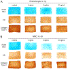
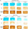
Tissue engineering is a promising approach for the repair of articular cartilage defects, with engineered constructs emerging that match native tissue properties. However, the inflammatory environment of the damaged joint might compromise outcomes, and this may be impacted by the choice of cell source in terms of their ability to operate anabolically in an inflamed environment. Here, we compared the response of engineered cartilage derived from native chondrocytes and mesenchymal stem cells (MSCs) to challenge by TNFα and IL-1β in order to determine if either cell type possessed an inherent advantage. Compositional (extracellular matrix) and functional (mechanical) characteristics, as well as the release of catabolic mediators (matrix metalloproteinases [MMPs], nitric oxide [NO]) were assessed to determine cell- and tissue-level changes following exposure to IL-1β or TNF-α. Results demonstrated that MSC-derived constructs were more sensitive to inflammatory mediators than chondrocyte-derived constructs, exhibiting a greater loss of proteoglycans and functional properties at lower cytokine concentrations. While MSCs and chondrocytes both have the capacity to form functional engineered cartilage in vitro, this study suggests that the presence of an inflammatory environment is more likely to impair the in vivo success of MSC-derived cartilage repair. © 2018 Orthopaedic Research Society. Published by Wiley Periodicals, Inc. J Orthop Res 36:2901-2910, 2018.
Keywords: cytokines; inflammation cartilage; matrix degradation cartilage; progenitors and stem cells cartilage; tissue engineering and repair cartilage.
© 2018 Orthopaedic Research Society. Published by Wiley Periodicals, Inc.
Figures


Similar articles
-
α-Melanocyte-stimulating-hormone (α-MSH) modulates human chondrocyte activation induced by proinflammatory cytokines.BMC Musculoskelet Disord. 2015 Jun 21;16:154. doi: 10.1186/s12891-015-0615-1. BMC Musculoskelet Disord. 2015. PMID: 26093672 Free PMC article.
-
Physioxia Has a Beneficial Effect on Cartilage Matrix Production in Interleukin-1 Beta-Inhibited Mesenchymal Stem Cell Chondrogenesis.Cells. 2019 Aug 20;8(8):936. doi: 10.3390/cells8080936. Cells. 2019. PMID: 31434236 Free PMC article.
-
Different response of human chondrocytes from healthy looking areas and damaged regions to IL1β stimulation under different oxygen tension.J Orthop Res. 2019 Jan;37(1):84-93. doi: 10.1002/jor.24142. Epub 2018 Oct 1. J Orthop Res. 2019. PMID: 30255592
-
Mechanical stimulation of mesenchymal stem cells: Implications for cartilage tissue engineering.J Orthop Res. 2018 Jan;36(1):52-63. doi: 10.1002/jor.23670. Epub 2017 Aug 11. J Orthop Res. 2018. PMID: 28763118 Review.
-
The role of cytokines in osteoarthritis pathophysiology.Biorheology. 2002;39(1-2):237-46. Biorheology. 2002. PMID: 12082286 Review.
Cited by
-
Mechano-activated biomolecule release in regenerating load-bearing tissue microenvironments.Biomaterials. 2021 Jan;265:120255. doi: 10.1016/j.biomaterials.2020.120255. Epub 2020 Oct 10. Biomaterials. 2021. PMID: 33099065 Free PMC article.
-
Orthopedic Surgeons' Perspectives on the Decision-Making Process for the Use of Bioprinter Cartilage Grafts: Web-Based Survey.Interact J Med Res. 2019 May 15;8(2):e14028. doi: 10.2196/14028. Interact J Med Res. 2019. PMID: 31094326 Free PMC article.
-
Total Meniscus Reconstruction Using a Polymeric Hybrid-Scaffold: Combined with 3D-Printed Biomimetic Framework and Micro-Particle.Polymers (Basel). 2021 Jun 8;13(12):1910. doi: 10.3390/polym13121910. Polymers (Basel). 2021. PMID: 34201327 Free PMC article.
-
Effects of interleukin 1β on long noncoding RNA and mRNA expression profiles of human synovial fluid derived mesenchymal stem cells.Sci Rep. 2022 May 19;12(1):8432. doi: 10.1038/s41598-022-12190-9. Sci Rep. 2022. PMID: 35589865 Free PMC article.
-
TNFα-Related Chondrocyte Inflammation Models: A Systematic Review.Int J Mol Sci. 2024 Oct 8;25(19):10805. doi: 10.3390/ijms251910805. Int J Mol Sci. 2024. PMID: 39409134 Free PMC article.
References
-
- Goldring MB, Osteoarthritis and cartilage: the role of cytokines. Curr Rheumatol Rep, 2000. 2(6): p. 459–65. - PubMed
-
- Little CB and Hunter DJ, Post-traumatic osteoarthritis: from mouse models to clinical trials. Nature Reviews Rheumatology, 2013. 9(8): p. 485–497. - PubMed
-
- Moutos FT, Freed LE, and Guilak F, A biomimetic three-dimensional woven composite scaffold for functional tissue engineering of cartilage. Nat Mater, 2007. - PubMed
Publication types
MeSH terms
Substances
Grants and funding
LinkOut - more resources
Full Text Sources
Other Literature Sources

