. 2018 May 29;14(5):e1007106.
doi: 10.1371/journal.ppat.1007106. eCollection 2018 May.

Polymorphic factor H-binding activity of CspA protects Lyme borreliae from the host complement in feeding ticks to facilitate tick-to-host transmission

Affiliations

Polymorphic factor H-binding activity of CspA protects Lyme borreliae from the host complement in feeding ticks to facilitate tick-to-host transmission

Thomas Hart et al. PLoS Pathog. .

Abstract

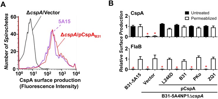
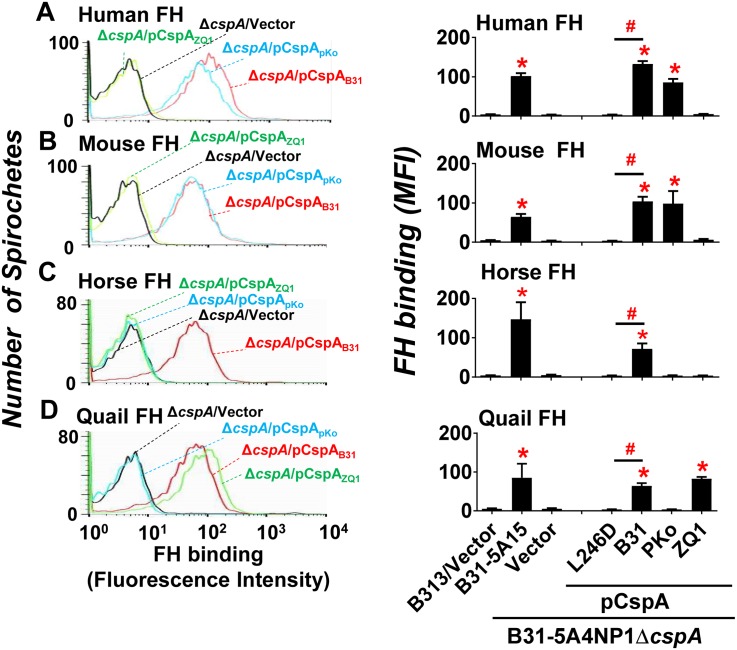
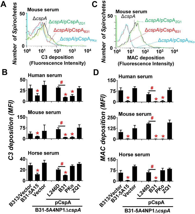
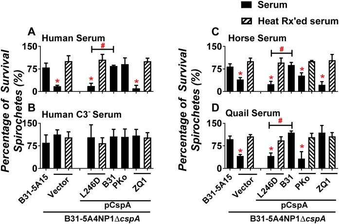
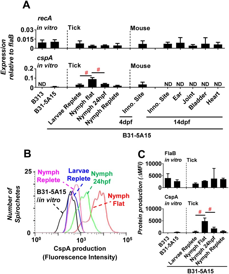
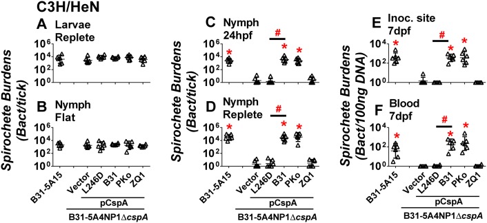
Borrelia burgdorferi sensu lato (Bbsl), the causative agent of Lyme disease, establishes an initial infection in the host's skin following a tick bite, and then disseminates to distant organs, leading to multisystem manifestations. Tick-to-vertebrate host transmission requires that Bbsl survives during blood feeding. Complement is an important innate host defense in blood and interstitial fluid. Bbsl produces a polymorphic surface protein, CspA, that binds to a complement regulator, Factor H (FH) to block complement activation in vitro. However, the role that CspA plays in the Bbsl enzootic cycle remains unclear. In this study, we demonstrated that different CspA variants promote spirochete binding to FH to inactivate complement and promote serum resistance in a host-specific manner. Utilizing a tick-to-mouse transmission model, we observed that a cspA-knockout B. burgdorferi is eliminated from nymphal ticks in the first 24 hours of feeding and is unable to be transmitted to naïve mice. Conversely, ectopically producing CspA derived from B. burgdorferi or B. afzelii, but not B. garinii in a cspA-knockout strain restored spirochete survival in fed nymphs and tick-to-mouse transmission. Furthermore, a CspA point mutant, CspA-L246D that was defective in FH-binding, failed to survive in fed nymphs and at the inoculation site or bloodstream in mice. We also allowed those spirochete-infected nymphs to feed on C3-/- mice that lacked functional complement. The cspA-knockout B. burgdorferi or this mutant strain complemented with cspA variants or cspA-L246D was found at similar levels as wild type B. burgdorferi in the fed nymphs and mouse tissues. These novel findings suggest that the FH-binding activity of CspA protects spirochetes from complement-mediated killing in fed nymphal ticks, which ultimately allows Bbsl transmission to mammalian hosts.


Conflict of interest statement

The authors have declared no competing interests exist.

Figures

Fig 1
Fig 1. Localization of CspA on the surface of B. burgdorferi.
Flow cytometry analysis of CspA localized on the surface of B. burgdorferi strains B31-5A15 (“B31-5A15”), B31-5A4NP1ΔcspA harboring the vector pBSV2G (“ΔcspA/Vector”), or this cspA mutant strain producing CspAB31 (“ΔcspA/pCspAB31”), CspAPKo (“ΔcspA/pCspAPKo”), CspAZQ1 (“ΔcspA/pCspAZQ1”), or CspAB31L246D (“ΔcspA/pCspAB31L246D”). (A) Representative histograms of flow cytometry analysis showing the levels of CspA surface production to indicated B. burgdorferi strains. (B) The production of CspA and FlaB (negative control) on the surface of indicated B. burgdorferi strains was detected by flow cytometry (see Materials and methods). Values are shown relative to the production levels of CspA or FlaB on the surface of permeabilized B. burgdorferi strain B31-5A15. Each bar represents the mean of four independent determinations ± the standard deviation. (*): indicates that relative surface production of the indicated proteins was significantly lower (P < 0.05 by one-way ANOVA with post hoc Bonferroni correction) than that of CspA or FlaB by B. burgdorferi strain B31-5A15.
Fig 2
Fig 2. CspA variants differ in their ability in promoting spirochetes binding to FH from different vertebrate animals.
B. burgdorferi strain B31-5A15 (“B31-5A15”), B31-5A4NP1ΔcspA harboring the vector pBSV2G (“ΔcspA/Vector”), or this cspA mutant strain producing CspAB31 (“ΔcspA/pCspAB31”), CspAPKo (“ΔcspA/pCspAPKo”), CspAZQ1 (“ΔcspA/pCspAZQ1”), or CspAB31L246D (“ΔcspA/pCspAB31L246D”), or B313 carrying the vector pBSV2G (“B313/Vector”, negative control) was incubated with FH from human, mouse, horse, or quail. The bacteria were stained with a sheep anti-FH polyclonal IgG (for the spirochetes incubated with human, mouse, or horse FH) or a mouse anti-FH monoclonal antibody VIG8 (for the spirochetes incubated with quail FH) followed by an Alexa 647-conjugated donkey anti-sheep IgG or goat anti-mouse IgG prior to being applied to flow cytometry analysis. (Left panel) Representative histograms of flow cytometry analysis showing the levels of FH from (A) human, (B) mouse, (C) horse, or (D) quail binding to indicated B. burgdorferi strains. (Right panel) The levels of B. burgdorferi binding to FH from (A) human, (B) mouse, (C) horse, or (D) quail were measured by flow cytometry and presented as mean fluorescence index (MFI). Each bar represents the mean of three independent determinations ± SEM. Significant differences (P < 0.05 by one-way ANOVA with post hoc Bonferroni correction) in the levels of FH binding relative to the B313/Vector (“*”) or between two strains relative to each other (“#”).
Fig 3
Fig 3. CspA variants differ in their ability to reduce the deposition of C3b or MAC from different vertebrate animals on the spirochete surface.
B. burgdorferi strain B31-5A15 (“B31-5A15”), B31-5A4NP1ΔcspA harboring the vector pBSV2G (“ΔcspA/Vector”), or this cspA mutant strain producing CspAB31 (“ΔcspA/pCspAB31”), CspAPKo (“ΔcspA/pCspAPKo”), CspAZQ1 (“ΔcspA/pCspAZQ1”), or CspAB31L246D (“ΔcspA/pCspAB31L246D”), or B313 carrying the vector pBSV2G (“B313/Vector”, negative control) was incubated with serum from human, mouse, or horse with a final concentration of 20%. The bacteria were stained with a guinea pig anti-C3 polyclonal IgG, a mouse anti-C5b-9 monoclonal antibody aE11 (for spirochetes incubated with human or horse serum), or a rabbit anti-C5b-9 polyclonal IgG (for spirochetes incubated with mouse serum) followed by an Alexa 647-conjugated goat anti-guinea pig IgG or goat anti-mouse IgG, or goat anti-rabbit IgG prior to being applied to flow cytometry analysis. Representative histograms of flow cytometry analysis showing the deposition levels of mouse (A) C3b or (C) MAC on the surface of indicated B. burgdorferi strains. The deposition levels of (B) C3b or (D) MAC of indicated animals on the surface of B. burgdorferi were measured by flow cytometry and presented as mean fluorescence index (MFI). Each bar represents the mean of three independent determinations ± SEM. Significant differences (P < 0.05 by one-way ANOVA with post hoc Bonferroni correction) in the deposition levels of C3b or MAC relative to the B313/Vector (“*”) or between two strains relative to each other (“#”).
Fig 4
Fig 4. CspA variants mediate distinct levels of spirochete survival in serum from different vertebrate animals.
B. burgdorferi strain B31-5A15 (“B31-5A15”), B31-5A4NP1ΔcspA harboring the vector pBSV2G (“ΔcspA/Vector”), or this cspA mutant strain producing CspAB31 (“ΔcspA/pCspAB31”), CspAPKo (“ΔcspA/pCspAPKo”), CspAZQ1 (“ΔcspA/pCspAZQ1”), or CspAB31L246D (“ΔcspA/pCspAB31L246D”) was incubated for four hours with untreated (filled bars) or heat-inactivated (HI, hatched bars) serum with a final concentration of 40%. These sera include (A) human serum or (B) C3-depleted human serum (Human C3- serum) or the serum from (C) horse or (D) quail. The number of motile spirochete was assessed microscopically. The percentage of survival for those B. burgdorferi strains was calculated using the number of mobile spirochetes at four hours post incubation normalized to that prior to the incubation with serum. Each bar represents the mean of three independent determinations ± SEM. Significant differences (P < 0.05 by one-way ANOVA with post hoc Bonferroni correction) in the percentage survival of spirochetes relative to the ΔcspA/Vector (“*”) or between two strains relative to each other (“#”).
Fig 5
Fig 5. B. burgdorferi expresses distinct levels of CspA at different stages of enzootic cycle.
C3H/HeN mice were infected with 105 B. burgdorferi strain B31-5A15. At 14 days post infection, the uninfected I. scapularis larval ticks were allowed to feed on these mice to repletion. After the replete larvae molted into nymphs, these B. burgdorferi-infected nymphs were allowed to feed on naïve C3H/HeN mice for different period of time or to repletion. The mice were euthanized at 7 or 14 days post feeding (“7dpf” or “14dpf”) to collect the inoculation site of skin (for mice at 7 and 14 days post feeding), ears, tibiotarsus joints, bladder, and heart (for mice at 14 days post feeding). RNA was extracted from replete larvae (“larvae replete”), flat nymphs (“nymph flat”), fed nymphs at 24 hours post feeding (“24hpf”) or replete nymphs (“nymph replete”) as well as in vitro cultured B. burgdorferi strain B313 (“B313”, negative control) or B31-5A15 (“B31-5A15”, positive control) in BSKII medium. (A) RNA was also extracted from mouse tissues including tick biting site (“inoc. site”), ears, tibiotarsus joints, bladder, and heart at 7 and/or 14 days post feeding. The extracted RNA was then used to determine the expression levels of cspA and constitutive expressed genes flaB and recA using qRT-PCR (see Materials and methods). The expression levels of (Top panel; negative control) recA and (Bottom panel) cspA are presented by normalizing the expression levels of flaB (negative control) (see Materials and methods). Each bar represents the mean of five independent determinations ± SEM (“#”). Significant differences (P < 0.05 by one-way ANOVA with post hoc Bonferroni correction) in the normalized expression levels of cspA between two conditions relative to each other. (B and C) The bacteria isolated from ticks or in vitro cultured B. burgdorferi strains were applied to flow cytometry. (B) Representative histograms of flow cytometry analysis showing the production levels of CspA in B. burgdorferi in the indicated environment. (C) The production of FlaB (Top panel; negative control) and CspA (Bottom panel) are presented as “ΔMFI”, the mean fluorescence index obtained from each of these strains subtracting that obtained from the strains stained only by the secondary antibody. Each bar represents the mean of three independent determinations ± SEM. Significant differences (P < 0.05 by one-way ANOVA with post hoc Bonferroni correction) in the production of CspA relative to the B313 cultured in vitro (“*”) or between two conditions relative to each other (“#”).
Fig 6
Fig 6. FH-binding ability of CspA promotes spirochete survival in nymphal ticks upon feeding.
C3H/HeN mice were infected with 105 B. burgdorferi strain B31-5A15 (“B31-5A15”), B31-5A4NP1ΔcspA harboring the vector pBSV2G (“ΔcspA/Vector”), or this cspA mutant strain producing CspAB31 (“ΔcspA/pCspAB31”), CspAPKo (“ΔcspA/pCspAPKo”), CspAZQ1 (“ΔcspA/pCspAZQ1”), or CspAB31L246D (“ΔcspA/pCspAB31L246D”). At 14 days post infection, the uninfected I. scapularis larval ticks were allowed to feed on each of these mice until they are replete. After the replete larvae molt into nymphs, those B. burgdorferi-infected nymphs were allowed to feed on naïve C3H/HeN mice to repletion. The bacterial loads in the (A) replete larvae (“larvae replete”), (B) flat nymphs (“nymph flat”), fed nymphs at (C) 24 hours post feeding (“nymph 24hpf”) or (D) replete nymphs (“nymph replete”), or (E) the site where nymphal ticks fed (“inoc. site”) or (F) blood at 7 days post nymph feeding (“blood 7dpf”) were determined by qPCR. The bacterial loads in mouse tissues or blood were normalized to 100 ng total DNA. Shown are the geometric mean of bacterial loads ± 95% confidence interval of six ticks or mice per group. Significant differences (P < 0.05 by one-way ANOVA with post hoc Bonferroni correction) in the spirochete burdens relative to the ΔcspA/Vector (“*”) or between two strains relative to each other (“#”).
Fig 7
Fig 7. CspA-mediated FH-binding activity facilitates spirochete evading complement present in fed nymphs resulting in nymph-to-mouse transmission.
C3H/HeN mice were infected with 105 B. burgdorferi strain B31-5A15 (“B31-5A15”), B31-5A4NP1ΔcspA harboring the vector pBSV2G (“ΔcspA/Vector”), or this cspA mutant strain producing CspAB31 (“ΔcspA/pCspAB31”), CspAPKo (“ΔcspA/pCspAPKo”), CspAZQ1 (“ΔcspA/pCspAZQ1”), or CspAB31L246D (“ΔcspA/pCspAB31L246D”). At 14 days post infection, the uninfected I. scapularis larval ticks were allowed to feed on each of these mice until they are replete. After the replete larvae molt into nymphs, those B. burgdorferi-infected nymphs were allowed to feed on naïve BALB/c or BALB/c C3-/- mice to repletion. The bacterial loads in the nymphs fed on BALB/c (Left panel) or BALB/c C3-/- (Right panel) for (A) 24 hours post feeding (“24hpf”) or (B) replete nymphs (“replete”), or at (C) the site where nymphs fed (“inoc. site”) on mice or (D) mouse blood at 7 days post nymph feeding (“7dpf”) were determined by qPCR. The bacterial loads in mouse tissues or blood were normalized to 100 ng total DNA. Shown are the geometric mean of bacterial loads ± 95% confidence interval of six ticks or mice per group. Significant differences (P < 0.05 by one-way ANOVA with post hoc Bonferroni correction) in the spirochete burdens relative to the ΔcspA/Vector (“*”) or between two strains relative to each other (“#”).
Fig 8
Fig 8. A proposed model showing CspA-mediated FH-binding activity promotes spirochete evasion of complement in feeding nymphs to facilitate tick-to-host transmission.
CspA of spirochetes binds to FH present in the host’s blood and interstitial fluid in feeding nymphs to facilitate spirochete escape from the complement-mediated killing. Such CspA-mediated immune evasion ultimately facilitates spirochete transmission from nymphs to hosts.

References

    1. Steere AC, Strle F, Wormser GP, Hu LT, Branda JA, Hovius JW, et al. Lyme borreliosis. Nat Rev Dis Primers. 2016;2:16090 doi: 10.1038/nrdp.2016.90 . - DOI - PMC - PubMed
    1. Radolf JD, Caimano MJ, Stevenson B, Hu LT. Of ticks, mice and men: understanding the dual-host lifestyle of Lyme disease spirochaetes. Nat Rev Microbiol. 2012;10(2):87–99. doi: 10.1038/nrmicro2714 . - DOI - PMC - PubMed
    1. Rosa PA, Tilly K, Stewart PE. The burgeoning molecular genetics of the Lyme disease spirochaete. Nat Rev Microbiol. 2005;3(2):129–43. doi: 10.1038/nrmicro1086 . - DOI - PubMed
    1. Steere AC, Coburn J, Glickstein L. The emergence of Lyme disease. J Clin Invest. 2004;113(8):1093–101. doi: 10.1172/JCI21681 . - DOI - PMC - PubMed
    1. Brisson D, Drecktrah D, Eggers CH, Samuels DS. Genetics of Borrelia burgdorferi. Annu Rev Genet. 2012;46:515–36. doi: 10.1146/annurev-genet-011112-112140 . - DOI - PMC - PubMed

Publication types

MeSH terms