Opal-like Multicolor Appearance of Self-Assembled Photonic Array
- PMID: 29842782
- PMCID: PMC6358003
- DOI: 10.1021/acsami.8b04912
Opal-like Multicolor Appearance of Self-Assembled Photonic Array
Abstract
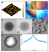
Molecular self-assembly of short peptide building blocks leads to the formation of various material architectures that may possess unique physical properties. Recent studies had confirmed the key role of biaromaticity in peptide self-assembly, with the diphenylalanine (FF) structural family as an archetypal model. Another significant direction in the molecular engineering of peptide building blocks is the use of fluorenylmethoxycarbonyl (Fmoc) modification, which promotes the assembly process and may result in nanostructures with distinctive features and macroscopic hydrogel with supramolecular features and nanoscale order. Here, we explored the self-assembly of the protected, noncoded fluorenylmethoxycarbonyl-β,β-diphenyl-Ala-OH (Fmoc-Dip) amino acid. This process results in the formation of elongated needle-like crystals with notable aromatic continuity. By altering the assembly conditions, arrays of spherical particles were formed that exhibit strong light scattering. These arrays display vivid coloration, strongly resembling the appearance of opal gemstones. However, unlike the Rayleigh scattering effect produced by the arrangement of opal, the described optical phenomenon is attributed to Mie scattering. Moreover, by controlling the solution evaporation rate, i.e., the assembly kinetics, we were able to manipulate the resulting coloration. This work demonstrates a bottom-up approach, utilizing self-assembly of a protected amino acid minimal building block, to create arrays of organic, light-scattering colorful surfaces.
Keywords: Fmoc modification; Mie scattering; amino acid self-assembly; biaromatic amino acid; colored surfaces; microspheres; opal-like; self-assembly.
Conflict of interest statement
The authors declare no competing financial interest.
Figures


References
-
- Zhang S. Fabrication of Novel Biomaterials through Molecular Self-Assembly. Nat Biotechnol. 2003;21:1171–1178. - PubMed
-
- Esin A, Baturin I, Nikitin T, Vasilev S, Salehli F, Shur VY, Kholkin AL. Pyroelectric Effect and Polarization Instability in Self-Assembled Diphenylalanine Microtubes. Appl Phys Lett. 2016;109 142902.
-
- Li Q, Jia Y, Dai L, Yang Y, Li J. Controlled Rod Nanostructured Assembly of Diphenylalanine and Their Optical Waveguide Properties. ACS Nano. 2015;9:2689–2695. - PubMed
-
- Yan X, Zhu P, Li J. Self-Assembly and Application of Diphenylalanine-Based Nanostructures. Chem Soc Rev. 2010;39:1877–1890. - PubMed
Grants and funding
LinkOut - more resources
Full Text Sources
Other Literature Sources

