Review
. 2018;64(s1):S567-S610.
doi: 10.3233/JAD-179941.

The Amyloid-β Oligomer Hypothesis: Beginning of the Third Decade

Affiliations
Review

The Amyloid-β Oligomer Hypothesis: Beginning of the Third Decade

Erika N Cline et al. J Alzheimers Dis. 2018.

Abstract

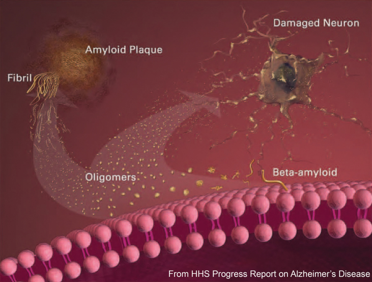
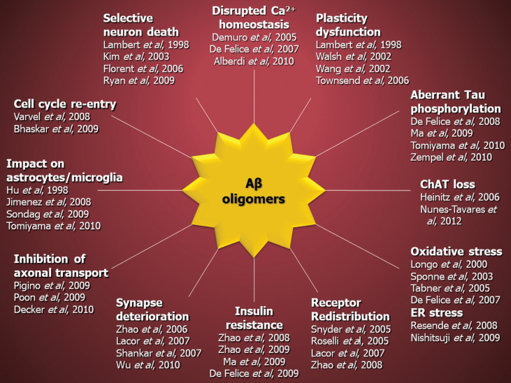
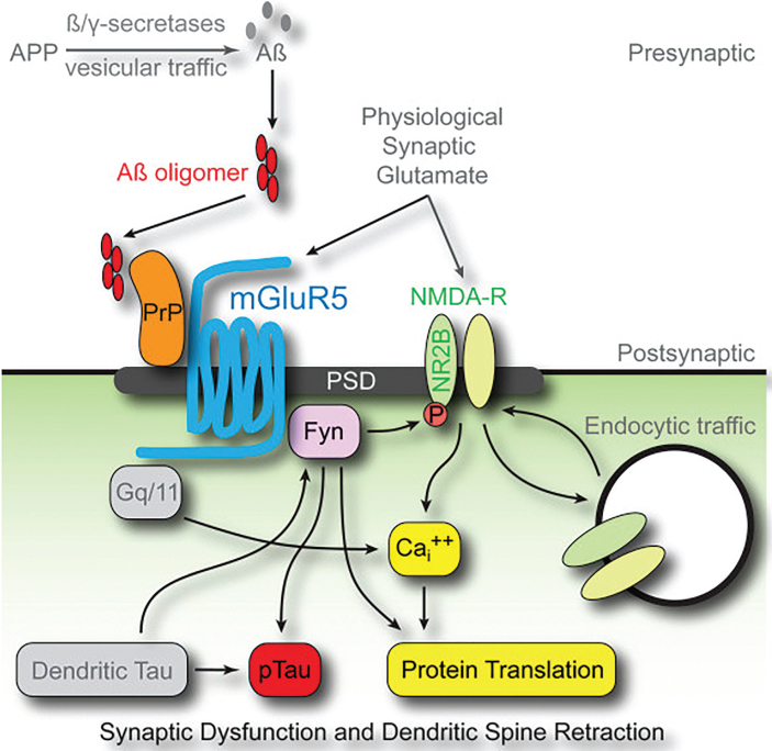
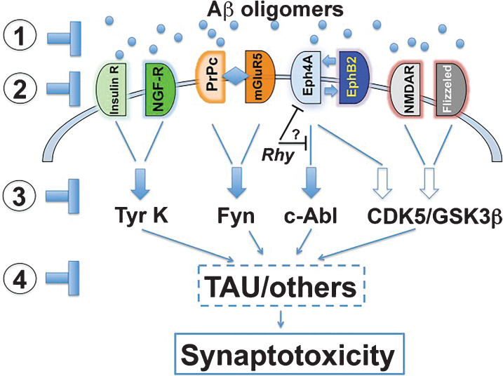
The amyloid-β oligomer (AβO) hypothesis was introduced in 1998. It proposed that the brain damage leading to Alzheimer's disease (AD) was instigated by soluble, ligand-like AβOs. This hypothesis was based on the discovery that fibril-free synthetic preparations of AβOs were potent CNS neurotoxins that rapidly inhibited long-term potentiation and, with time, caused selective nerve cell death (Lambert et al., 1998). The mechanism was attributed to disrupted signaling involving the tyrosine-protein kinase Fyn, mediated by an unknown toxin receptor. Over 4,000 articles concerning AβOs have been published since then, including more than 400 reviews. AβOs have been shown to accumulate in an AD-dependent manner in human and animal model brain tissue and, experimentally, to impair learning and memory and instigate major facets of AD neuropathology, including tau pathology, synapse deterioration and loss, inflammation, and oxidative damage. As reviewed by Hayden and Teplow in 2013, the AβO hypothesis "has all but supplanted the amyloid cascade." Despite the emerging understanding of the role played by AβOs in AD pathogenesis, AβOs have not yet received the clinical attention given to amyloid plaques, which have been at the core of major attempts at therapeutics and diagnostics but are no longer regarded as the most pathogenic form of Aβ. However, if the momentum of AβO research continues, particularly efforts to elucidate key aspects of structure, a clear path to a successful disease modifying therapy can be envisioned. Ensuring that lessons learned from recent, late-stage clinical failures are applied appropriately throughout therapeutic development will further enable the likelihood of a successful therapy in the near-term.

Keywords: Alzheimer’s disease; amyloid-β peptide; diagnostics; etiology; model systems; oligomers; prions; receptors; structure-function; tau; therapeutics.


Figures

Fig.1
Fig.1
AβOs, not Aβ monomers or fibrils, instigate the neuron damage leading to dementia. Following cleavage from the membrane, Aβ peptides aggregate to form AβOs, some of which further aggregate to fibrils and some of which instigate the neuron damage leading to dementia. Reprinted with Jannis Productions permissions from the “Progress Report on Alzheimer’s Disease 2004-2005” (ed. AB Rodgers), NIH Publication Number: 05-5724. Digital images produced by Stacy Jannis and Rebekah Fredenburg of Jannis Productions [455].
Fig.2
Fig.2
AβOs instigate multiple facets of AD-neuropathology. Observed in various culture and animal models. Reprinted by permission from Springer Nature: Acta Neuropathol, 129(2): 183-206, “Amyloid beta oligomers in Alzheimer’s disease pathogenesis, treatment, and diagnosis” by Viola KL and Klein WL. Copyright 2015 Springer Nature [200].
Fig.3
Fig.3
Single injection (30μg) of an AβO-specific antibody ameliorates cognitive deficits in AD mice for at least 40 days. 5xFAD Tg mice and their wild-type (WT) littermates (6 months of age) were evaluated by Object Recognition Tasks before and after (40 days) a single injection (30μg) of a humanized AβO-specific antibody (anti-AβO) or non-specific human IgG (hIgG). First, locomotor activity was assessed while mice were allowed to habituate to the testing field (Habituation). Assessments were the number of times the mice crossed grids in the field (Crossings, light gray) and the number of times mice put their hind paws on the walls of the field (Rearings, purple), with no differences between WT and 5xFAD mice. Next, the test objects (F1 and F2) were introduced to the mice in the Training session. All mice showed normal exploratory behavior, defined by 50% exploration of each object, as both objects are equal and new to the mice. The ability of mice to remember object placement was then tested 24 hours after the Training session in a hippocampal (HP)-dependent task. Another 24 hours later, the ability of mice to remember the object was tested in a cortical (CT)-dependent task. Only the WT mice were able to recognize the familiar object (F1) from the Training session, as evidenced by >50% exploration of the displaced (D, pink) or new (N, light blue) object. The 5xFAD mice failed to recognize F1 in both tasks. When re-evaluated 40 days post-antibody injection in a HP-dependent task, only the 5xFAD mice that received the AβO antibody recovered their ability to recognize object F1. These data support the hypothesis that AβOs induce memory dysfunction in AD (Bicca and Klein, unpublished).
Fig.4
Fig.4
AβOs can be divided into two classes based on their temporal, spatial, and structural relationships to amyloid plaques as well as their ability to cause memory dysfunction. Type 1 AβOs (aka “peak 1” or HMW) are thought to be associated with memory impairment, while type 2 AβOs (aka “peak 2” or LMW) are not. Only type 2 AβOs are associated with amyloid plaques. Reprinted from “Quaternary Structure Defines a Large Class of Amyloid-beta Oligomers Neutralized by Sequestration” by Liu P, Reed MN, Kotilinek LA, Grant MK, Forster CL, Qiang W, Shapiro SL, Reichl JH, Chiang AC, Jankowsky JL, Wilmot CM, Cleary JP, Zahs KR, and Ashe KH. This was published in Cell Rep, 2015, 11(11): 1760-1771, under the terms of the Creative Commons Attribution Non-Commercial No Derivatives License (CC BY NC ND) https://creativecommons.org/licenses/by-nc-nd/4.0/ [119].
Fig.5
Fig.5
Only high-molecular weight AβOs are capable of binding cultured hippocampal neurons. Synthetic AβOs were divided into high and low molecular weight populations using 50 kDa molecular weight cutoff ultrafiltration (A-B) or size exclusion chromatography (D-F) and incubated with cultured hippocampal neurons. Only high-molecular weight AβOs bind neurons (A, E); no binding of low-molecular weight AβOs was evident (B, F). Scale bar = 40μm. Reprinted from “Synaptic targeting by Alzheimer’s-related amyloid beta oligomers” by Lacor PN, Buniel MC, Chang L, Fernandez SJ, Gong Y, Viola KL, Lambert MP, Velasco PT, Bigio EH, Finch CE, Krafft GA, and Klein WL. This was published in J Neurosci, 2004, 24(45): 10191-10200, copyright 2004; permission conveyed through Copyright Clearance Center, Inc. [16].
Fig.6
Fig.6
PrPc mediates AβO toxicity through mGluR5, Fyn kinase, and NMDARs. Downstream consequences of the pathway include calcium dyshomeostasis, tau hyperphosphorylation, and synaptic dysfunction and loss. Reprinted from “Fyn kinase inhibition as a novel therapy for Alzheimer’s disease” by Nygaard HB, van Dyck CH, and Strittmater SM. This was published in Alzheimers Res Ther, 2014, 6(1): 8, under the terms of the Creative Commons Attribution License (CC BY) [227].
Fig.7
Fig.7
AβOs induce membrane re-distribution of NKAα3 subunit resulting its downregulation and excessive Ca++ buildup. A hypothesized early event in AβO-induced neuronal damage is binding to NKAα3 on neuronal membranes, causing restructuring of the NKAα3 docking station into toxic clusters of membrane proteins. Ultimately, this results in downregulation of NKAα3 on the neuronal surface and buildup of toxic Ca++. Adapted and reprinted from “Alzheimer’s Toxic Amyloid Beta Oligomers: Unwelcome Visitors to the Na/K ATPase alpha3 Docking Station” by DiChiara T, DiNunno N, Clark J, Bu RL, Cline EN, Rollins MG, Gong Y, Brody DL, Sligar SG, Velasco PT, Viola KL, and Klein WL. This was published in Yale J Biol Med, 2017, 90(1): 45-61, under the terms of the Creative Commons Attribution Non-Commercial No Derivatives License (CC BY NC ND) https://creativecommons.org/licenses/by-nc-nd/4.0/ [199].
Fig.8
Fig.8
A cumulative hypothesis for the development of sporadic AD. From De Felice, sporadic AD is hypothesized to be the result of the cumulative impact over a life-time of injuries to the brain and peripheral organs that results in increased AβO levels. Reprinted from “Alzheimer’s disease and insulin resistance: translating basic science into clinical applications” by De Felice FG. This was published in JClin Invest, 2013, 123(2): 531-539, under Free access [322].
Fig.9
Fig.9
Putative therapeutic targets of the AβO pathogenic cascade. Including: 1) AβOs themselves; 2) AβO receptors; 3) signaling pathways; or 4) downstream effectors such as tau. Reprinted with permission of PNAS from “Toward a unified therapeutics approach targeting putative amyloid-beta oligomer receptors” by Overk CR and Masliah E. This was published in Proc Natl Acad Sci U S A, 2014, 111(38): 13680-13681 [392].
Fig.10
Fig.10
Mechanisms of Aβ-targeting phase III drugs in AD clinical trials. Drugs inhibiting AβO formation (A) or downstream consequences of toxic AβOs (B). Reprinted from “Alzheimer’s disease drug development pipeline: 2017” by Cummings J, Garam L, Mortsdorf T, Ritter A, and Zhong K. This was published in Alzheimers Dementia (N Y), 2017, 3(3): 367-384 [393], under the terms of the Creative Commons Attribution Non-Commercial No Derivatives License (CC BY NC ND) https://creativecommons.org/licenses/by-nc-nd/4.0/) [393].

References

    1. Frackowiak J, Zoltowska A, Wisniewski HM (1994) Non-fibrillar beta-amyloid protein is associated with smooth muscle cells of vessel walls in Alzheimer disease, J Neuropathol Exp Neurol 53, 637–645. - PubMed
    1. Oda T, Pasinetti GM, Osterburg HH, Anderson C, Johnson SA, Finch CE (1994) Purification and characterization of brain clusterin, Biochem Biophys Res Commun 204, 1131–1136. - PubMed
    1. Oda T, Wals P, Osterburg HH, Johnson SA, Pasinetti GM, Morgan TE, Rozovsky I, Stine WB, Snyder SW, Holzman TF, Krafft GA, Finch CE (1995) Clusterin (Apoj) alters the aggregation of amyloid beta-peptide (Aβ(1-42)) and forms slowly sedimenting Aβ complexes that cause oxidative stress, Exp Neurol 136, 22–31. - PubMed
    1. Hayden EY, Teplow DB (2013) Amyloid beta-protein oligomers and Alzheimer’s disease, Alzheimers Res Ther 5, 60. - PMC - PubMed
    1. Selkoe DJ, Hardy J (2016) The amyloid hypothesis of Alzheimer’s disease at 25 years, EMBO Mol Med 8, 595–608. - PMC - PubMed

Publication types

Substances